. 2022 Mar 15:13:830234.
doi: 10.3389/fpsyt.2022.830234. eCollection 2022.

Autism Spectrum Disorder in Children Is Not Associated With Abnormal Autonomic Nervous System Function: Hypothesis and Theory

Affiliations

Autism Spectrum Disorder in Children Is Not Associated With Abnormal Autonomic Nervous System Function: Hypothesis and Theory

Ashley Barbier et al. Front Psychiatry. .

Abstract

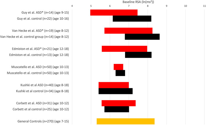
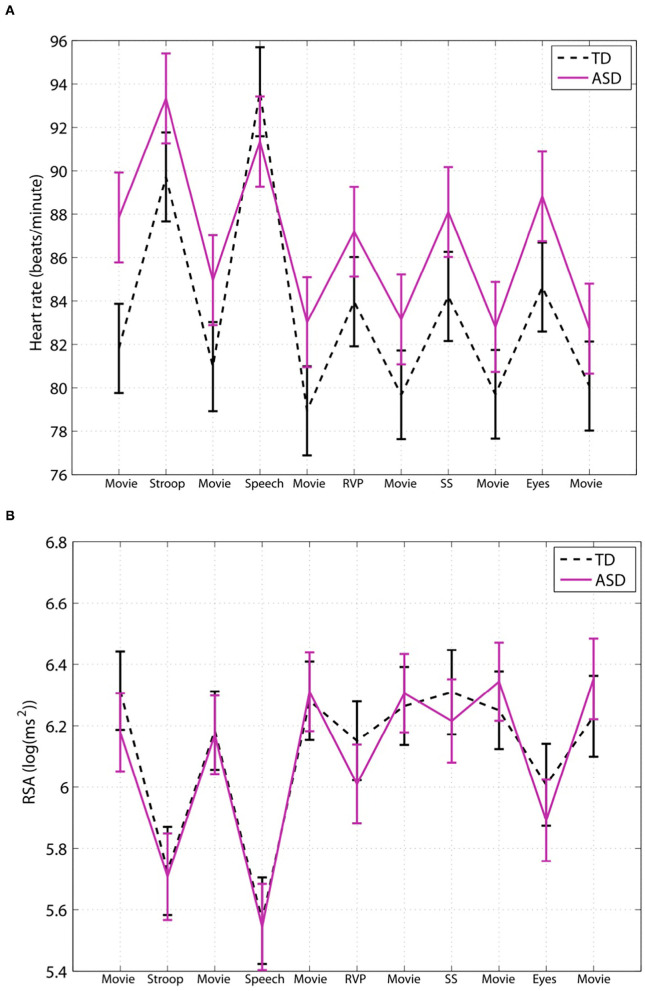
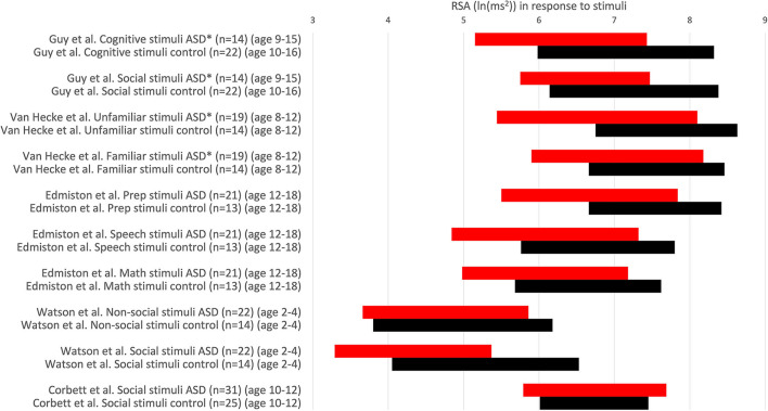
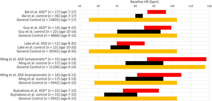
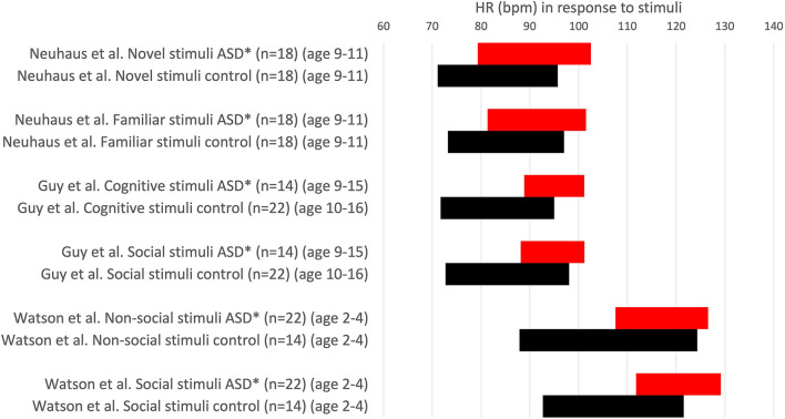
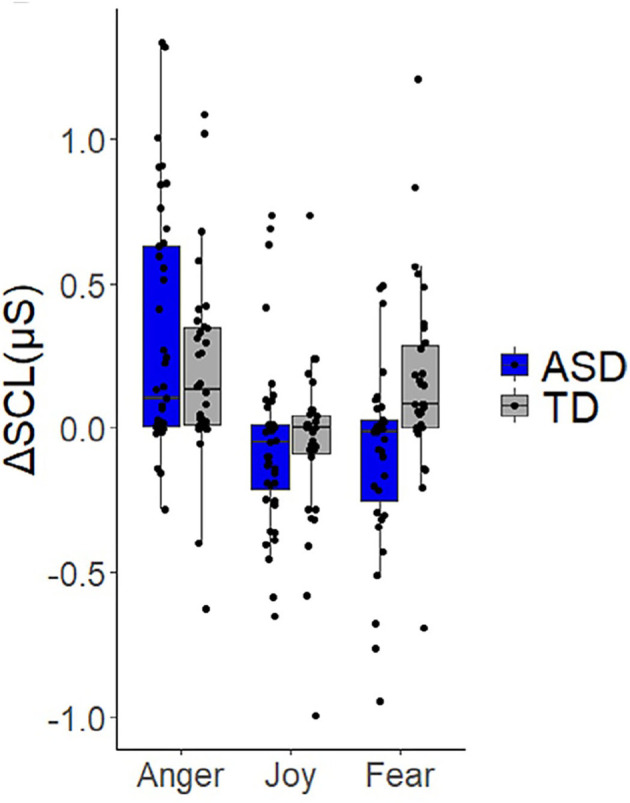
The quest to understand the pathophysiology of autism spectrum disorder (ASD) has led to extensive literature that purports to provide evidence for autonomic dysfunction based on heart rate and heart rate variability (HRV), in particular respiratory sinus arrhythmia (RSA), a measure of parasympathetic functioning. Many studies conclude that autism is associated with vagal withdrawal and sympathetic hyperactivation based on HRV and electrodermal analyses. We will argue that a critical analysis of the data leads to the hypothesis that autonomic nervous system dysfunction is not a dominant feature of autism. Most children with ASD have normal parasympathetic baseline values and normal autonomic responses to social stimuli. The existing HRV and electrodermal data cannot lead to the conclusion of an over-excitation of the sympathetic nervous system. A small subgroup of ASD children in experimental settings has relatively low RSA values and relatively high heart rates. The data suggest that this is likely associated with a relatively high level of anxiety during study conditions, associated with co-morbidities such as constipation, or due to the use of psychoactive medication. Many studies interpret their data to conform with a preferred hypothesis of autonomic dysfunction as a trait of autism, related to the polyvagal theory, but the HRV evidence is to the contrary. HRV analysis may identify children with ASD having autonomic dysfunction due to co-morbidities.

Keywords: autism (ASD); autonomic nervous system; electrodermal activity; gastrointestinal disorders; heart rate variability (HRV); parasympathetic and sympathetic reactivity; respiratory sinus arrhythmia (RSA).


Conflict of interest statement

The authors declare that the research was conducted in the absence of any commercial or financial relationships that could be construed as a potential conflict of interest.

Figures

Figure 1
Figure 1
RSA is expressed in comparative studies with an ASD group (red) and typically developing children (control, black). RSA ± 1SD; *P < 0.05 compared to in-study controls. Others: no significant difference. Note that comparisons cannot be absolute because of differences in measuring RSA. Muscatello et al. and Corbett et al. used ln(HF power) with the HF range of 0.12–0.40 Hz, Kuski et al. (27) used 0.24–1.04 Hz. Edmiston et al. (23) used 0.15–0.40 Hz. Guy et al. (22) report that the amplitude of RSA was calculated as the natural logarithm of the extracted variance for each successive 30-s epoch within 12–1 Hz (probably 0.12–1 Hz). Vaughan van Hecke et al. (24) chose the natural logarithm of the variance of the band-pass series from HF, 0.12–1 Hz. At the bottom, the orange control values are derived from a study that examined RSA over time in 270 children (28); we used a range based on their average values ± 1 SD from ages 7–15, obtaining a normal range of 5.3–8.4 ln(ms2).
Figure 2
Figure 2
Children with ASD have a normal baseline HRV parameter and a normal autonomic response to social stimuli. This figure is taken from a study by Kushki et al. (27); they studied autonomic regulation in children with autism while performing tasks that elicit anxiety, attention, response inhibition and social cognition. Expressed are heart rate (A) and RSA (B). The authors conclude that children with ASD show overall autonomic hyperarousal and selective atypical reactivity to social tasks. (A) The average baseline heart rate of the children with ASD was 88 bpm, which is not indicative of cardiac dysfunction nor an overactive sympathetic nervous system; it cannot be interpreted to show that children with ASD show hyperarousal. (B) There were no statistical differences in baseline RSA nor general group differences. Evaluating each task, there were no differences in RSA reactivity in the Stroop, public speaking or rapid visual information processing tasks. The reading the mind in the eyes task also did not show a significant difference except when the medication group was excluded. However, both the control group and the ASD children showed a normal strong decrease in RSA, and there is no evidence that this difference is clinically significant. (TD), n = 34, and ASD children, n = 40. “Movie”: considered resting baseline; “Stroop”: eliciting a stress reaction; “Speech”: public speaking considered anxiety eliciting; “RVP”: rapid visual information processing, eliciting sustained attention; “SS”: stop-signal task, testing response inhibition; “Eyes”: reading the mind in the eyes, testing social cognition. This figure labels RSA to be log(ms2) however the values indicate that RSA is likely ln(ms2).
Figure 3
Figure 3
RSA values in response to stimuli. RSA values are expressed comparing the response to stimuli in children with ASD (red) and a control group (black). RSA ± 1SD. *P < 0.05. Others: no significant difference. RSA is expressed in ln(ms2).
Figure 4
Figure 4
Heart rate values (HR ± 1SD) at baseline. HR expressed in comparative studies with an ASD group (red) and a group with typically developing children (control, black) and normal HR values in a large of children taken from Ostchega et al. (orange) (45). *P < 0.05. Others: no significant difference.
Figure 5
Figure 5
Heart rate values (HR ± 1SD) in response to stimuli. HR is expressed in comparative studies with an ASD group (red) and typically developing children (control, black). *P < 0.05. Others: no significant difference.
Figure 6
Figure 6
Children do not show a significant difference in sympathetic response to stimuli. Expression of skin conductance as a measure of activity in the sympathetic nervous system. From Vernetti et al. (92). Note the wide range of values in both the control and ASD groups. Eliciting anger, frustration or joy evoked a similar increase in sympathetic activity in the ASD and control group. In response to a fear-inducing stimulus, the control group exhibited an increase while the ASD group exhibited a decrease in sympathetic activity. However, the scatter plots show that most children with ASD and controls have a response that centers around 0, and most values in the ASD group fall within the range of control values. Hence a correct conclusion is that some children with ASD have a decreased arousal response to fear stimuli. The authors did not find a statistically significant difference in baseline sympathetic values and no correlation between the degree of sympathetic responses and the severity of autism. They conclude that toddlers with ASD should not be labeled as “dysregulated” or “upregulated” with respect to autonomic functioning.

References

    1. Szatmari P. The causes of autism spectrum disorders. BMJ. (2003) 326:173–4. 10.1136/bmj.326.7382.173 - DOI - PMC - PubMed
    1. American Association of Psychiatrists. What is Autism Spectrum Disorder? American Association of Psychiatrists (2021). Available online at: https://www.psychiatry.org/patients-families/autism/what-is-autism-spect... (accessed February 20, 2022).
    1. Centers for Disease Control Prevention; Autism Spectrum Disorder. (2021). Available online at: https://www.cdc.gov/ncbddd/autism/facts.html (accessed Febraury 20, 2022).
    1. Lord C, Pickles A, McLennan J, Rutter M, Bregman J, Folstein S, et al. . Diagnosing autism: analyses of data from the Autism Diagnostic Interview. J Autism Dev Disord. (1997) 27:501–17. 10.1023/A:1025873925661 - DOI - PubMed
    1. Porges SW, Macellaio M, Stanfill SD, McCue K, Lewis GF, Harden ER, et al. . Respiratory sinus arrhythmia and auditory processing in autism: modifiable deficits of an integrated social engagement system. Int J Psychophysiol. (2013) 88:261–70. 10.1016/j.ijpsycho.2012.11.009 - DOI - PMC - PubMed

LinkOut - more resources