. 2022 Mar 14:13:817229.
doi: 10.3389/fneur.2022.817229. eCollection 2022.

Do Muscle Changes Contribute to the Neurological Disorder in Spastic Paresis?

Affiliations

Do Muscle Changes Contribute to the Neurological Disorder in Spastic Paresis?

Maud Pradines et al. Front Neurol. .

Abstract

Background: At the onset of stroke-induced hemiparesis, muscle tissue is normal and motoneurones are not overactive. Muscle contracture and motoneuronal overactivity then develop. Motor command impairments are classically attributed to the neurological lesion, but the role played by muscle changes has not been investigated.

Methods: Interaction between muscle and command disorders was explored using quantified clinical methodology-the Five Step Assessment. Six key muscles of each of the lower and upper limbs in adults with chronic poststroke hemiparesis were examined by a single investigator, measuring the angle of arrest with slow muscle stretch (XV1) and the maximal active range of motion against the resistance of the tested muscle (XA). The coefficient of shortening CSH = (XN-XV1)/XN (XN, normally expected amplitude) and of weakness CW = (XV1-XA)/XV1) were calculated to estimate the muscle and command disorders, respectively. Composite CSH (CCSH) and CW (CCW) were then derived for each limb by averaging the six corresponding coefficients. For the shortened muscles of each limb (mean CSH > 0.10), linear regressions explored the relationships between coefficients of shortening and weakness below and above their median coefficient of shortening.

Results: A total of 80 persons with chronic hemiparesis with complete lower limb assessments [27 women, mean age 47 (SD 17), time since lesion 8.8 (7.2) years], and 32 with upper limb assessments [18 women, age 32 (15), time since lesion 6.4 (9.3) years] were identified. The composite coefficient of shortening was greater in the lower than in the upper limb (0.12 ± 0.04 vs. 0.08 ± 0.04; p = 0.0002, while the composite coefficient of weakness was greater in the upper limb (0.28 ± 0.12 vs. 0.15 ± 0.06, lower limb; p < 0.0001). In the lower limb shortened muscles, the coefficient of weakness correlated with the composite coefficient of shortening above the 0.15 median CSH (R = 0.43, p = 0.004) but not below (R = 0.14, p = 0.40).

Conclusion: In chronic hemiparesis, muscle shortening affects the lower limb particularly, and, beyond a threshold of severity, may alter descending commands. The latter might occur through chronically increased intramuscular tension, and thereby increased muscle afferent firing and activity-dependent synaptic sensitization at the spinal level.

Keywords: chronic hemiparesis; clinical extensibility; muscle disorder; quantified assessment; spastic cocontraction; spastic myopathy; stretch-sensitive paresis; synaptic sensitization.


Conflict of interest statement

The authors declare that the research was conducted in the absence of any commercial or financial relationships that could be construed as a potential conflict of interest.

Figures

Figure 1
Figure 1
Flow diagram Flow diagram for the lower limb (A) and upper limb (B) data collection.
Figure 2
Figure 2
Clinical assessment of active movement XA. (A) Hamstrings: patient supine, lower limb lying straight on the table. The clinician asks for a hip flexion keeping the knee extended. Axis of rotation: lateral condyle. The angle XA is measured between the two lines lateral condyle-external malleolus and lateral condyle-acromion (through projection, parallel to the line greater trochanter-acromion). (B) Shoulder extensors: patient seated, upper limb alongside the body. The clinician asks for a shoulder flexion keeping the elbow extended. Axis of rotation: acromion. The angle XA is measured between the two lines acromion-olecranon and acromion-GT.
Figure 3
Figure 3
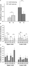
Degree of muscle shortening and of motor command impairment in chronic hemiparesis. (A) Coefficients of impairment of the Composite score of the upper and lower limbs. (B) Coefficient of Shortening of each of the investigated muscles. (C) Coefficient of Weakness of each of the investigated muscles. SE, shoulder extensor; SS, subscapularis; EF, elbow flexors; PQ, pronatus quadratus; WF, wrist flexors; FF, finger flexors; SO, soleus; GN, gastrocnemius; GM, gluteus maximus; HS, hamstrings; VA, vastus; RF, rectus femoris. **p < 0.01; ***p < 0.001.
Figure 4
Figure 4
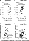
Relationship between passive (XV1) and active movements (XA) for the upper limb (A), for the lower limb (B). Composite XV1, mean XV1 of the six investigated muscles; Composite XA, mean XA of the six investigated muscles. Relationship between the mean Coefficient of Shortening and the mean Coefficient of Weakness (Composite Scores) in the upper limb (C), in the lower limb (D), respectively.
Figure 5
Figure 5
Relationship between the muscular (shortening) and the neurological (weakness) disorders for the shortened muscle groups of the lower limb (sample of the muscle groups with mean CSH > 0.10, i.e., soleus, gastrocnemius, gluteus maximus, and rectus femoris) (A–C) and for the soleus muscle (D–F). Left, the entire sample (A,D); middle, coefficients of shortening below the median (B,E); right, coefficients of shortening beyond the median (C,F).
Figure 6
Figure 6
Respective contribution of the muscular and the neurological disorders to functional impairments. The coefficient of impairment refers to the coefficient of shortening or the coefficient of weakness, as indicated in the legend. The composite coefficient is the mean of the individual coefficients for each of the six muscles of the lower limb (A); and of the upper limb (B).

References

    1. Dietz V, Quintern J, Berger W. Electrophysiological studies of gait in spasticity and rigidity. Evidence that altered mechanical properties of muscle contribute to hypertonia. Brain. (1981) 104:431–49. 10.1093/brain/104.3.431 - DOI - PubMed
    1. Dietz V, Sinkjaer T. Spastic movement disorder: impaired reflex function and altered muscle mechanics. Lancet Neurol. (2007) 6:725–33. 10.1016/S1474-4422(07)70193-X - DOI - PubMed
    1. O'Dwyer NJ, Ada L, Neilson PD. Spasticity and muscle contracture following stroke. Brain. (1996) 119:1737–49. 10.1093/brain/119.5.1737 - DOI - PubMed
    1. Gracies JM. Pathophysiology of spastic paresis. Part I. Paresis and soft tissue contracture. Muscle Nerve. (2005) 31:535–51. 10.1002/mus.20284 - DOI - PubMed
    1. Gracies JM. Pathophysiology of spastic paresis. Part II. The emergence of muscle overactivity. Muscle Nerve. (2005) 31:552–71. 10.1002/mus.20285 - DOI - PubMed

LinkOut - more resources