. 2022 Mar 15:13:826920.
doi: 10.3389/fendo.2022.826920. eCollection 2022.

Pituitary Gonadotropin Gene Expression During Induced Onset of Postsmolt Maturation in Male Atlantic Salmon: In Vivo and Tissue Culture Studies

Affiliations

Pituitary Gonadotropin Gene Expression During Induced Onset of Postsmolt Maturation in Male Atlantic Salmon: In Vivo and Tissue Culture Studies

Diego Crespo et al. Front Endocrinol (Lausanne). .

Abstract

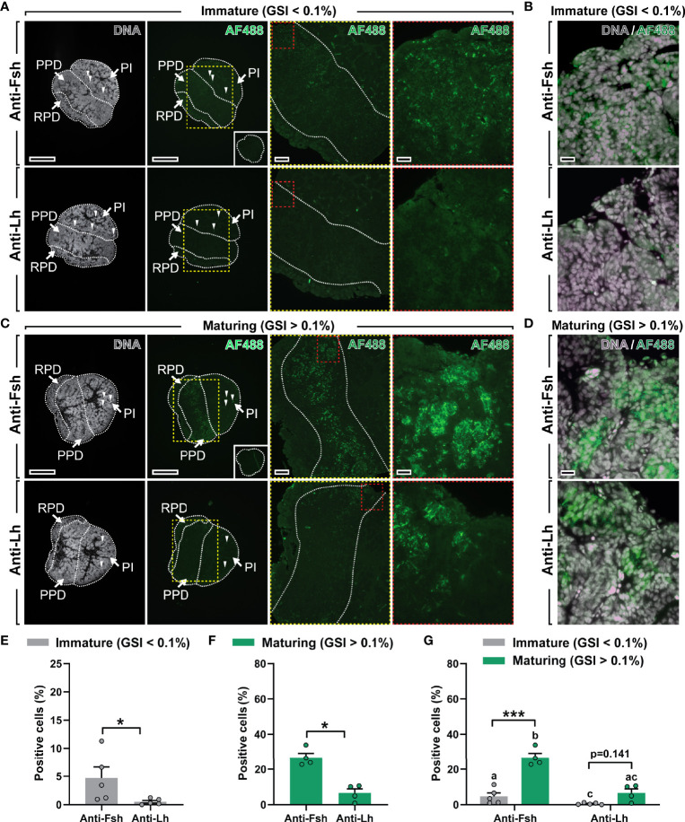
Precocious male maturation causes reduced welfare and increased production costs in Atlantic salmon (Salmo salar) aquaculture. The pituitary produces and releases follicle-stimulating hormone (Fsh), the gonadotropin triggering puberty in male salmonids. However, little is known about how Fsh production is regulated in Atlantic salmon. We examined, in vivo and ex vivo, transcriptional changes of gonadotropin-related genes accompanying the initial steps of testis maturation, in pituitaries of males exposed to photoperiod and temperature conditions promoting maturation (constant light and 16°C). Pituitary fshb, lhb and gnrhr2bba transcripts increased in vivo in maturing males (gonado-somatic index > 0.1%). RNA sequencing (RNAseq) analysis using pituitaries from genetically similar males carrying the same genetic predisposition to mature, but differing by responding or not responding to stimulatory environmental conditions, revealed 144 differentially expressed genes, ~2/3rds being up-regulated in responders, including fshb and other pituitary hormones, steroid-related and other puberty-associated transcripts. Functional enrichment analyses confirmed gene involvement in hormone/steroid production and gonad development. In ex vivo studies, whole pituitaries were exposed to a selection of hormones and growth factors. Gonadotropin-releasing hormone (Gnrh), 17β-estradiol (E2) and 11-ketotestosterone (11-KT) up-regulated gnrhr2bba and lhb, while fshb was up-regulated by Gnrh but down-regulated by 11-KT in pituitaries from immature males. Also pituitaries from maturing males responded to Gnrh and sex steroids by increased gnrhr2bba and lhb transcript levels, but fshb expression remained unchanged. Growth factors (inhibin A, activin A and insulin-like growth factor 1) did not change gnrhr2bba, lhb or fshb transcript levels in pituitaries either from immature or maturing males. Additional pituitary ex vivo studies on candidates identified by RNAseq showed that these transcripts were preferentially regulated by Gnrh and sex steroids, but not by growth factors, and that Gnrh/sex steroids were less effective when incubating pituitaries from maturing males. Our results suggest that a yet to be characterized mechanism up-regulating fshb expression in the salmon pituitary is activated in response to stimulatory environmental conditions prior to morphological signs of testis maturation, and that the transcriptional program associated with this mechanism becomes unresponsive or less responsive to most stimulators ex vivo once males had entered pubertal developmental in vivo.

Keywords: Atlantic salmon; follicle-stimulating hormone; pituitary; puberty; transcriptomics.


Conflict of interest statement

The authors declare that the research was conducted in the absence of any commercial or financial relationships that could be construed as a potential conflict of interest.

Figures

Figure 1
Figure 1
Evaluation of gonadal and pituitary responses to non-stimulatory and stimulatory conditions. (A, B) Gonado-somatic index (GSI; A) and in vivo expression levels of selected pituitary genes (fshb, lhb, gnrhr2bba; B) in immature postsmolt males exposed to non-stimulatory conditions (12 hours dark/12 hours light, and 16°C) for 16 days. (C–F) GSI (C), representative testis histology (D), fshb, lhb, gnrhr2bba expression levels (E) and 11-ketotestosterone (11-KT) plasma levels (F) in immature and maturing males after exposure to stimulatory conditions (constant light and 16°C) for 16 days. In (A, C, F), data are shown as mean ± SEM (N = 18-96; **p < 0.01; ***p < 0.001) and, in (B, E), shown as mean ± SEM (N = 9-35; *p < 0.05; ***p < 0.001; different letters denote significant differences between groups) and expressed relative to the fshb mRNA abundance. n/f, not found. AU, arbitrary units. In (D), boxes identify the testis tissue areas shown at higher magnification, white arrowheads indicate representative groups of type B spermatogonia. Scale bar, 100 µm.
Figure 2
Figure 2
Characterization of a specific antiserum for Atlantic salmon Fsh. (A) Coomassie blue protein staining of the single-chain Fsh protein (rFsh-B) and of a pituitary homogenate. (B) Representative immunoblots of rFsh-B using Fsh antiserum (left panel) and pre-immune serum (as negative control; right panel). (C) Representative immunoblots of a pituitary homogenate using anti-Fsh serum (left panel) and anti-Fsh serum preabsorbed with rFsh-B (as negative control; right panel). In all blots, molecular mass markers (kDa) are shown on the left.
Figure 3
Figure 3
Localization and quantification of Fsh and Lh cells in immature and maturing male salmon pituitaries. (A–D) Immunolocalization of Fsh and Lh proteins in serial sections (sagittally oriented; anterior to the left) of immature and maturing salmon pituitaries using the Atlantic salmon Fsh antiserum generated in this study and an anti-(coho salmon)Lh antibody previously validated (18). Right panels in (A) and C show pituitary tissue magnified from the marked areas (yellow and red dashed lines; scale bar = 200 µm and 50 µm, respectively). Scale bar in low magnification pictures = 500 µm. Negative control (pre-immune serum) for Fsh immunostaining showed no specific staining (insets in right lower panels in (A, C). Arrowheads indicate red blood autofluorescence. Further magnified areas of pituitary tissue of immature (B) and maturing (D) male salmon do not seem to show clear Fsh/Lh co-localization in serial sections (scale bar = 20 µm). Propidium iodide (in grey) was used as nuclear counterstain. GSI, gonado-somatic index; AF488, Alexa Fluor 488; PPD, proximal pars distalis; PI, pars intermedia; RPD, rostral pars distalis. (E–G) Quantification of the percentage of Fsh- and Lh-positive cells in immature (E) and maturing (F) pituitaries, and combined (G). Data are shown as mean ± SEM (N = 4-5; *p < 0.05; ***p < 0.001), and expressed relative to the total number of cells. In (G), different letters indicate significant differences between groups.
Figure 4
Figure 4
Gene expression profiling of pituitaries from male postsmolts about to enter puberty. (A–C) To select pituitary samples for RNAseq, sibling salmon males with low genetical variance were sampled at day 7 and 11 after exposure to stimulatory conditions (constant light and 16°C) and different puberty-associated parameters evaluated. Analysis of fshb expression (A) and 11-ketotestosterone [11-KT; (B)] levels revealed two groups of fish showing low (non-responders) or high (responders) levels for both parameters, while gonado-somatic indices (GSI; C) were unaffected. Data are shown as mean ± SEM (N = 5; **p < 0.01; ***p < 0.001). ns, no significant differences between groups. (D) Total numbers of up- and down-regulated genes (DEGs) identified by RNAseq (N = 5; p-adjusted < 0.001). (E, F) Regulated KEGG pathways (E) and Gene Ontology (F) terms in pituitaries of males responding to stimulatory conditions. KEGG pathways represented by at least 3 DEGs and ratio of regulated genes higher than 2.5 were considered for the analysis. (G) Selected DEGs identified by KEGG and GO analyses grouped by their function. Fold change values are shown with a red or blue background indicating up- or down-regulation, respectively.
Figure 5
Figure 5
Ex vivo effects of potential regulators on selected pituitary genes. (A, B) Expression levels of fshb, lhb and gnrhr2bba in pituitaries collected from immature postsmolt males exposed to non-stimulatory conditions [12 hours dark/12 hours light, and 16°C; (A)] and immature and maturing postsmolt males exposed to stimulatory conditions [constant light and 16°C; (B)] for 16 days, and subsequently incubated ex vivo for 9 days in the presence of various potential regulators of pituitary gene expression. Results are shown as mean fold change ± SEM (N = 3-6; *p < 0.05; **p < 0.01; ***p < 0.001) and expressed relative to the control basal condition, which is set at 1 (dashed line).
Figure 6
Figure 6
Ex vivo effects of different ligands on selected pituitary genes identified by RNAseq. (A–D) Expression levels of steroid (cyp19a1b, esr1), neurotransmission (tac1-like) and pituitary hormone (smtlb) genes in pituitaries collected from immature postsmolts exposed to standard photoperiod conditions and 16°C for 16 days, and subsequently incubated ex vivo for 9 days in the presence of various ligands. (E–L) The same set of candidate genes was analyzed in immature (E–H) and maturing (I–L) pituitary tissue after exposure to stimulatory conditions (constant light and 16°C) for 16 days, and subsequently incubated ex vivo for 9 days in the presence of the same ligands. Data are shown as mean fold change ± SEM (N = 4-11; *p < 0.05; **p < 0.01; ***p < 0.001) and expressed relative to the basal control group, which is set at 1 (dashed line).
Figure 7
Figure 7
Schematic illustration summarizing the regulation of pituitary Fsh/fshb expression and production at the, experimentally induced, onset of puberty in male Atlantic salmon. Described effects are indicated by solid lines, while dashed lines denote no experimental evidence reported here but previously demonstrated in other studies. Yellow lines highlight effects that are potentially limited in time: in vivo response to stimulatory conditions blunts both, the stimulatory effect of Gnrh on fshb and gnrhr2bba, and the inhibitory effect of 11-KT on fshb transcript levels. Gnrh, gonadotropin-releasing hormone; Fsh, follicle-stimulating hormone; Lh, luteinizing hormone; 11-KT, 11-ketotestosterone; T, testosterone; E2, 17β-estradiol; Tac1-like, protachykinin-like; gnrhr2bba, gonadotropin-releasing hormone receptor 2bba; cyp19a1b, brain aromatase; esr1, estrogen receptor 1.

Similar articles

Cited by

References

    1. Edwards W, Raetzman LT. Complex Integration of Intrinsic and Peripheral Signaling Is Required for Pituitary Gland Development. Biol Reprod (2018) 99(3):504–13. doi: 10.1093/biolre/ioy081 - DOI - PMC - PubMed
    1. Perez-Castro C, Renner U, Haedo MR, Stalla GK, Arzt E. Cellular and Molecular Specificity of Pituitary Gland Physiology. Physiol Rev (2012) 92(1):1–38. doi: 10.1152/physrev.00003.2011 - DOI - PubMed
    1. Coss D. Regulation of Reproduction via Tight Control of Gonadotropin Hormone Levels. Mol Cell Endocrinol (2018) 463:116–30. doi: 10.1016/j.mce.2017.03.022 - DOI - PMC - PubMed
    1. Fontaine R, Ciani E, Haug TM, Hodne K, Ager-Wick E, Baker DM, et al. . Gonadotrope Plasticity at Cellular, Population and Structural Levels: A Comparison Between Fishes and Mammals. Gen Comp Endocrinol (2020) 287:113344. doi: 10.1016/j.ygcen.2019.113344 - DOI - PubMed
    1. Trudeau VL. Neuroendocrine Control of Reproduction in Teleost Fish: Concepts and Controversies. Annu Rev Anim Biosci (2022) 10:107–30. doi: 10.1146/annurev-animal-020420-042015 - DOI - PubMed

LinkOut - more resources