Selective Depletion of Adult GFAP-Expressing Tanycytes Leads to Hypogonadotropic Hypogonadism in Males
- PMID: 35370973
- PMCID: PMC8966543
- DOI: 10.3389/fendo.2022.869019
Selective Depletion of Adult GFAP-Expressing Tanycytes Leads to Hypogonadotropic Hypogonadism in Males
Abstract
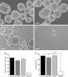
In adult mammals, neural stem cells are localized in three neurogenic regions, the subventricular zone of the lateral ventricle (SVZ), the subgranular zone of the dentate gyrus of the hippocampus (SGZ) and the hypothalamus. In the SVZ and the SGZ, neural stem/progenitor cells (NSPCs) express the glial fibrillary acidic protein (GFAP) and selective depletion of these NSPCs drastically decreases cell proliferation in vitro and in vivo. In the hypothalamus, GFAP is expressed by α-tanycytes, which are specialized radial glia-like cells in the wall of the third ventricle also recognized as NSPCs. To explore the role of these hypothalamic GFAP-positive tanycytes, we used transgenic mice expressing herpes simplex virus thymidine kinase (HSV-Tk) under the control of the mouse Gfap promoter and a 4-week intracerebroventricular infusion of the antiviral agent ganciclovir (GCV) which kills dividing cells expressing Tk. While GCV significantly reduced the number and growth of hypothalamus-derived neurospheres from adult transgenic mice in vitro, it causes hypogonadotropic hypogonadism in vivo. The selective death of dividing tanycytes expressing GFAP indeed results in a marked decrease in testosterone levels and testicular weight, as well as vacuolization of the seminiferous tubules and loss of spermatogenesis. Additionally, GCV-treated GFAP-Tk mice show impaired sexual behavior, but no alteration in food intake or body weight. Our results also show that the selective depletion of GFAP-expressing tanycytes leads to a sharp decrease in the number of gonadotropin-releasing hormone (GnRH)-immunoreactive neurons and a blunted LH secretion. Overall, our data show that GFAP-expressing tanycytes play a central role in the regulation of male reproductive function.
Keywords: GnRH; adult neural stem/progenitor cells; glial fibrillary acidic protein; hypothalamus; reproduction; sexual behavior; tanycytes.
Copyright © 2022 Butruille, Batailler, Cateau, Sharif, Leysen, Prévot, Vaudin, Pillon and Migaud.
Conflict of interest statement
The authors declare that the research was conducted in the absence of any commercial or financial relationships that could be construed as a potential conflict of interest.
Figures


Similar articles
-
Reductions in hypothalamic Gfap expression, glial cells and α-tanycytes in lean and hypermetabolic Gnasxl-deficient mice.Mol Brain. 2016 Apr 14;9:39. doi: 10.1186/s13041-016-0219-1. Mol Brain. 2016. PMID: 27080240 Free PMC article.
-
The predominant neural stem cell isolated from postnatal and adult forebrain but not early embryonic forebrain expresses GFAP.J Neurosci. 2003 Apr 1;23(7):2824-32. doi: 10.1523/JNEUROSCI.23-07-02824.2003. J Neurosci. 2003. PMID: 12684469 Free PMC article.
-
Glial fibrillary acidic protein-expressing neural progenitors give rise to immature neurons via early intermediate progenitors expressing both glial fibrillary acidic protein and neuronal markers in the adult hippocampus.Neuroscience. 2010 Mar 10;166(1):241-51. doi: 10.1016/j.neuroscience.2009.12.026. Epub 2009 Dec 16. Neuroscience. 2010. PMID: 20026190
-
Glial fibrillary acidic protein-expressing cells in the neurogenic regions in normal and injured adult brains.J Neurosci Res. 2007 Sep;85(12):2783-92. doi: 10.1002/jnr.21257. J Neurosci Res. 2007. PMID: 17394257 Review.
-
Hypothalamic Neurogenesis as an Adaptive Metabolic Mechanism.Front Neurosci. 2017 Apr 5;11:190. doi: 10.3389/fnins.2017.00190. eCollection 2017. Front Neurosci. 2017. PMID: 28424582 Free PMC article. Review.
Cited by
-
Immunomodulatory and Anti-inflammatory effect of Neural Stem/Progenitor Cells in the Central Nervous System.Stem Cell Rev Rep. 2023 May;19(4):866-885. doi: 10.1007/s12015-022-10501-1. Epub 2023 Jan 17. Stem Cell Rev Rep. 2023. PMID: 36650367 Review.
-
Investigation of the Effects of Metallic Nanoparticles on Fertility Outcomes and Endocrine Modification of the Hypothalamic-Pituitary-Gonadal Axis.Int J Mol Sci. 2023 Jul 20;24(14):11687. doi: 10.3390/ijms241411687. Int J Mol Sci. 2023. PMID: 37511445 Free PMC article.
-
Potential role of tanycyte-derived neurogenesis in Alzheimer's disease.Neural Regen Res. 2025 Jun 1;20(6):1599-1612. doi: 10.4103/NRR.NRR-D-23-01865. Epub 2024 Jun 26. Neural Regen Res. 2025. PMID: 38934388 Free PMC article.
-
In Preclinical Epilepsy, GLUT1 and GFAP Dysregulation in Cells Surrounding the Third Ventricle, Including Tanycytes, Is Differentially Restored with Ketogenic Diet Treatment.Nutrients. 2025 May 28;17(11):1824. doi: 10.3390/nu17111824. Nutrients. 2025. PMID: 40507093 Free PMC article.
-
Candidate genes for infertility: an in-silico study based on cytogenetic analysis.BMC Med Genomics. 2022 Aug 2;15(1):170. doi: 10.1186/s12920-022-01320-x. BMC Med Genomics. 2022. PMID: 35918717 Free PMC article.
References
-
- Morshead CM, Garcia AD, Sofroniew MV, van der Kooy D. The Ablation of Glial Fibrillary Acidic Protein-Positive Cells From the Adult Central Nervous System Results in the Loss of Forebrain Neural Stem Cells But Not Retinal Stem Cells. Eur J Neurosci (2003) 18(1):76–84. doi: 10.1046/j.1460-9568.2003.02727.x - DOI - PubMed
Publication types
MeSH terms
Substances
LinkOut - more resources
Full Text Sources
Miscellaneous

