. 2022 Mar 15:13:832989.
doi: 10.3389/fimmu.2022.832989. eCollection 2022.

Induction of IDO1 and Kynurenine by Serine Proteases Subtilisin, Prostate Specific Antigen, CD26 and HtrA: A New Form of Immunosuppression?

Affiliations

Induction of IDO1 and Kynurenine by Serine Proteases Subtilisin, Prostate Specific Antigen, CD26 and HtrA: A New Form of Immunosuppression?

Felix I L Clanchy et al. Front Immunol. .

Abstract

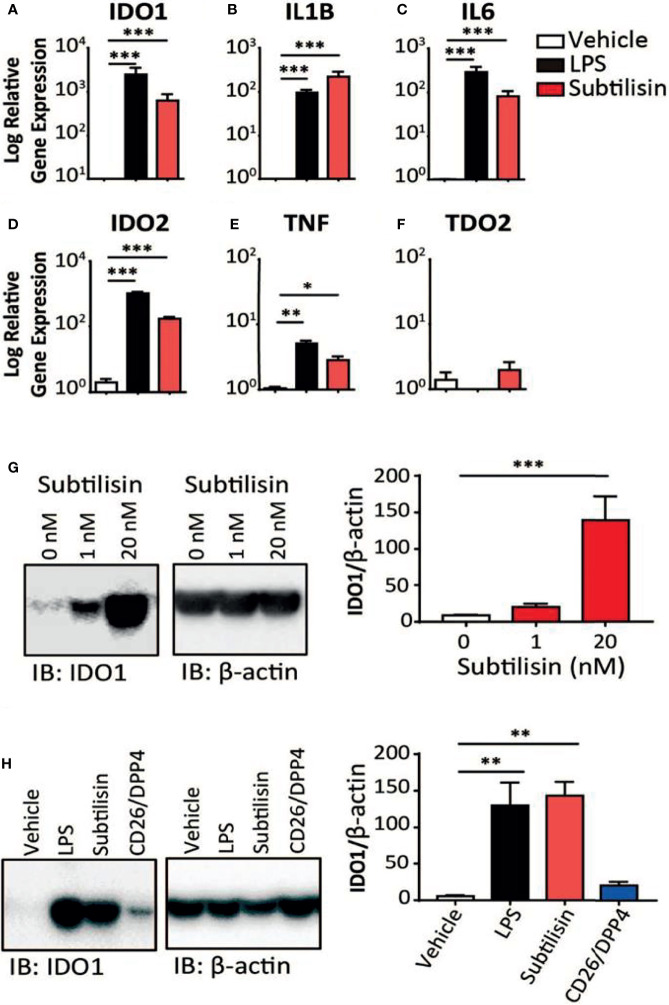
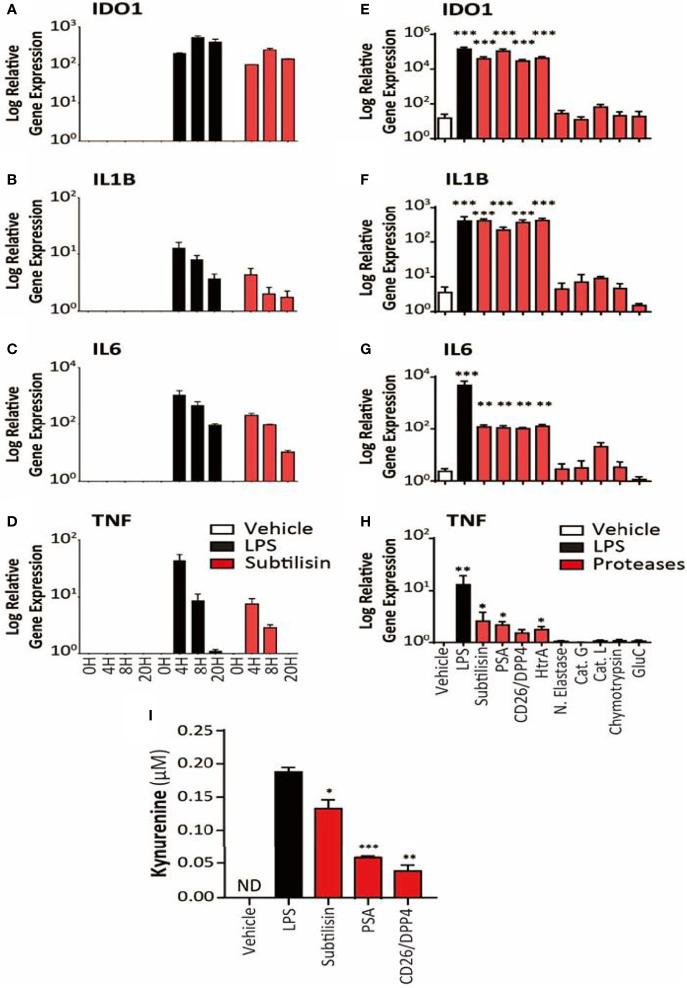
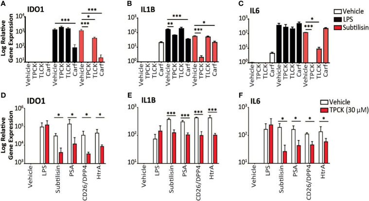
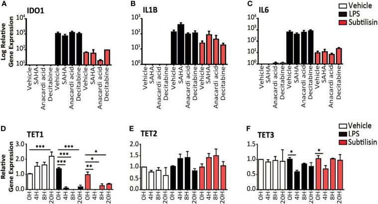
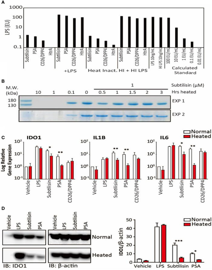
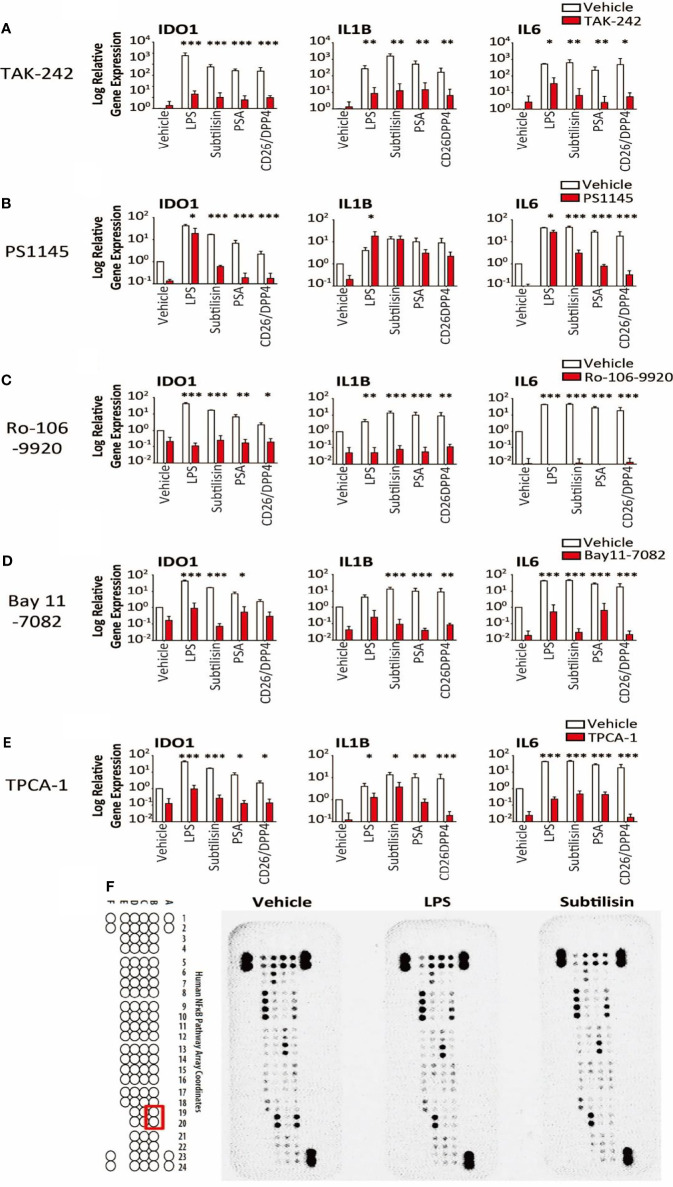
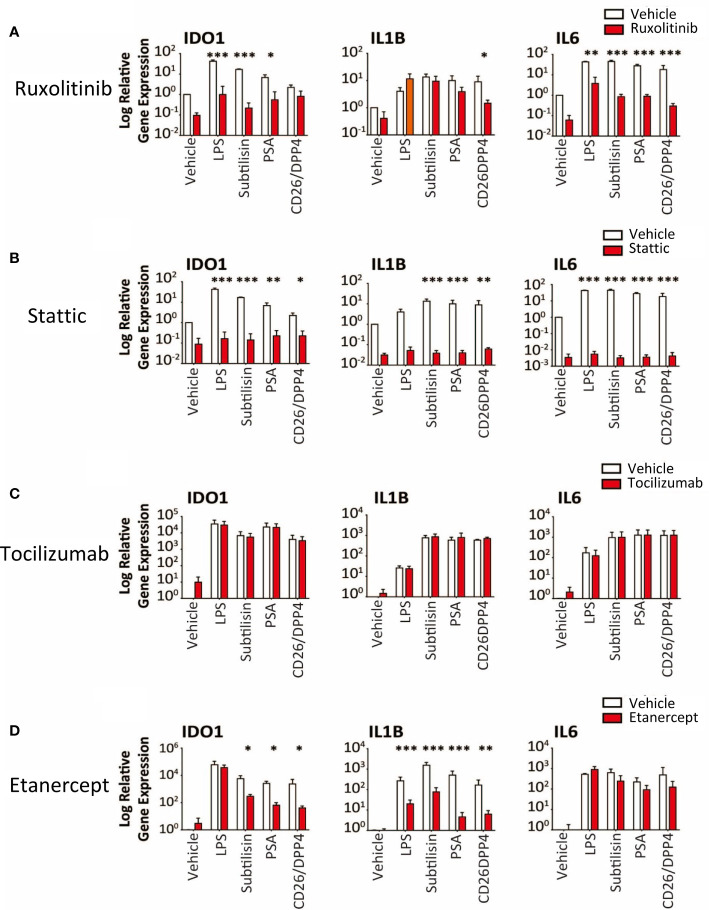
Several serine proteases have been linked to autoimmune disorders and tumour initiation although the mechanisms are not fully understood. Activation of the kynurenine pathway enzyme indoleamine-2,3-dioxygenase (IDO1) modulates cellular activity in the brain, tolerogenesis in the immune system and is a major checkpoint in cancer development. We now report that IDO1 mRNA and IDO1 protein expression (generating kynurenine) are induced in human monocyte-derived macrophages by several chymotryptic serine proteases with direct links to tumorigenesis, including Prostate Specific Antigen (PSA), CD26 (Dipeptidyl-peptidase-4, CD26/DPP-4), High Temperature Requirement protein-A (HtrA), and the bacterial virulence factor subtilisin. These proteases also induce expression of the pro-inflammatory cytokine genes IL1B and IL6. Other serine proteases tested: bacterial glu-C endopeptidase and mammalian Pro-protein Convertase Subtilase-Kexin-3 (PCSK3, furin), urokinase plasminogen activator (uPA), cathepsin G or neutrophil elastase, did not induce IDO1, indicating that the reported effects are not a general property of all serine proteases. The results represent a novel mechanism of activating immunosuppressive IDO1 and inducing kynurenine generation which, together with the production of inflammatory cytokines, would contribute to tumour initiation and progression, providing a new target for drug development. In addition, the proteasomal S20 serine protease inhibitor carfilzomib, used in the treatment of myeloma, prevented the induction of IDO1 and cytokine gene expression, potentially contributing to its clinical anti-cancer activity.

Keywords: CD26; DPP4; HtrA; IDO1; PSA; Prostate Specific Antigen; kynurenine; serine proteases.


Conflict of interest statement

The authors declare that the research was conducted in the absence of any commercial or financial relationships that could be construed as a potential conflict of interest.

Figures

Figure 1
Figure 1
Gene induction by subtilisin. LPS (10 ng/mL) and subtilisin (20 nM; 60 ng/mL) induce the expression of IDO1 (A), IL1B (B), IL6 (C), IDO2 (D), TNF (E) and TDO2 (F) in human monocyte-derived macrophages (MDMs) (logarithmic scale). (G) Western blots of MDMs from three different donors shows the expression of IDO1 protein is also induced at 1 nM or 20 nM subtilisin; the bar chart shows the quantified blot density as the ratio of IDO1 to β-actin. (H) CD26/DPP4 induces IDO1 but to a lesser extent than LPS or subtilisin; the bar chart shows the quantified blot density as the ratio of IDO1 to β-actin. Charts show the mean ± s.e.m. for (n ≧ 3 donors). The number of experiments (n) was 3 except panels A (n = 18), B (n = 15), C (n = 12) and F (n = 6). *P < 0.05, **P < 0.01; ***P < 0.001.
Figure 2
Figure 2
Time course and activity of serine proteases on human MDMs. The left-side panels (logarithmic scales) illustrate (A) the apparent maximal induction of IDO1 after 8 h incubation with LPS (10 ng/mL) or subtilisin (20 nM); (B) peak induction before 4 h of IL1B and (C) IL6; (D) a relatively rapid induction of TNF expression with a maximum before 4 h declining to baseline by 20 h. The right-side panels indicate the effects of proteases on (E) IDO1 (F) IL1B, (G) IL6 and (H) TNF expression by vehicle, LPS (10 ng/mL), subtilisin (20 nM), PSA (100 nM), CD26/DPP4 (10 nM), HtrA (10 nM), neutrophil elastase (25 μg/mL), cathepsin G (25 μg/mL), cathepsin L (25 μg/mL), chymotrypsin (25 μg/mL) and glu-C (10 nM). Panel (I) shows the concentration of kynurenine in the culture supernatant of cells exposed to LPS, subtilisin, PSA and CD26/DPP4. Charts show the mean ± s.e.m. (n ≧ 3 donors). The number of experiments (n) was 3 (for A-D) or n ≧ 3 (for E-I). *P < 0.05, **P < 0.01; ***P < 0.001.
Figure 3
Figure 3
Inhibition of serine protease activity. (A–C) The charts illustrate the expression relative to baseline (logarithmic scale) of (A) IDO1, (B) IL1B, (C) IL6 and the effects of vehicle (open bars), 10 ng/mL LPS (black bars) or 20 nM subtilisin (red/shaded bars). The inhibitors tested were TPCK (100 μM), TLCK (100 μM) and carfilzomib 20 nM (Carf). (D–F) The charts show a comparison of the effects of TPCK (30 μM) on the induction of IDO1 (D), IL1B (E) and IL6 (F) by LPS (10 ng/mL), subtilisin (20 nM), PSA (100 nM), CD26/DPP4 (10 nM) and HtrA (10 nM). Expression is shown for vehicle (open bars) and TPCK (30 μM) (red/shaded bars) treatments. Charts show the mean ± s.e.m. (n ≧ 3 donors). *P < 0.05, **P < 0.01; ***P < 0.001.
Figure 4
Figure 4
Epigenetic modulators and gene expression. (A–C) Charts of the gene expression (logarithmic axis) relative to baseline of (A) IDO1, (B) IL1B and (C) IL6 in MDMs exposed to vehicle (open bars), 10 ng/mL LPS (black bars) or 20 nM subtilisin (red/shaded bars). The data shows the effects of 10 μM SAHA (an inhibitor of histone deacetylase), 10 μM anacardic acid (an inhibitor of histone acetylase) and 2 μM decitabine (an inhibitor or DNA methyltransferase). (D–F) Logarithmic expression relative to baseline of TET1 (D), TET2 (E) and TET3 (F) in cells exposed to vehicle (open bars), 10 ng/mL LPS (black bars) or 20 nM subtilisin (red/shaded bars). Charts show the mean ± s.e.m. (n ≧ 3 donors). *P < 0.05, **P < 0.01; ***P < 0.001.
Figure 5
Figure 5
LPS concentrations and the effects of heating on gene expression. (A). Samples of the four active serine proteases were analysed for endotoxin contamination. In bars 1-4, when compared against LPS 10 ng/mL, subtilisin (20 nM) is associated with < 1% LPS, while PSA (100 nM), CD26/DPP4 (10 nM) and HtrA (10 nM) have even lower amounts. Bars 5-8 show that the serine proteases do not affect the LPS signal, confirming that they do not interfere with the assay procedure. Bars 9-12 and 13-16 reveal that heating the proteases at 70°C for 1 h did not alter the protease signals (–12) or the combined protease and LPS signals (–16), confirming that LPS was not affected by the heat treatment and was still not affected by the proteases. This conclusion is strengthened by the absence of heat on LPS alone (bars 17-18). Bars 19-23 show a calibration for the absolute concentration of LPS in this assay. (B) A Coomassie Blue stained gel of collagen in two separate experiments shows that the undigested protein in the absence of subtilisin (0 μM) is reduced at 0.1 μM and abolished at 1 and 10 μM. After heating subtilisin at 70°C for 0.5 h or longer, protease activity has been substantially reduced and is not able to degrade the collagen. (C) Quantified data for the heating experiments. Bar charts show the relative expression of genes IDO1, IL1B and IL6 by LPS (10 ng/mL), subtilisin (20 nM), PSA (100 nM) and CD26/DPP4 (10 nM) under normal experimental conditions (open bars) and after heating at 70°C for 1 h (red/shaded bars). Heating removes much of the activity of subtilisin and PSA on all three genes. (D) Representative Western blot shows that the more heat-sensitive proteases (20 nM subtilisin and 100 nM PSA) induce less IDO1 protein when heated at 70°C for 1 h. The bar chart shows the quantified ratio of IDO1 to β-actin density. Charts show the mean ± s.e.m. (n ≧ 3 donors). *P < 0.05, **P < 0.01; ***P < 0.001.
Figure 6
Figure 6
Transduction pathways affected by serine proteases. The logarithmic relative expression of IDO1, IL1B and IL6 induced by vehicle, LPS (10 ng/mL), subtilisin (20 nM), PSA (100 nM) and CD26/DPP4 (10 nM) in the presence of vehicle (open bars) or test agents (red/shaded bars): (A) TAK-242 (10 μM), (B) PS1145 (200 μM), (C) Ro-106-9920 (10 μM), (D) Bay11-7082 (10 μM) or (E) TPCA-1 (10 μM). Charts show the mean ± s.e.m. (n ≧ 3 donors). *P < 0.05, **P < 0.01; ***P < 0.001. (F) illustrates a proteomics array based on NFκB transduction pathways exposed to vehicle, LPS (10 ng/mL) or subtilisin (20 nM) for 0.5 h. LPS and subtilisin induced a loss of B19 and B20 corresponding to IκBε. Bars show the mean ± s.e.m. (n ≧ 3). *P < 0.05, **P < 0.01; ***P < 0.001.
Figure 7
Figure 7
Transduction pathways affected by serine proteases. The logarithmic expression of IDO1, IL1B and IL6 induced by vehicle, LPS (10 ng/mL), subtilisin (20 nM), PSA (100 nM) and CD26/DPP4 (10 nM) in the presence of vehicle (open bars) or test agents (red/shaded bars): (A) Ruxolitinib (20 μM), (B) stattic (10 μM), (C) tocilizumab (20 μg/mL), (D) etanercept (20 μg/mL). Bars show the mean ± s.e.m. The number of experiments (n) was 3 in all cases except panels C and D, where n = 6. *P < 0.05, **P < 0.01; ***P < 0.001.

Similar articles

Cited by

References

    1. Badawy AAB. Kynurenine Pathway of Tryptophan Metabolism: Regulatory and Functional Aspects. Intern J Tryptophan Res (2017) 10:AR1178646917691938. doi: 10.1177/1178646917691938 - DOI - PMC - PubMed
    1. Badawy AAB. Kynurenic and Quinolinic Acids: The Main Players of the Kynurenine Pathway and Opponents in Inflammatory Disease. Med Hypoth (2018) 118:129–38. doi: 10.1016/j.mehy.2018.06.021 - DOI - PubMed
    1. Stone TW, Darlington LG. Endogenous Kynurenines as Targets for Drug Discovery and Development. Nat Rev Drug Disc (2002) 1:609–20. doi: 10.1038/nrd870 - DOI - PubMed
    1. Cribbs AP, Kennedy A, Penn H, Read JE, Amjadi P, Green P, et al. . Treg Cell Function in Rheumatoid Arthritis is Compromised by CTLA-4 Promoter Methylation Resulting in a Failure to Activate the Indoleamine 2,3-Dioxygenase Pathway. Arthritis Rheumatol (2014) 66:2344–54. doi: 10.1002/art.38715 - DOI - PubMed
    1. Huang YS, Ogbechi J, Clanchy F, Williams RO, Stone TW. Kynurenine Metabolites in Peripheral Disorders and Neuroinflammation. Front Immunol (2020) 11:art.388:art.388. doi: 10.3389/fimmu.2020.00388 - DOI - PMC - PubMed

Publication types