

High-speed high-resolution data collection on a 200 keV cryo-TEM
- PMID: 35371504
- PMCID: PMC8895008
- DOI: 10.1107/S2052252522000069
High-speed high-resolution data collection on a 200 keV cryo-TEM
Abstract
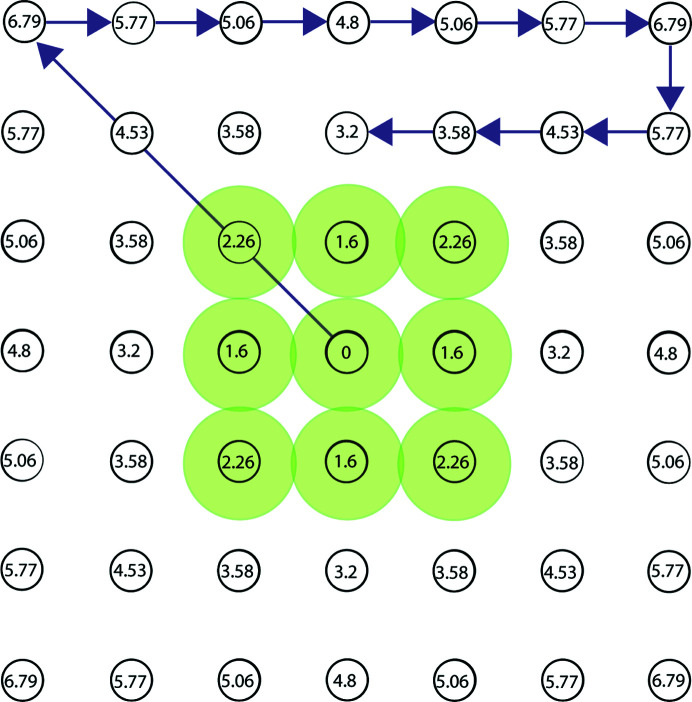
Limitations to successful single-particle cryo-electron microscopy (cryo-EM) projects include stable sample generation, production of quality cryo-EM grids with randomly oriented particles embedded in thin vitreous ice and access to microscope time. To address the limitation of microscope time, methodologies to more efficiently collect data on a 200 keV Talos Arctica cryo-transmission electron microscope at speeds as fast as 720 movies per hour (∼17 000 per day) were tested. In this study, key parameters were explored to increase data collection speed including: (1) using the beam-image shift method to acquire multiple images per stage position, (2) employing UltrAufoil TEM grids with R0.6/1 hole spacing, (3) collecting hardware-binned data and (4) adjusting the image shift delay factor in SerialEM. Here, eight EM maps of mouse apoferritin at 1.8-1.9 Å resolution were obtained in the analysis with data collection times for each dataset ranging from 56 min to 2 h. An EM map of mouse apoferritin at 1.78 Å was obtained from an overnight data collection at a speed of 500 movies per hour and subgroup analysis performed, with no significant variation observed in data quality by image shift distance and image shift delay. The findings and operating procedures detailed herein allow for rapid turnover of single-particle cryo-EM structure determination.
Keywords: 200 keV cryo-TEM; beam image shift; cryo-electron microscopy; direct-electron detector.
© Peck, Fay and Strauss 2022.
Figures


Similar articles
-
High-resolution cryo-EM using a common LaB6 120-keV electron microscope equipped with a sub-200-keV direct electron detector.Sci Adv. 2025 Jan 3;11(1):eadr0438. doi: 10.1126/sciadv.adr0438. Epub 2025 Jan 3. Sci Adv. 2025. PMID: 39752481 Free PMC article.
-
High-resolution cryo-EM using beam-image shift at 200 keV.IUCrJ. 2020 Oct 29;7(Pt 6):1179-1187. doi: 10.1107/S2052252520013482. eCollection 2020 Nov 1. IUCrJ. 2020. PMID: 33209328 Free PMC article.
-
Routine Collection of High-Resolution cryo-EM Datasets Using 200 KV Transmission Electron Microscope.J Vis Exp. 2022 Mar 16;(181). doi: 10.3791/63519. J Vis Exp. 2022. PMID: 35377368 Review.
-
Fabrication of carbon films with ∼ 500nm holes for cryo-EM with a direct detector device.J Struct Biol. 2014 Jan;185(1):42-7. doi: 10.1016/j.jsb.2013.11.002. Epub 2013 Nov 21. J Struct Biol. 2014. PMID: 24269484
-
Setting up and operating a cryo-EM laboratory.Q Rev Biophys. 2021 Jan 8;54:e2. doi: 10.1017/S003358352000013X. Q Rev Biophys. 2021. PMID: 33413714 Review.
Cited by
-
Molecular dissection of the glutamine synthetase-GlnR nitrogen regulatory circuitry in Gram-positive bacteria.Nat Commun. 2022 Jul 1;13(1):3793. doi: 10.1038/s41467-022-31573-0. Nat Commun. 2022. PMID: 35778410 Free PMC article.
-
Design and structural validation of peptide-drug conjugate ligands of the kappa-opioid receptor.Nat Commun. 2023 Dec 6;14(1):8064. doi: 10.1038/s41467-023-43718-w. Nat Commun. 2023. PMID: 38052802 Free PMC article.
-
Miffi: Improving the accuracy of CNN-based cryo-EM micrograph filtering with fine-tuning and Fourier space information.J Struct Biol. 2024 Jun;216(2):108072. doi: 10.1016/j.jsb.2024.108072. Epub 2024 Feb 29. J Struct Biol. 2024. PMID: 38431179 Free PMC article.
-
Structure-guided design of a peripherally restricted chemogenetic system.Cell. 2024 Dec 26;187(26):7433-7449.e20. doi: 10.1016/j.cell.2024.11.001. Epub 2024 Dec 3. Cell. 2024. PMID: 39631393
-
Ligand recognition and allosteric modulation of the human MRGPRX1 receptor.Nat Chem Biol. 2023 Apr;19(4):416-422. doi: 10.1038/s41589-022-01173-6. Epub 2022 Oct 27. Nat Chem Biol. 2023. PMID: 36302898
References
-
- Cao, C., Kang, H. J., Singh, I., Chen, H., Zhang, C., Ye, W., Hayes, B. W., Liu, J., Gumpper, R. H., Bender, B. J., Slocum, S. T., Krumm, B. E., Lansu, K., McCorvy, J. D., Kroeze, W. K., English, J. G., DiBerto, J. F., Olsen, R. H. J., Huang, X.-P., Zhang, S., Liu, Y., Kim, K., Karpiak, J., Jan, L. Y., Abraham, S. N., Jin, J., Shoichet, B. K., Fay, J. F. & Roth, B. L. (2021). Nature, 600, 170–175. - PMC - PubMed
Grants and funding
LinkOut - more resources
Full Text Sources
