. 2022 Apr 1;13(2):540-551.
doi: 10.14336/AD.2021.1008. eCollection 2022 Apr.

Aging Impairs the Cellular Interplay between Myeloid Cells and Mesenchymal Cells during Skin Healing in Mice

Affiliations

Aging Impairs the Cellular Interplay between Myeloid Cells and Mesenchymal Cells during Skin Healing in Mice

Saeid Amini-Nik et al. Aging Dis. .

Abstract

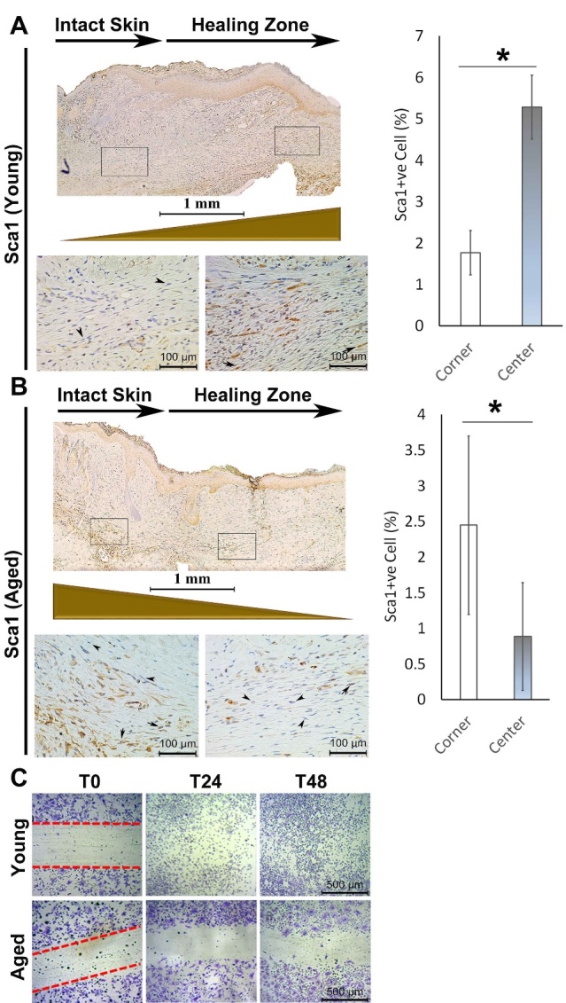
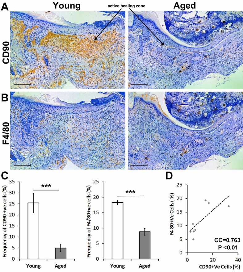
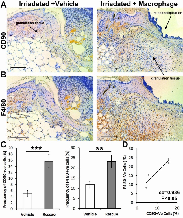
Impaired wound healing is a major issue in the elderly population and is associated with substantial health and economic burden, which is exponentially increasing with the growing aging population. While the underlying pathobiology of disturbed skin healing by aging is linked to several genetic and epigenetic factors, little is known about the cell-cell interaction during the wound healing process in aged individuals, particularly the mesenchymal stem cell (MSCs)-macrophages axis. In this study, by using a thermal injury animal model in which we compared the wound healing process of adult and young mice, we found that the insufficient pool of MSCs in adult animals are deficient in migrating to the wound bed and instead are restricted to the wound edge. We identified a deficiency of a CD90-positive MSC subpopulation in the wounds of adult animals, which is positively correlated with the number of F4/80+ macrophages. In vitro, we found that CD90+ cells preferentially adhere to the myeloid cells forming doublet cells. Thus, our findings highlight that in adult mice subjected to a thermal injury, impaired wound healing is likely mediated by a disturbed cellular interplay between myeloid cells and mesenchymal cells.

Keywords: burns; elderly; macrophages; mesenchymal stem cells; thermal injury; wound healing.


Figures

Figure 1.
Figure 1.
Adult mice show deficient skin healing post thermal injury. Trichrome staining of the edge and the center of the burned area shows superior granulation tissue and healing in the skin of young mice in comparison with adult mice (right). Note the complete lack of granulation tissue, particularly in the center of the damage zone of the adult animal (n=8). Decreased wound length (mm) in adult relative to young mice (left) (n=5-7). (*P< 0.001, error bars are showing 95% confidence intervals). Areas of impaired re-epithelialization and poor granulation are indicated in adult (aged) tissue and marked with an arrow.
Figure 2.
Figure 2.
Sca1+ mesenchymal stem cells are restricted to the junction of intact skin in injured adult mice and are enriched in the active healing area in young mice. (A) In young animals, the number of Sca1+ mesenchymal cells is significantly less at the junction of intact skin in comparison with the active healing area toward the center of the wound two weeks post-injury (B) In the adult animal, the number of Sca1+ mesenchymal cells is significantly higher at the junction of intact skin (left call out box) in comparison with the active healing area (right call out box) toward the center of the wound (n=5). (C) In vitro scratch assay of BM-MSCs isolated from young vs. adult animals show migration deficiency in the adult group (*P < 0.05, error bars are showing 95% confidence intervals). Arrows indicate Sca1+ cells present in murine dermis.
Figure 3.
Figure 3.
Adult mice have a decreased number of both F4/80+ macrophages, and CD90+ mesenchymal cells to the wound are compared to young mice. (A) Immunohistochemical staining of the healing skin two weeks post-injury shows a large population of CD90+ cells spanning from the junction of intact skin to the active healing zone (active healing zone marked with an arrow). This CD90+ cell population is significantly decreased in the adult mouse both at the junction and active healing area. (B) Immunohistochemical stain of F4/80 shows a decreased number of macrophages in the area of active healing in adult mice compared to young mice. (C) Quantification of the IHC stain shows significantly decreased frequency of CD90+ and F4/80+ cells in adult animals. (D) There is a significant correlation between frequency of CD90+ and F4/80+ cells in the wound sites of young animals (n=8, *P < 0.05, ***P <0.001).
Figure 4.
Figure 4.
Topical addition of macrophages in irradiated young mice increases the number of CD90+ cells within the active wound area and rescues wound healing. (A,C) There is deficient wound healing in irradiated young mice with the vehicle, illustrated by the decreased amount of granulation tissue (labeled with an arrow). There is a decreased proportion of macrophages and CD90+ cells. (B,D) Applying macrophages, locally, increased granulation tissue formation and improved re-epithelialization (labeled with arrows), associated with a statistically significant increase in the proportion of macrophages and CD90+ cells. (C) There is a significant correlation between the number of macrophages and CD90+ cells in the active wound area (n=4, **P< 0.01, *** P<0.001, error bars are showing 95% confidence intervals).
Figure 5.
Figure 5.
Macrophages may preferentially adhere to the CD90+ vs. CD90-ve MSCs. (A) Dapi (blue) and CD90 (red) immunofluorescence was performed after the addition of EYFP+ macrophages from young mice to a Boname marrow-derived MSCs. Macrophages preferentially adhere to CD90+ cells as compared to CD90-ve cells. (B) Macrophage (EYFP+ cells) and mesenchymal stem cell interaction were examined using flow cytometry staining for CD90. There is a significant increase in the percentage of doublet cells within the CD90+ cell population. There is also an increased proportion of EYFP+ cells within the CD90+ doublet cell population, suggesting a stronger interaction between CD90+ cells and macrophages than CD90- cells and macrophages (n=3 and ** P < 0.01, *** P <0.001, 95% confidence intervals).

References

    1. Jeschke MG, Chinkes DL, Finnerty CC, Kulp G, Suman OE, Norbury WB, et al.. (2008). Pathophysiologic response to severe burn injury. Ann Surg, 248:387-401. - PMC - PubMed
    1. Jeschke MG, Gauglitz GG, Kulp GA, Finnerty CC, Williams FN, Kraft R, et al.. (2011). Long-term persistance of the pathophysiologic response to severe burn injury. PLoS One, 6:e21245. - PMC - PubMed
    1. Amini-Nik S, Yousuf Y, Jeschke MG (2017). Scar management in burn injuries using drug delivery and molecular signaling: Current treatments and future directions. Adv Drug Deliv Rev. - PMC - PubMed
    1. Bringham PA, McLoughlin E (1996). Burn incidence and medical care use in the United States: estimates, trends and data sources. J Burn Care Rehabil, 17:95-107. - PubMed
    1. (WHO) OWH. 2002. A graphical overview of the global burden of injuries. The Injury Chart Book. Geneva.

LinkOut - more resources