

Oral Lipoma Resembling Popeye's Pipe: A Case Report
- PMID: 35371685
- PMCID: PMC8938201
- DOI: 10.7759/cureus.22350
Oral Lipoma Resembling Popeye's Pipe: A Case Report
Abstract
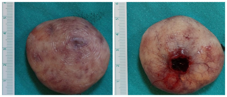
Lipomas are benign neoplasms of mesenchymal origin. Although they are frequently seen in other parts of the body, they are rare in the oral cavity. In the oral cavity, they most often develop from buccal mucosa. They tend to grow slowly, so they may remain asymptomatic for a long time and go unnoticed. Lipomas in the oral cavity may cause deterioration in chewing-speaking and esthetic problems over time, depending on the increase in their size. The most reliable imaging method for differential diagnosis is magnetic resonance imaging. Complete excision of the lipoma is essential for treatment. In this study, a case of an unusual oral lipoma, causing nutrition-speaking difficulties in a geriatric male patient is presented.
Keywords: buccal area; excision; geriatrics; lipoma; oral cavity.
Copyright © 2022, Akın et al.
Conflict of interest statement
The authors have declared that no competing interests exist.
Figures


References
-
- Intraoral lipoma at an unusual site: a rare presentation. Thakur M, Kundoor VK, Maloth KN, Nayanala VA. J Dent Allied Sci. 2017;6:98–100.
-
- Lipoma of the oral and maxillofacial region: site and subclassification of 125 cases. Furlong MA, Fanburg-Smith JC, Childers EL. Oral Surg Oral Med Oral Pathol Oral Radiol. 2004;98:441–450. - PubMed
Publication types
LinkOut - more resources
Full Text Sources