A Case of Syncope Due to Intracardiac Leiomyomatosis
- PMID: 35371771
- PMCID: PMC8965043
- DOI: 10.7759/cureus.22666
A Case of Syncope Due to Intracardiac Leiomyomatosis
Abstract
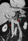
We present a case of a 46-year-old female presenting with syncope. Echocardiography initially showed a right atrial mass. Further evaluation revealed a mass arising from the fundus of the uterus, with a tumor thrombus in the left gonadal vein, extending into the left renal vein and through the inferior vena cava (IVC) into the right heart across the tricuspid valve. She was managed with surgical resection, and postoperative pathology was consistent with intravenous leiomyomatosis (IVL). IVL is a rare uterine smooth muscle cell neoplasm which extends into the venous system. Gynecological tumors are often overlooked in differential diagnosis for atrial masses. A benign tumor like fibroid, in rare circumstances, can extend into the right side of the heart causing dynamic obstruction to outflow tract, thus increasing mortality. The objective of this article is to present such a case and highlight the broad differentials of atrial masses, including IVL.
Keywords: atrial mass; intracardiac leiomyomatosis; intravenous leiomyomatosis; leiomyoma; syncope.
Copyright © 2022, Thapa et al.
Conflict of interest statement
The authors have declared that no competing interests exist.
Figures


References
-
- Tumors of the heart. A 20-year experience with a review of 12,485 consecutive autopsies. Lam KY, Dickens P, Chan AC. https://europepmc.org/article/med/8215825?utm_medium=email&utm_source=tr.... Arch Pathol Lab Med. 1993;117:1027–1031. - PubMed
-
- Intravenous leiomyomatosis of the uterus: a clinicopathologic study of 18 cases, with emphasis on early diagnosis and appropriate treatment strategies. Du J, Zhao X, Guo D, Li H, Sun B. Hum Pathol. 2011;42:1240–1246. - PubMed
-
- Mesenchymal tumors of the uterus. V. Intravenous leiomyomatosis: a clinical and pathologic study of 14 cases. Norris HJ, Parmley T. Cancer. 1975;36:2164–2178. - PubMed
-
- Intravenous leiomyomatosis: two cases with different routes of tumor extension. Lam PM, Lo KW, Yu MY, Wong WS, Lau JY, Arifi AA, Cheung TH. J Vasc Surg. 2004;39:465–469. - PubMed
Publication types
LinkOut - more resources
Full Text Sources
