

NUT Carcinoma: Clinicopathologic Features, Molecular Genetics and Epigenetics
- PMID: 35372003
- PMCID: PMC8966081
- DOI: 10.3389/fonc.2022.860830
NUT Carcinoma: Clinicopathologic Features, Molecular Genetics and Epigenetics
Abstract
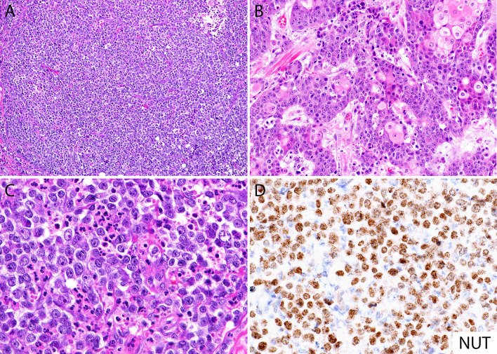
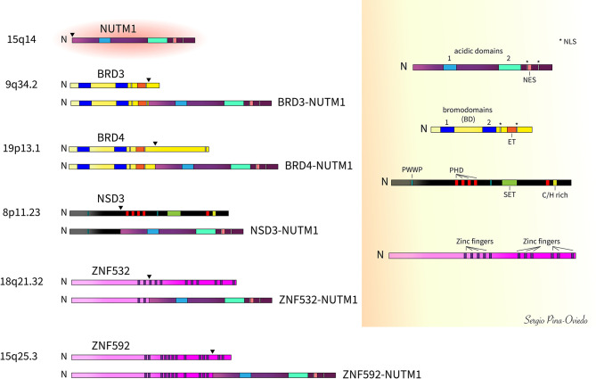
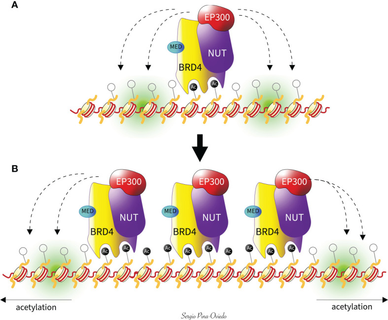
Nuclear protein in testis (NUT) carcinoma is a rare, highly aggressive, poorly differentiated carcinoma occurring mostly in adolescents and young adults. This tumor usually arises from the midline structures of the thorax, head, and neck, and exhibits variable degrees of squamous differentiation. NUT carcinoma is defined by the presence of a NUTM1 (15q14) rearrangement with multiple other genes. In about 70-80% of the cases, NUTM1 is involved in a balanced translocation with the BRD4 gene (19p13.12), leading to a BRD4-NUTM1 fusion oncogene. Other variant rearrangements include BRD3-NUTM1 fusion (~15-20%) and NSD3-NUTM1 fusion (~6%), among others. The diagnosis of NUT carcinoma requires the detection of nuclear expression of the NUT protein by immunohistochemistry. Additional methods for diagnosis include the detection of a NUTM1 rearrangement by fluorescence in situ hybridization or by reverse transcriptase PCR. NUT carcinoma is usually underrecognized due to its rarity and lack of characteristic histological features. Therefore, the goal of this review is to provide relevant recent information regarding the clinicopathologic features of NUT carcinoma, the role of the multiple NUTM1 gene rearrangements in carcinogenesis, and the impact of understanding these underlying molecular mechanisms that may result in the development of possible novel targeted therapies.
Keywords: BET inhibitors; BRD-NUTM1; HDAC inhibitors; NSD3; NUT carcinoma; NUT midline carcinoma; zinc finger proteins.
Copyright © 2022 Moreno, Saluja and Pina-Oviedo.
Conflict of interest statement
The authors declare that the research was conducted in the absence of any commercial or financial relationships that could be construed as a potential conflict of interest.
Figures
Similar articles
-
BRD3-NUTM1-expressing NUT carcinoma of lung on endobronchial ultrasound-guided transbronchial needle aspiration cytology, a diagnostic pitfall.Diagn Cytopathol. 2022 Feb;50(2):E47-E53. doi: 10.1002/dc.24885. Epub 2021 Oct 21. Diagn Cytopathol. 2022. PMID: 34672128
-
Thyroid Carcinoma with NSD3::NUTM1 Fusion: a Case with Thyrocyte Differentiation and Colloid Production.Endocr Pathol. 2022 Jun;33(2):315-326. doi: 10.1007/s12022-021-09700-2. Epub 2022 Jan 8. Endocr Pathol. 2022. PMID: 34997561 Free PMC article.
-
NUT Carcinoma of the Submandibular Gland: A Case at This Uncommon Site with Review of the Literature.Head Neck Pathol. 2022 Sep;16(3):881-892. doi: 10.1007/s12105-021-01400-x. Epub 2021 Dec 14. Head Neck Pathol. 2022. PMID: 34905155 Free PMC article. Review.
-
Demystified molecular pathology of NUT midline carcinomas.J Clin Pathol. 2010 Jun;63(6):492-6. doi: 10.1136/jcp.2007.052902. Epub 2008 Jun 13. J Clin Pathol. 2010. PMID: 18552174 Review.
-
Misleading Germ Cell Phenotype in Pulmonary NUT Carcinoma Harboring the ZNF532-NUTM1 Fusion.Am J Surg Pathol. 2022 Feb 1;46(2):281-288. doi: 10.1097/PAS.0000000000001774. Am J Surg Pathol. 2022. PMID: 34232599 Free PMC article.
Cited by
-
Suspected NUT carcinoma progressing on pembrolizumab, carboplatin, and paclitaxel as first-line treatment: a case report.Ann Med Surg (Lond). 2023 Dec 2;86(2):1061-1065. doi: 10.1097/MS9.0000000000001501. eCollection 2024 Feb. Ann Med Surg (Lond). 2023. PMID: 38333300 Free PMC article.
-
Nuclear protein in testis carcinoma of the lung.Transl Oncol. 2023 Apr;30:101640. doi: 10.1016/j.tranon.2023.101640. Epub 2023 Feb 11. Transl Oncol. 2023. PMID: 36780749 Free PMC article. Review.
-
Primary cutaneous NUT adnexal carcinoma: morphologic, genetic and methylation analysis of seven new cases with comparison to extracutaneous NUT carcinoma and NUTM1-rearranged porocarcinoma.Histopathology. 2025 Sep;87(3):375-387. doi: 10.1111/his.15444. Epub 2025 Mar 24. Histopathology. 2025. PMID: 40125640 Free PMC article.
-
Clinical imaging of primary pulmonary nucleoprotein of the testis carcinoma.Front Med (Lausanne). 2023 Jan 4;9:1083206. doi: 10.3389/fmed.2022.1083206. eCollection 2022. Front Med (Lausanne). 2023. PMID: 36687409 Free PMC article.
-
NUT Carcinoma of the Lung:A Case report and Literature Analysis.Front Oncol. 2022 Jul 12;12:890338. doi: 10.3389/fonc.2022.890338. eCollection 2022. Front Oncol. 2022. PMID: 35903693 Free PMC article.
References
-
- French CA, Miyoshi I, Kubonishi I, Grier HE, Perez-Atayde AR, Fletcher JA. BRD4-NUT Fusion Oncogene: A Novel Mechanism in Aggressive Carcinoma. Cancer Res (2003) 63(2):304–7. - PubMed
-
- French CA, Ramirez CL, Kolmakova J, Hickman TT, Cameron MJ, Thyne ME, et al. . BRD-NUT Oncoproteins: A Family of Closely Related Nuclear Proteins That Block Epithelial Differentiation and Maintain the Growth of Carcinoma Cells. Oncogene (2008) 27(15):2237–42. doi: 10.1038/sj.onc.1210852 - DOI - PubMed
Publication types
LinkOut - more resources
Full Text Sources