. 2022 Mar 17:12:836242.
doi: 10.3389/fcimb.2022.836242. eCollection 2022.

Matrix Metalloproteinase 2 and 9 Enzymatic Activities are Selectively Increased in the Myocardium of Chronic Chagas Disease Cardiomyopathy Patients: Role of TIMPs

Affiliations

Matrix Metalloproteinase 2 and 9 Enzymatic Activities are Selectively Increased in the Myocardium of Chronic Chagas Disease Cardiomyopathy Patients: Role of TIMPs

Monique Andrade Baron et al. Front Cell Infect Microbiol. .

Abstract

Chronic Chagas disease (CCC) is an inflammatory dilated cardiomyopathy with a worse prognosis compared to other cardiomyopathies. We show the expression and activity of Matrix Metalloproteinases (MMP) and of their inhibitors TIMP (tissue inhibitor of metalloproteinases) in myocardial samples of end stage CCC, idiopathic dilated cardiomyopathy (DCM) patients, and from organ donors. Our results showed significantly increased mRNA expression of several MMPs, several TIMPs and EMMPRIN in CCC and DCM samples. MMP-2 and TIMP-2 protein levels were significantly elevated in both sample groups, while MMP-9 protein level was exclusively increased in CCC. MMPs 2 and 9 activities were also exclusively increased in CCC. Results suggest that the balance between proteins that inhibit the MMP-2 and 9 is shifted toward their activation. Inflammation-induced increases in MMP-2 and 9 activity and expression associated with imbalanced TIMP regulation could be related to a more extensive heart remodeling and poorer prognosis in CCC patients.

Keywords: Chagas disease; MMP; cardiac remodeling; cardiomyopathy; fibrosis; heart failure; metalloproteinases.


Conflict of interest statement

The authors declare that the research was conducted in the absence of any commercial or financial relationships that could be construed as a potential conflict of interest.

Figures

Figure 1
Figure 1
Histopathological features of myocardial samples. (A) Slides of hematoxylin–eosin (H&E) and picrosirius red stained myocardial sections of representative patients with CCC, DCM and individuals without cardiomyopathies (Controls). Representative heart sections were shown with magnification: ×20. Myocardial hypertrophy characterized by fiber and nuclear enlargement is evident in the CCC and DCM groups. Diffuse mononuclear or lymphocytic myocarditis is present only in the CCC group. Interstitial fibrosis stained in red with picrosirius red is present in the CCC and DCM groups. (B) The severity of fibrosis was quantified by collagen area fraction (%) in DCM and CCC group exhibit a significant increase in the deposition of collagen. Groups were compared by a non-parametrical test (Mann–Whitney Rank Sum Test) with GraphPad Prism software (version 6.0; GraphPad). Results were expressed as mean ± SD. *P-values were considered significant if p-value (0.05) corrected for multiple comparisons by Bonferroni’s method (corrected p-value = 0.05/3 = 0.0166).
Figure 2
Figure 2
MMP-2 and MMP-9 mRNA and protein expression and activity in myocardium tissue from control, DCM and CCC samples. Myocardial expression of MMP-2 and MMP-9 mRNA. Real-time PCR analysis of mRNA expression in control (n = 6), DCM (n = 12) and CCC (n = 17) myocardium. The expression was calculated as the mean ± SEM for each group as individual data points using the relative expression (fold change over control) by the 2−ΔΔCt method, normalized with the endogenous gene RPL0, as described in the Materials and Methods section. (A) Relative expression of MMP-2 mRNA and MMP-9 mRNA. (B) Western blotting image showing protein of MMP-2 and MMP-9 in extracts from myocardium samples from the control, DCM and CCC (n = 5 in each group). The densitometric values of each protein for each sample were normalized by the values of Beta actin, as described in the Materials and Methods section. Densitometry analysis of the MMP-2. Densitometry analysis of the MMP-9. (C) Zimography image showing activity of MMP-2 and MMP-9 in extracts from myocardium samples from control, DCM and CCC (n = 5 in each group). (D) Densitometry analysis of the activated MMP-2 (66 kD band) and activated MMP-9 (88 kD band) results. Groups were compared by a non-parametrical test (Mann–Whitney Rank Sum Test) with GraphPad Prism software (version 6.0; GraphPad). Results were expressed as mean ± SD. *P-values were considered significant if p-value (0.05) corrected for multiple comparisons by Bonferroni’s method (corrected p-value = 0.05/3 = 0.0166). Primary antibodies (with their respective dilution) against the following proteins were used: MMP-2 (mouse monoclonal, 1:1,000, Abcam, UK); MMP-3 (rabbit polyclonal, 1:500, Abcam, UK); MMP-8 (rabbit polyclonal, 1:500, UK); MMP-9 (rabbit polyclonal, 1:1,000, UK); MMP-12 (rabbit polyclonal, 1:1,000, UK); MMP-13 (rabbit polyclonal, 1:1,000, UK); EMMPRIN (mouse monoclonal, 1:500, Santa Cruz, Biotechnology, USA); TIMP-1 (1:1,000); TIMP-2 (1:1,000); TIMP-3 (goat plyclonal,1:500, Santa Cruz Biotechnology, USA); TIMP-4 (goat polyclonal,1:500, Santa Cruz Biotechnology, USA); Reck (mouse polyclonal,1:1,000, Abcam, UK). Anti- beta-actin antibody (mouse monoclonal, 1:2,000, Sigma, USA), was used to detect beta-actin, used as protein loading control. All antibodies were diluted in TBST and incubated at 4°C overnight. After washing twice over 30 min with TBST, each membrane was incubated with compatible secondary antibodies horseradish peroxidase conjugate (goat anti-rabbit, rabbit anti-goat or goat anti-mouse, 1:10,000, Calbiochem, USA) for 2 h at room temperature.
Figure 3
Figure 3
Expression of MMP-3, MMP-8, MMP-12, MMP-13 and EMMPRIN in heart tissue from CCC and DCM samples. Myocardial expression of MMP-12, MMP-13 and EMMPRIN mRNA. Real-time PCR analysis of mRNA expression in control (n = 6), DCM (n = 12) and CCC (n = 17) myocardium. The expression was calculated as the mean ± SD for each group as individual data points using the relative expression (fold change over control) by 2−ΔΔCt method, normalized with the endogenous gene RPL0, as described in the Materials and Methods section. (A) Relative expression of MMP-12 mRNA. (B) Relative expression of MMP-13 mRNA. (C) Relative expression of EMMPRIN mRNA. The expression of mRNA of MMP-3 and MMP-8 was undetectable in all samples tested (data no show). (D) Densitometry analysis of the MMP-3, MMP-8, MMP-12, MMP-13, and EMMPRIN. Groups were compared by a non-parametrical test (Mann–Whitney Rank Sum Test) with GraphPad Prism software (version 6.0; GraphPad). Results were expressed as mean ± SD. *P-values were considered significant if p-value (0.05) corrected for multiple comparisons by Bonferroni’s method (corrected p-value = 0.05/3 = 0.0166).
Figure 4
Figure 4
Tissue inhibitors of MMPs (TIMPs) mRNA expression and protein expression in heart tissue from CCC and DCM samples. Real-time PCR analysis of mRNA expression in control (n = 6), DCM (n = 12) and CCC (n = 17) myocardium. The expression was calculated as the mean ± SD for each group as individual data points using the relative expression (fold change over control) by the 2−ΔΔCt method, normalized with the endogenous gene RPL0, as described in the Materials and Methods section. (A) Relative expression of TIMP-1 mRNA; TIMP-2 mRNA; TIMP-3 mRNA; TIMP-4 mRNA. (B) Western blotting image showing protein of the 21 kDa TIMP-2 bands in extracts from myocardium samples from the control, DCM and CCC (n = 5 in each group). The densitometric values of TIMP-2 protein for each sample were normalized by the values of 42 kD Beta actin band, as described in the Materials and Methods section. Groups were compared by a non-parametrical test (Mann–Whitney Rank Sum Test) with GraphPad Prism software (version 6.0; GraphPad). Results were expressed as mean ± SD. *P-values were considered significant if p-value (0.05) corrected for multiple comparisons by Bonferroni’s method (corrected p-value = 0.05/3 = 0.0166).
Figure 5
Figure 5
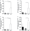
Ratios of MMP2/TIMP in heart tissue from CCC and DCM samples. Activity of MMP-2/expression protein TIMP stoichiometric ratios were calculated for samples CCC, DCM and control. (A) MMP-2/TIMP-1. (B) MMP-2/TIMP-2. (C) MMP-2/TIMP-3. (D) MMP-2/TIMP-4. *P-values were considered significant if p-value (0.05) corrected for multiple comparisons by Bonferroni’s method (corrected p-value = 0.05/3 = 0.0166).
Figure 6
Figure 6
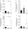
Ratios of MMP9/TIMP in heart tissue from CCC and DCM samples. Activity of MMP-9/expression protein TIMP stoichiometric ratios were calculated for samples CCC, DCM and control (A) MMP-9/TIMP-1. (B) MMP-9/TIMP-2. (C) MMP-9/TIMP-3. (D) MMP-9/TIMP-4. Groups were compared by a non-parametrical test (Mann–Whitney Rank Sum Test) with GraphPad Prism software (version 6.0; GraphPad). Results were expressed as mean ± SD and arbitrary units. *P-values were considered significant if p-value (0.05) corrected for multiple comparisons by Bonferroni’s method (corrected p-value = 0.05/3 = 0.0166).

Similar articles

Cited by

References

    1. Apte S. S., Parks W. C. (2015). Metalloproteinases: A Parade of Functions in Matrix Biology and an Outlook for the Future. Matrix Biol. 44–46:1-6. doi: 10.1016/j.matbio.2015.04.005 - DOI - PubMed
    1. Bautista-Lopez N. L., Morillo C. A., Lopez-Jaramillo P., Quiroz R., Luengas C., Silva S. Y., et al. . (2013). Matrix Metalloproteinases 2 and 9 as Diagnostic Markers in the Progression to Chagas Cardiomyopathy. Am. Heart J. 165 (4), 558–566. doi: 10.1016/j.ahj.2013.01.001 - DOI - PubMed
    1. Benchimol-Barbosa P. R., Barbosa-Filho J. (2009). Mechanical Cardiac Remodeling and New-Onset Atrial Fibrillation in Long-Term Follow-Up of Subjects With Chronic Chagas' Disease. Braz. J. Med. Biol. Res. 42, 251–262. doi: 10.1590/S0100-879X2009000300006 - DOI - PubMed
    1. Bocchi E. A., Guimaraes G., Tarasoutshi F., Spina G., Mangini S., Bacal F. (2009). Cardiomyopathy, Adult Valve Disease and Heart Failure in South America. Heart 95, 181–189. doi: 10.1136/hrt.2008.151225 - DOI - PubMed
    1. Bradham W. S., Bozkurt B., Gunasinghe H., Mann D., Spinale F. G. (2002). Tumor Necrosis Factor-Alpha and Myocardial Remodeling in Progression of Heart Failure: A Current Perspective. Cardiovasc. Res. 53, 822–830. doi: 10.1016/S0008-6363(01)00503-X - DOI - PubMed

Publication types

MeSH terms