Review
. 2022 Mar 24:2022:6797745.
doi: 10.1155/2022/6797745. eCollection 2022.

Anatomical Engineering and 3D Printing for Surgery and Medical Devices: International Review and Future Exponential Innovations

Affiliations
Review

Anatomical Engineering and 3D Printing for Surgery and Medical Devices: International Review and Future Exponential Innovations

José Cornejo et al. Biomed Res Int. .

Abstract

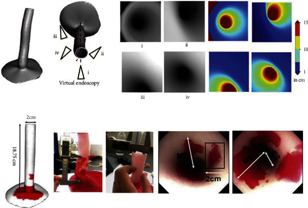
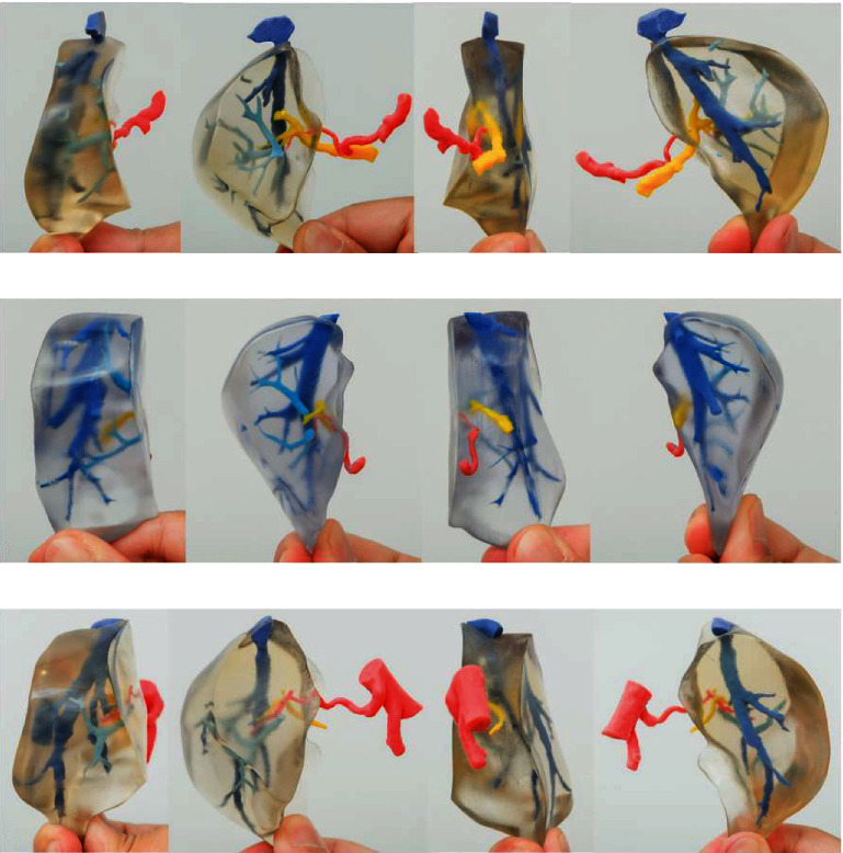
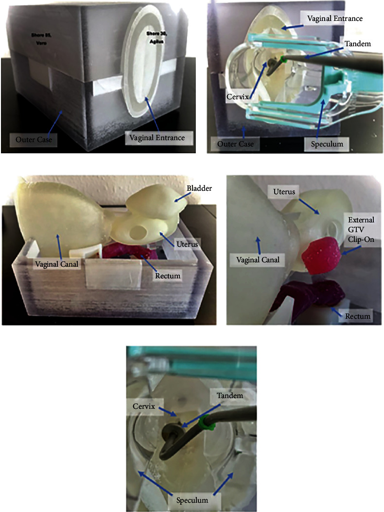
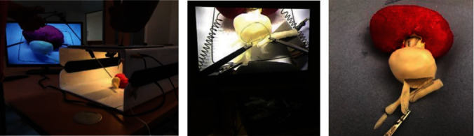
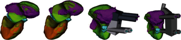
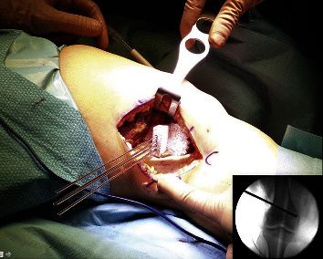
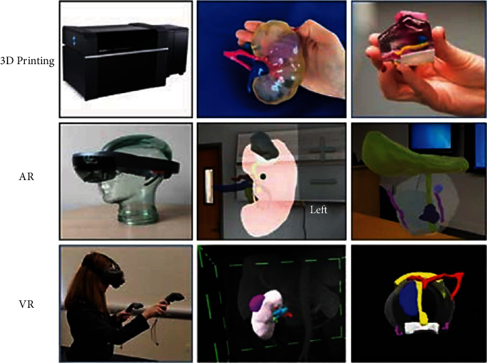
Three-dimensional printing (3DP) has recently gained importance in the medical industry, especially in surgical specialties. It uses different techniques and materials based on patients' needs, which allows bioprofessionals to design and develop unique pieces using medical imaging provided by computed tomography (CT) and magnetic resonance imaging (MRI). Therefore, the Department of Biology and Medicine and the Department of Physics and Engineering, at the Bioastronautics and Space Mechatronics Research Group, have managed and supervised an international cooperation study, in order to present a general review of the innovative surgical applications, focused on anatomical systems, such as the nervous and craniofacial system, cardiovascular system, digestive system, genitourinary system, and musculoskeletal system. Finally, the integration with augmented, mixed, virtual reality is analyzed to show the advantages of personalized treatments, taking into account the improvements for preoperative, intraoperative planning, and medical training. Also, this article explores the creation of devices and tools for space surgery to get better outcomes under changing gravity conditions.


Conflict of interest statement

The authors declare no conflicts of interest.

Figures

Figure 1
Figure 1
Computerized tomography (CT) of coronal, transverse, and sagittal planes that shows the orbital floor fracture. 3D-printed CT model is shown with the appropriate mesh, and this technique is used to provide a better approach in the treatment of skull fractures. Used with permission from the author Dr. Javier Asensio-Salazar.
Figure 2
Figure 2
Postprinting result. (a, b) The printed brain can be combined with the tumor print (red) in order to establish the relationships of the adjacent anatomic structures. (c) The tumor can be painted to determine the separation from the brain parenchyma. This is a cost-effective procedure that can help to improve the three-dimensional visualization of the brain tumors to improve the management [71]. Used with permission from Elsevier.
Figure 3
Figure 3
Simulated 3D-printed flexible, intact-heart model used in the surgical planning for complex total cavopulmonary connection. LPA: left pulmonary artery; RPA: right pulmonary artery; RSVC: right superior vena cava; IVC: inferior vena cava; RA: right atrium; Ao: aorta; LA: left atrium; LV: left ventricle; RV: right ventricle; SVC: superior vena cava [117]. Used with permission from Elsevier.
Figure 4
Figure 4
3D modeling processes for 3D aortic model. (a) Individual modeling of the central line of graft using the patient's computed tomography (CT) images. (b) 3D computerized models for the graft guides consisting of the visualization (left) and the marking (right). (c) 3D printed guides. (d) Intraoperative view of the 3D printed guides in the TAAA. SMA: superior mesenteric artery; CA: celiac artery; RA: renal artery; TAAA: thoracoabdominal aortic aneurysm [137]. Used with permission from Elsevier.
Figure 5
Figure 5
(a) Visualization of 3D depth maps models of the esophagus. Techniques to determine the localization, length, and depth of the Barret's lesions through the endoscopy camera. (b) 3D printed model of the esophagus that shows the measurements for the lesions (C and M) and also the endoscopy video frames are shown [163]. Used with permission from Elsevier.
Figure 6
Figure 6
Four aspects of 3D print MSG with portal venous variations. (a)–(c) Shows the different anatomical variations of the portal vein in relation to the medial segmental graft. 3D: three-dimensional; MSG: medial segment graft; P4: portal vein to the medial segment [184]. Used with permission from Elsevier.
Figure 7
Figure 7
(a) Closed 3D printed model. (b) The speculum entering the vaginal canal. (c) 3D printed intrapelvic organs and its anatomical position. (d) Presence of 3D printed gross tumor attached to the uterine body. (e) Tandem used in brachytherapy procedures is inserted through the speculum and placed inside the cervix and uterine canal. These phantom models help in teaching physicians the process of intracavitary procedures in cervical cancers [201]. Used with permission from Elsevier.
Figure 8
Figure 8
Pyeloplasty is a surgical procedure performed in cases of ureteropelvic junction (UPJ) obstruction. (a)–(c) For surgical training, the 3D-printed models are placed within laparoscopic consoles to recreate and performed the pyeloplasty procedure [226]. Used with permission from Elsevier.
Figure 9
Figure 9
Computer-assisted preoperative planning of a scaphoid fracture. (a, b) Green: scaphoid and lunate bones of the hand. Light blue: proximal scaphoid fragment. Violet: distal scaphoid fragment. (a) Scaphoid fragments before the reduction. (b) Scaphoid fragments after the reduction. (c, d) 3D-printed K-wires are placed in to reduce the two scaphoid fragments in order to have a better sealing of the bone [232]. Used with permission from Elsevier.
Figure 10
Figure 10
Surgical correction of valgus knees through the use of osteotomy procedure, which consist in the removal or insertion of a wedge of bone near a damaged cartilage in order to provide a well-distributed weight area over the affected knee. Four 3D-printed Kirschner wires are inserted through the guide. Depth and orientation checked under fluoroscopy [249]. Used with permission from Elsevier.
Figure 11
Figure 11
Examples of 3D printing (top), AR (middle), and VR (bottom) technologies. Top row: Stratasys J750 Digital Anatomy Printer; 3D printed kidney tumor model with the kidney in clear, collecting system (semi-transparent), lesion (purple), renal artery (pink), renal vein (light blue), and collecting system (dark blue); 3D printed prostate cancer model with the prostate clear, lesion—blue, neurovascular bundles (yellow), rectal wall (white), bladder neck, and urethra (pink). Middle row: HoloLens-AR headset; AR kidney tumor model shown projected in a room with the kidney (pink), tumor (gray), artery (red), vein (blue), and collecting system (yellow); prostate cancer model shown projected in a room with the prostate (transparent), lesions (blue), neurovascular bundles (purple), bladder neck, and collecting system (yellow). Bottom row: person wearing HTC Vive VR headset; kidney tumor model; prostate cancer model configuration colours as at the middle row picture, also with arterial supply (red) [274]. Used with permission from Elsevier.
Figure 12
Figure 12
3D printed instruments for space surgery applications [290]. Used with permission from Elsevier.

References

    1. Gibson I., Rosen D., Stucker B. Additive Manufacturing Technologies . New York, NY: Springer; 2015. Applications for additive manufacture; pp. 451–474.
    1. Abedin-Nasab M. H. Handbook of Robotic and Image-Guided Surgery . Elsevier; 2019.
    1. Cornejo J., Cornejo-Aguilar J. A., Perales-Villarroel J. P. Innovaciones internacionales en robótica médica para mejorar el manejo del paciente en Perú. Revista de la Facultad de Medicina Humana . 2019;19(4):105–113. doi: 10.25176/RFMH.v19i4.2349. - DOI
    1. Desai J. P. Encyclopedia Of Medical Robotics, The (In 4 Volumes) World Scientific; 2018.
    1. Cornejo J., Cornejo-Aguilar J. A., Palomares R. Biomedik surgeon: surgical robotic system for training and simulation by medical students in Peru. 2019 International Conference on Control of Dynamical and Aerospace Systems (XPOTRON); 2019; Arequipa, Peru. pp. 1–4. - DOI

LinkOut - more resources