

POCUS for Nephrologists: Basic Principles and a General Approach
- PMID: 35372985
- PMCID: PMC8785785
- DOI: 10.34067/KID.0002482021
POCUS for Nephrologists: Basic Principles and a General Approach
Abstract
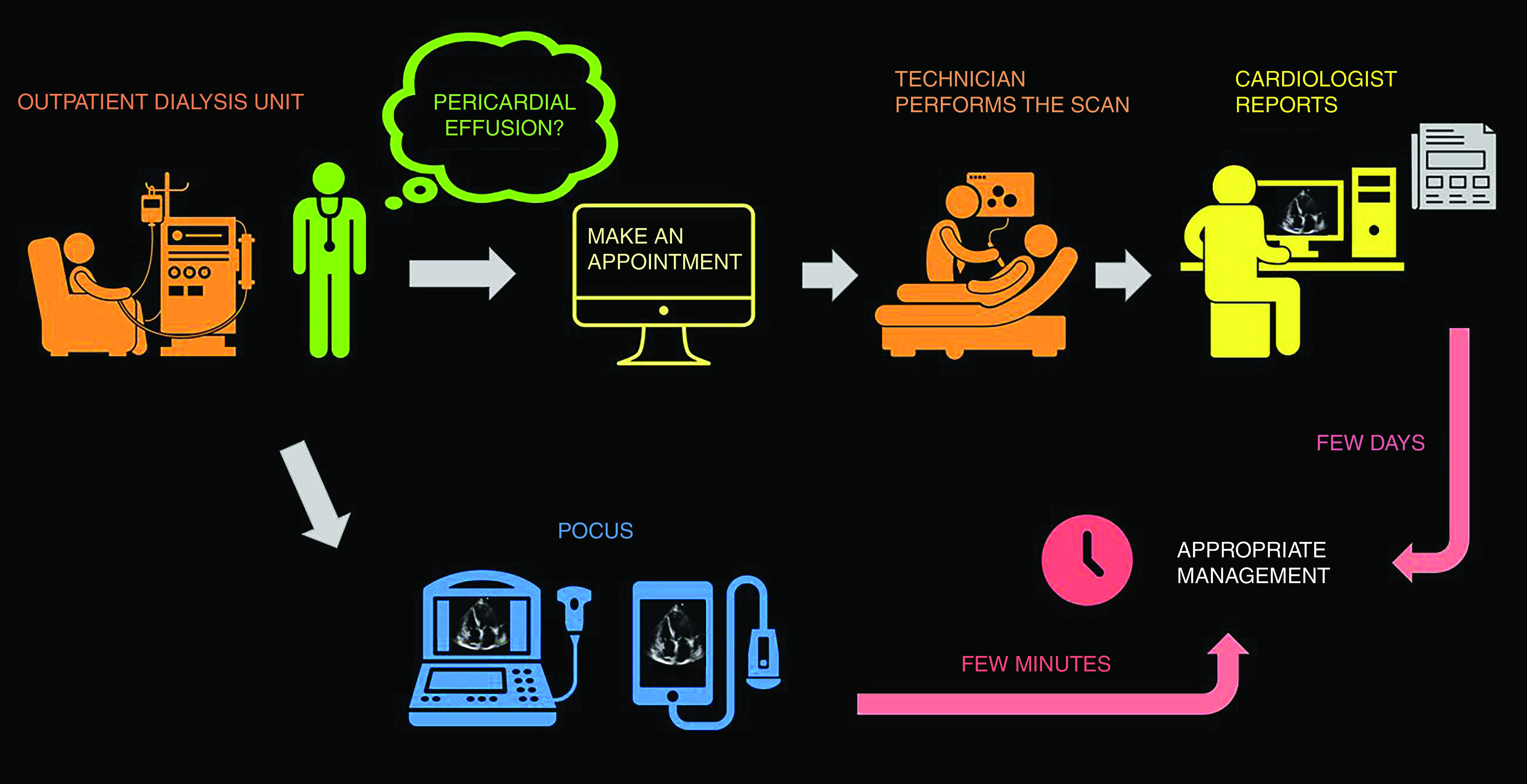
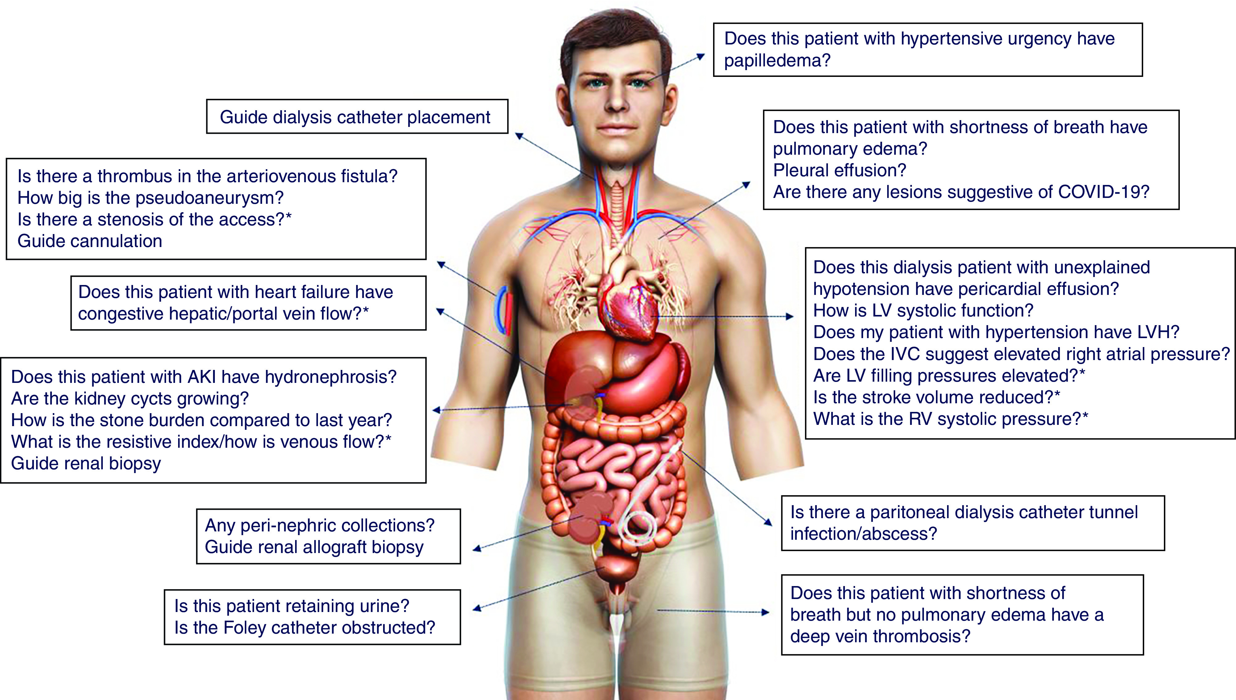
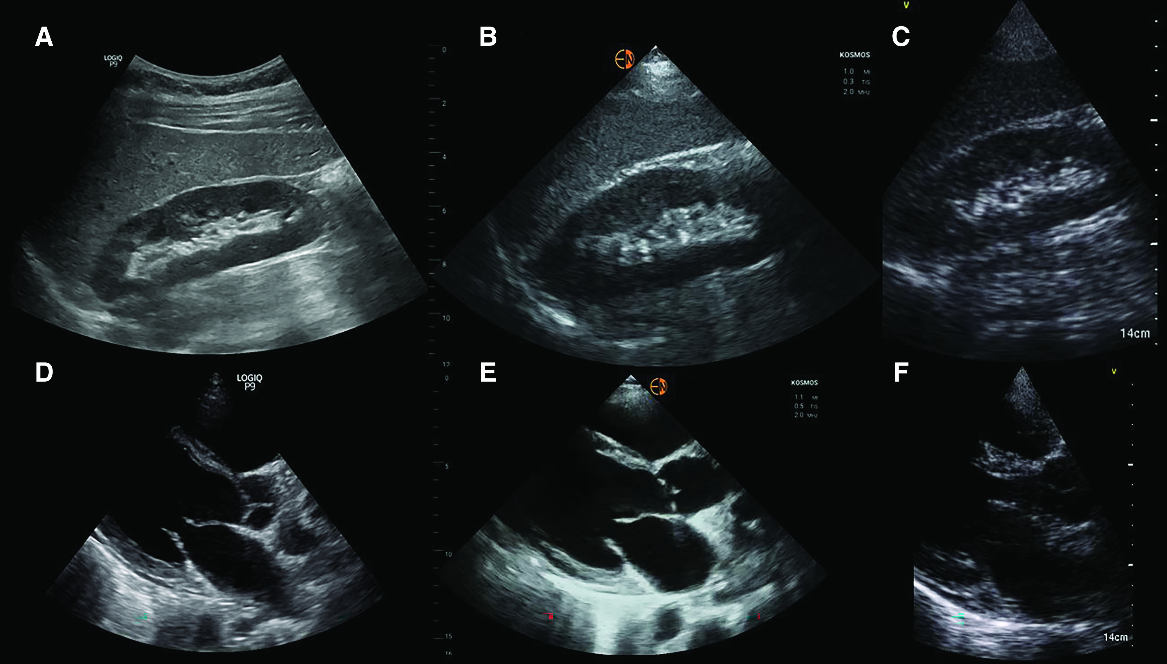
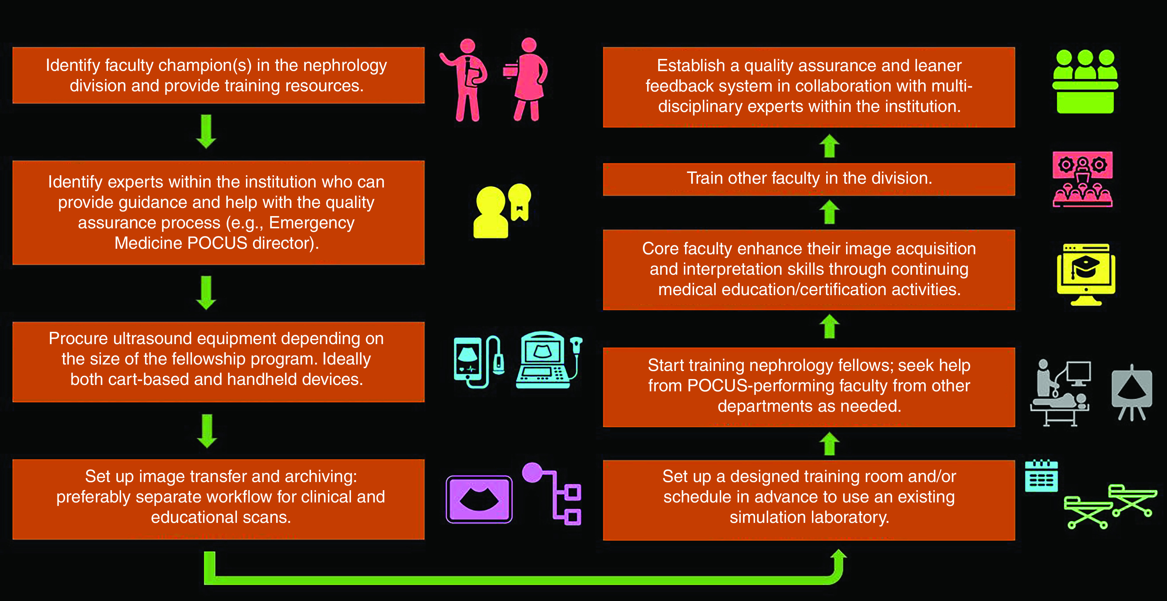
Point-of-care ultrasonography (POCUS) has evolved as a valuable adjunct to physical examination in the recent past and various medical specialties have embraced it. However, POCUS training and scope of practice remain relatively undefined in nephrology. The utility of diagnostic POCUS beyond kidney and vascular access is under-recognized. Assessment of fluid status is a frequent dilemma faced by nephrologists in day-to-day practice where multiorgan POCUS can enhance the sensitivity of conventional physical examination. POCUS also reduces fragmentation of care, facilitates timely diagnosis, and expedites management. Although the need for further imaging studies is obviated in selected patients, POCUS is not meant to serve as an alternative to consultative imaging. In addition, the utility of POCUS depends on the skills and experience of the operator, which in turn depend on the quality of training. In this review, we discuss the rationale behind nephrologists performing POCUS, discuss patient examples to illustrate the basic principles of focused ultrasonography, and share our experience-based opinion about developing a POCUS training program at the institutional level.
Keywords: POCUS; clinical nephrology; educational personnel; nephrologists; nephrology; physical examination; point of care ultrasound; sonography; training.
Copyright © 2021 by the American Society of Nephrology.
Conflict of interest statement
All authors have nothing to disclose.
Figures
References
-
- Narula J, Chandrashekhar Y, Braunwald E: Time to add a fifth pillar to bedside physical examination: Inspection, palpation, percussion, auscultation, and insonation. JAMA Cardiol 3: 346–350, 2018 - PubMed
-
- Accreditation Council for Graduate Medical Education: ACGME Program Requirements for Graduate Medical Education in Emergency Medicine. 2020. Available at: https://www.acgme.org/Portals/0/PFAssets/ProgramRequirements/110_Emergen.... Accessed May 4, 2021
-
- LoPresti CM, Jensen TP, Dversdal RK, Astiz DJ: Point-of-care ultrasound for internal medicine residency training: A position statement from the Alliance of Academic Internal Medicine. Am J Med 132: 1356–1360, 2019 - PubMed
-
- Sohaey R, Di Salvo DN, Bluth EI, Lockhart ME, Cohen HL, Pellerito JS, Baltarowich OH, Nisenbaum HL, Coleman BG: Medical student ultrasound education: The radiology chair weighs in. Ultrasound Q 37: 3–9, 2021 - PubMed
-
- Bahner DP, Goldman E, Way D, Royall NA, Liu YT: The state of ultrasound education in U.S. medical schools: Results of a national survey. Acad Med 89: 1681–1686, 2014 - PubMed
Publication types
MeSH terms
LinkOut - more resources
Full Text Sources
Miscellaneous
