. 2020 Dec 22;2(2):355-364.
doi: 10.34067/KID.0004772020. eCollection 2021 Feb 25.

Proximal Tubular Oxidative Metabolism in Acute Kidney Injury and the Transition to CKD

Affiliations

Proximal Tubular Oxidative Metabolism in Acute Kidney Injury and the Transition to CKD

Jennifer A Schaub et al. Kidney360. .

Abstract

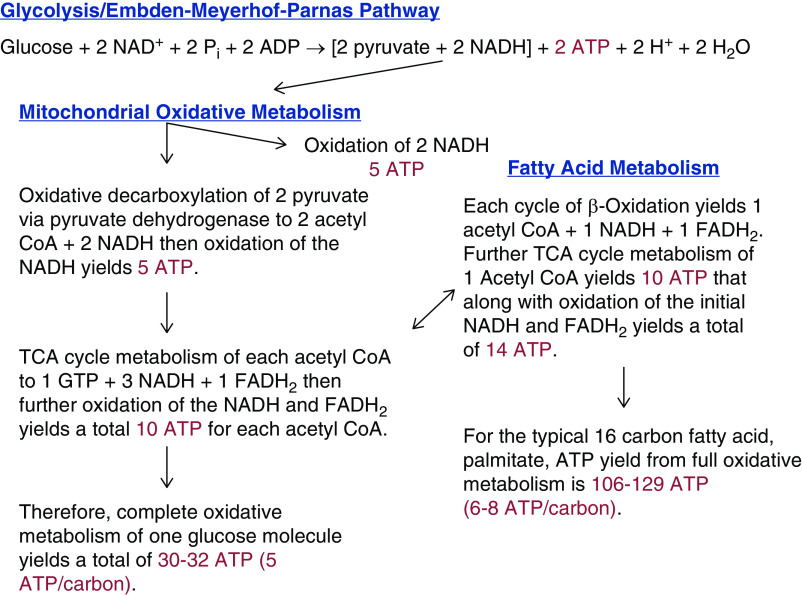
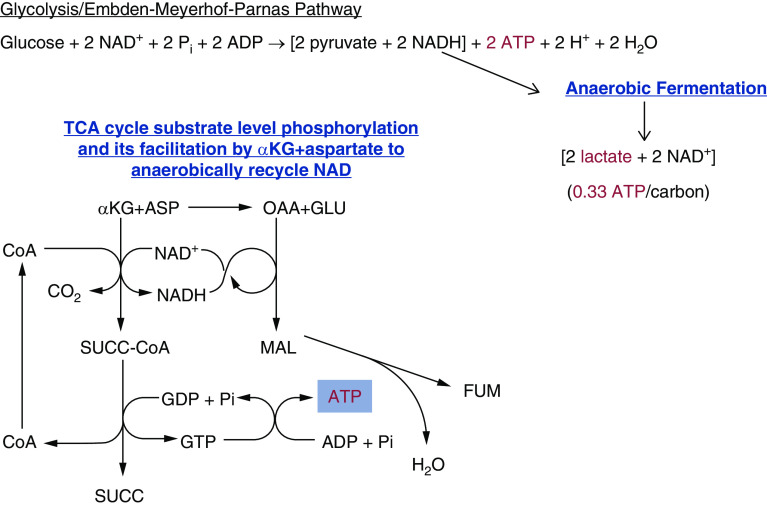
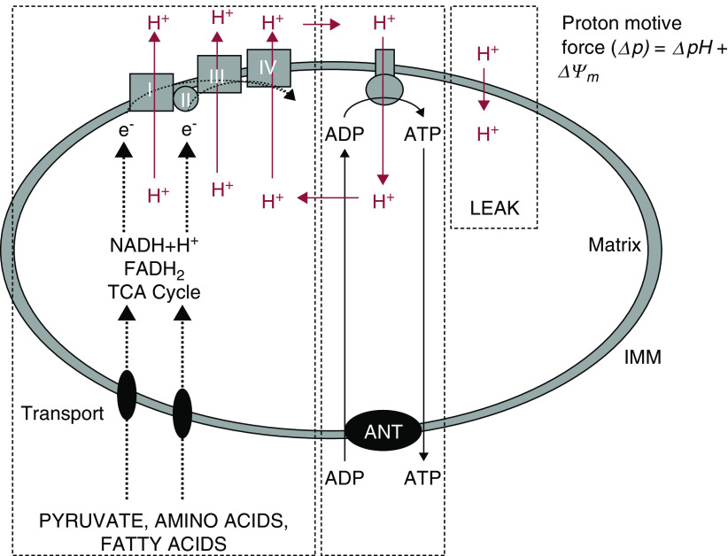
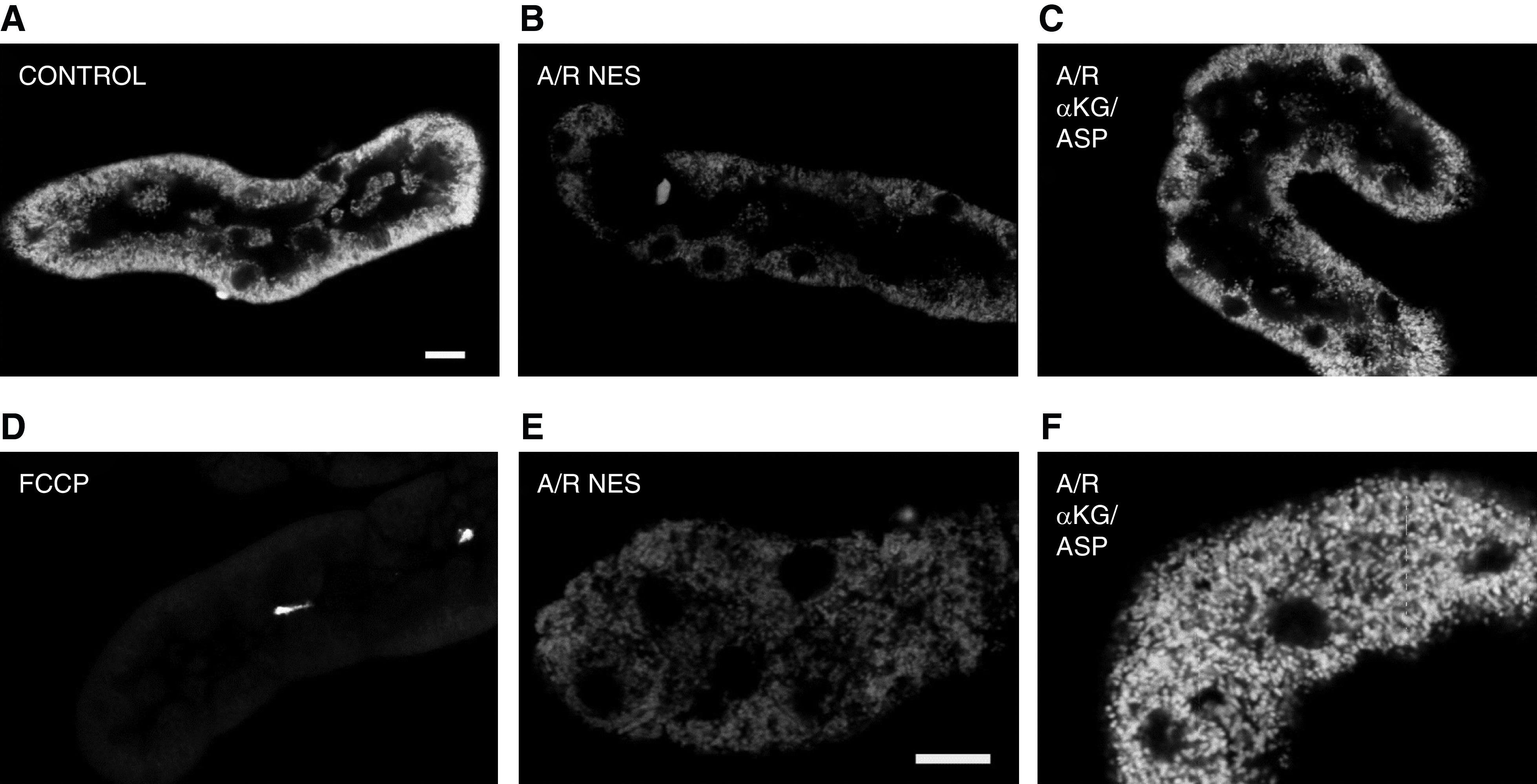
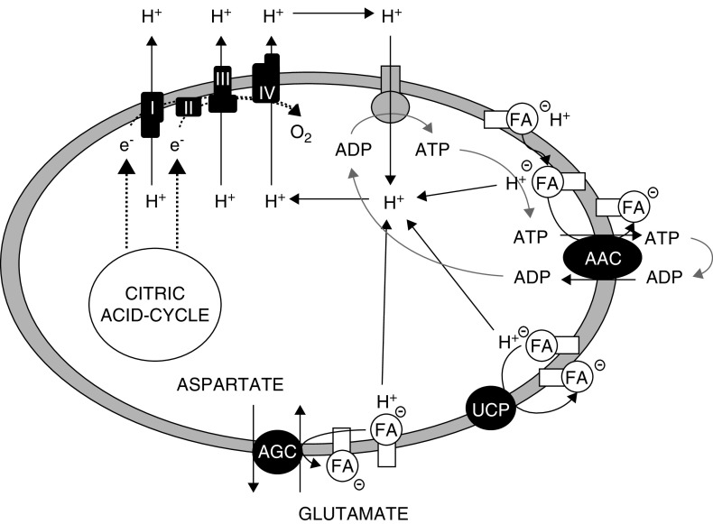
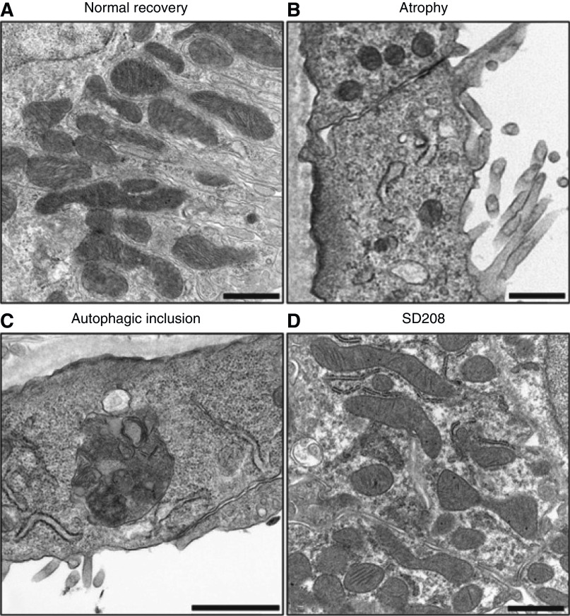
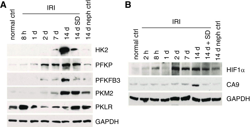
The proximal tubule relies on oxidative mitochondrial metabolism to meet its energy needs and has limited capacity for glycolysis, which makes it uniquely susceptible to damage during AKI, especially after ischemia and anoxia. Under these conditions, mitochondrial ATP production is initially decreased by several mechanisms, including fatty acid-induced uncoupling and inhibition of respiration related to changes in the shape and volume of mitochondria. Glycolysis is initially insufficient as a source of ATP to protect the cells and mitochondrial function, but supplementation of tricarboxylic acid cycle intermediates augments anaerobic ATP production, and improves recovery of mitochondrial oxidative metabolism. Incomplete recovery is characterized by defects of respiratory enzymes and lipid metabolism. During the transition to CKD, tubular cells atrophy but maintain high expression of glycolytic enzymes, and there is decreased fatty acid oxidation. These metabolic changes may be amenable to a number of therapeutic interventions.

Keywords: AKI; CKD; aTP; acute kidney injury and ICU nephrology; basic science; glycolysis; metabolism; mitochondria; tricarboxylic acid cycle.


Conflict of interest statement

M.A. Venkatachalam reports honoraria from Surrozen. All remaining authors have nothing to disclose.

Figures

Figure 1.
Figure 1.
Pathways of aerobic tubule ATP production. Glycolysis of one glucose molecule yields two pyruvates and two molecules of ATP. In the TCA cycle, further aerobic metabolism of these pyruvates yields an additional 30–32 ATPs or five ATPs/carbon. Amino acids (via transamination) and carboxylic acids are aerobically metabolized in this fashion. Fatty acids enter the TCA cycle via β-oxidation and yield a net of 6–8 ATPs per carbon. ATP yields are corrected for estimated efficiency as discussed in refs. (–7). CoA, coenzyme A; FADH2, reduced flavin adenine dinucleotide; NADH, reduced NAD; TCA, tricarboxylic acid.
Figure 2.
Figure 2.
Pathways of anaerobic tubule ATP production. Under anaerobic conditions, pyruvate undergoes anaerobic fermentation and generates lactate and only two ATPs. A limited amount of ATP can also be generated anaerobically during TCA cycle substrate level phosphorylation. ADP, adenosine diphosphate; αKG, α-ketoglutarate; ASP, aspartate; CoA, Coenzyme A; FADH2, reduced flavin adenine dinucleotide; FUM, fumarate; GDP, guanosine diphosphate; GTP, guanosine triphosphate; MAL, malate; NADH, reduced NAD; OAA, oxaloacetate; Pi, inorganic phosphate; SUCC, succinate; TCA, tricarboxylic acid.
Figure 3.
Figure 3.
Mitochondrial energy transduction. NADH and FADH2 deliver electrons to the electron transport chain on the inner mitochondrial membrane where they are progressively transferred to oxygen. Hydrogen ions are pumped out of the mitochondrial matrix to generate an electrochemical potential, which powers the F1F0-ATPAse on the inner mitochondrial membrane to drive phosphorylation of ADP to ATP. ADP, adenosine diphosphate; FADH2, reduced flavin adenine dinucleotide; IMM, inner mitochondrial membrane; NADH, reduced NAD; TCA, tricarboxylic acid. Adapted from ref. (17).
Figure 4.
Figure 4.
Differential expression of glycolytic and gluconeogenic enzymes along the nephron. (A) Expression of enzymes involved in glycolysis. (B) Expression of enzymes involved in gluconeogenesis. GL, glomerulus; PCT, early proximal convoluted tubule; PCT2, late proximal convoluted tubule; PST, proximal straight tubule; TL, loop of Henle; MAL, medullary thick ascending limb; CAL, cortical ascending limb; CT, distal convoluted tubule; CCT, cortical collecting tubule; MCT, medullary collecting tubule. Figure is from ref. (23) with permission.
Figure 5.
Figure 5.
Tricarboxylic acid cycle intermediate supplementation improves mitochondrial membrane potential of isolated proximal tubules. Isolated proximal tubules showing mitochondrial tetramethylrhodamine methyl ester (TMRM) fluorescence, which detects changes in mitochondrial membrane potential. (A) In control samples, mitochondria in the basal portion of tubule cells are brightly stained, indicating high membrane potential. (B) After anoxia/reperfusion (A/R) with no extra substrates (NES), TMRM fluorescence is decreased indicating decreased mitochondrial membrane potential. (C) Supplementation of α-ketoglutarate + aspartate (αKG/ASP) during only reoxygenation improves mitochondrial fluorescence. (D) In the presence of carbonyl fluoride-m-chlorophenylhydrazone (FCCP), a mitochondrial uncoupler that completely eliminates membrane potential, there is virtually no fluorescence. (E) High magnification image of A/R with NES. (F) High magnification image of A/R with αKG/ASP. Sizing bars are 10 μm. Figure is from from ref. (13) with permission.
Figure 6.
Figure 6.
Fatty acid cycling across the inner mitochondrial membrane decrease membrane potential and uncouples oxidative phosphorylation. Fatty acids (FA) enter the mitochondrial matrix via nonionic diffusion and deliver their protons to the matrix, which short-circuits the normal path of proton re-entry via the ATPase. Fatty acid ions can then be recycled out of the matrix by membrane carriers including ADP/ATP carrier (AAC), glutamate-aspartate transport (AGC) and uncoupling proteins (UCP). Figure is from ref. (34) with permission.
Figure 7.
Figure 7.
Mitochondrial pathology in proximal tubules during the AKI to CKD transition. Electron micrographs showing (A) normal recovery, (B) atrophy with loss and simplification of mitochondria (C) autophagolysosome associated with atrophy and (D) TGF-β receptor antagonist, SD-208, improves mitochondrial number and morphology. Figure is from ref. (46) with permission.
Figure 8.
Figure 8.
Changes in glycolysis enzymes and hypoxia markers during transition to CKD after ischemia-reperfusion injury. (A) Western blot showing increases in glycolytic enzyme protein expression during progression to CKD after ischemia/reperfusion injury (IRI). (B) Western blot showing increases in hypoxia markers after IRI. HK2, Hexokinase 2; PFKP, Phosphofructokinase platelet isoform; PFKFB3, 6-phosphofructo-2-kinase/fructose-2,6-bisphosphate-3; PKM2, Pyruvate kinase M2; PKLR, pyruvate kinase liver/RBC isoform; GAPDH, glyceraldehyde-3-phosphate dehydrogenase; CA9, carbonic anhydrase 9; SD, SD208, a TGF-β receptor antagonist. Nephrectomy control 14 days after sham left-kidney ischemia (14 days neph ctrl). Figure is from ref. (46) with permission.

References

    1. Mehr A, Tran MT, Ralto KM, Leaf DE, Washco V, Messmer J, Lerner A, Kher A, Kim SH, Khoury CC, Herzig SJ, Trovato ME, Simon-Tillaux N, Lynch MR, Thadhani RI, Clish CB, Khabbaz KR, Rhee EP, Waikar SS, Berg AH, Parikh SM: De novo NAD+ biosynthetic impairment in acute kidney injury in humans. Nat Med 24: 1351–1359, 2018. 10.1038/s41591-018-0138-z - DOI - PMC - PubMed
    1. Szeto HH: Pharmacologic approaches to improve mitochondrial function in AKI and CKD. J Am Soc Nephrol 28: 2856–2865, 2017. 10.1681/ASN.2017030247 - DOI - PMC - PubMed
    1. Cameron RB, Beeson CC, Schnellmann RG: Development of therapeutics that induce mitochondrial biogenesis for the treatment of acute and chronic degenerative diseases. J Med Chem 59: 10411–10234, 2016. 10.1021/acs.jmedchem.6b00669 - DOI - PMC - PubMed
    1. Romano AH, Conway T: Evolution of carbohydrate metabolic pathways. Res Microbiol 147: 448–455, 1996. 10.1016/0923-2508(96)83998-2 - DOI - PubMed
    1. Schmidt-Rohr K: Oxygen is the high-energy molecule powering complex multicellular life: Fundamental corrections to traditional bioenergetics. ACS Omega 5: 2221–2233, 2020. 10.1021/acsomega.9b03352 - DOI - PMC - PubMed

Publication types

MeSH terms