. 2022 May;36(5):e24393.
doi: 10.1002/jcla.24393. Epub 2022 Apr 4.

Hsa_circ_0060927 participates in the regulation of Caudatin on colorectal cancer malignant progression by sponging miR-421/miR-195-5p

Affiliations

Hsa_circ_0060927 participates in the regulation of Caudatin on colorectal cancer malignant progression by sponging miR-421/miR-195-5p

Juan Chen et al. J Clin Lab Anal. 2022 May.

Abstract

Background: Caudatin is extracted from radix cynanchi bungei and has an inhibitory effect on cancer progression. The study aims to reveal the impacts of hsa_circ_0060927 on Caudatin-mediated colorectal cancer (CRC) development and the underneath mechanism.

Methods: The expression levels of hsa_circ_0060927, microRNA-421 (miR-421) and miR-195-5p were detected by quantitative real-time reverse transcription-polymerase chain reaction. The protein expression was analyzed by Western blot or immunohistochemistry assay. Cell viability and proliferation were analyzed by 3-(4,5)-dimethylthiahiazo (-z-y1)-3,5-di-phenytetrazoliumromide or 5-Ethynyl-29-deoxyuridine assay. Cell apoptosis was quantified by flow cytometry analysis. Cell migration and invasion were investigated by transwell assay. The putative associations among hsa_circ_0060927, miR-421 and miR-195-5p were predicted by the starbase online database, and identified by dual-luciferase reporter, RNA pull-down and RNA immunoprecipitation (RIP) assays. The impacts of Caudatin treatment on tumor growth in vivo were revealed by a xenograft tumor model assay.

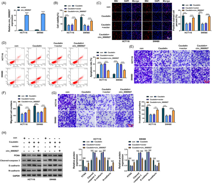
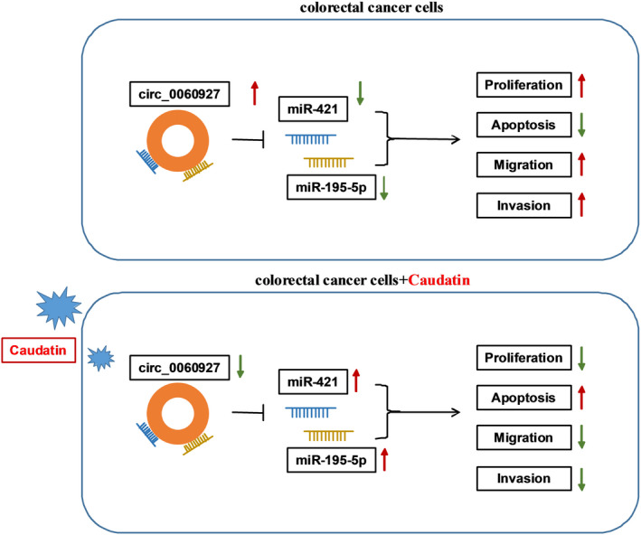
Results: Hsa_circ_0060927 expression was significantly upregulated, whereas miR-421 and miR-195-5p were downregulated in CRC tissues and cells compared with control groups. Hsa_circ_0060927 expression was closely associated with lymph node metastasis and tumor-node-metastasis stage. Caudatin treatment significantly decreased hsa_circ_0060927 expression but increased miR-421 and miR-195-5p expression. Caudatin exposure suppressed CRC cell proliferation, migration and invasion, and induced cell apoptosis; however, hsa_circ_0060927 overexpression hindered these impacts. Additionally, hsa_circ_0060927 was associated with miR-421/miR-195-5p. Depletion of miR-421 or miR-195-5p attenuated the influences of hsa_circ_0060927 silencing on CRC development. Furthermore, Caudatin treatment repressed tumor growth in vivo.

Conclusion: Caudatin inhibited CRC cell malignancy through the hsa_circ_0060927/miR-421/miR-195-5p pathway, which provided a potential therapeutic agent for CRC.

Keywords: Caudatin; colorectal cancer; hsa_circ_0060927; miR-195-5p; miR-421.


Conflict of interest statement

The authors declare that they have no competing interests.

Figures

FIGURE 1
FIGURE 1
Caudatin exposure suppressed cell proliferation while induced cell apoptosis in CRC cells. (A and B) The effects of Caudatin (0, 25, 50 and 100 μM) treatment on the viability of HCT116 and SW480 cells were determined by MTT assay. (C) The impact of 50 μM Caudatin on HCT116 and SW480 cell proliferation was determined by EDU assay. (D) Flow cytometry analysis was performed to analyze the influence of 50 μM Caudatin on the apoptosis of HCT116 and SW480 cells. (E) Western blot analysis was employed to detect the effects of 50 μM Caudatin treatment on the protein levels of PCNA and Cleaved‐caspase 3 in HCT116 and SW480 cells. Con: 0 μM. *p < 0.05, **p < 0.01 and ***p < 0.001. CRC, Colorectal cancer; EDU, 5‐Ethynyl‐29‐deoxyuridine; MTT, 3‐(4,5)‐dimethylthiahiazo (‐z‐y1)‐3,5‐di‐phenytetrazoliumromide; PCNA, proliferating cell nuclear antigen
FIGURE 2
FIGURE 2
Caudatin treatment hindered the metastasis of HCT116 and SW480 cells. (A and B) The impacts of 50 μM Caudatin on the migration and invasion of HCT116 and SW480 cells were disclosed by transwell assay. (C) The influences of 50 μM Caudatin on the protein levels of E‐cadherin and N‐cadherin were revealed by Western blot in HCT116 and SW480 cells. **p < 0.01 and ***p < 0.001
FIGURE 3
FIGURE 3
Caudatin treatment decreased hsa_circ_0060927 expression. (A and B) Hsa_circ_0060927 expression was detected by qRT‐PCR in CRC tissues (n = 68), paracancerous non‐cancerous colorectal tissues (n = 68) and FHC, HCT116 and SW480 cells. (C and D) The stability of hsa_circ_0060927 was determined by RNase R treatment assay. (E and F) The effect of 50 μM Caudatin treatment on hsa_circ_0060927 expression was determined by qRT‐PCR in HCT116 and SW480 cells. ***p < 0.001. CRC, Colorectal cancer
FIGURE 4
FIGURE 4
Caudatin inhibited CRC progression by modulating hsa_circ_0060927 expression. (A) The overexpression efficiency of hsa_circ_0060927 was detected by qRT‐PCR in HCT116 and SW480 cells. (B and C) MTT and EDU assays were performed to investigate the impacts of Caudatin treatment and hsa_circ_0060927 overexpression on the viability and proliferation of HCT116 and SW480 cells. (D) Flow cytometry analysis was employed to reveal the impacts of Caudatin treatment and hsa_circ_0060927 overexpression on the apoptosis of HCT116 and SW480 cells. (E–G) The influences of Caudatin exposure and ectopic hsa_circ_0060927 expression on HCT116 and SW480 cell migration and invasion were revealed by transwell assay. (H) Western blot was carried out to determine the impacts of Caudatin treatment and hsa_circ_0060927 overexpression on the protein levels of PCNA, Cleaved‐caspase 3, E‐cadherin and N‐cadherin in HCT116 and SW480 cells. **p < 0.01 and ***p < 0.001. CRC, Colorectal cancer; EDU, 5‐Ethynyl‐29‐deoxyuridine; MTT, 3‐(4,5)‐dimethylthiahiazo (‐z‐y1)‐3,5‐di‐phenytetrazoliumromide; PCNA, proliferating cell nuclear antigen
FIGURE 5
FIGURE 5
Hsa_circ_0060927 was associated with miR‐421 and miR‐195‐5p in CRC cells. (A) Cytoplasmic and nuclear RNA analysis was performed to detect the expression levels of GAPDH, U6 and hsa_circ_0060927. (B) The efficiency of si‐circ_0060927 in reducing has_circ_0060927 expression was determined by qRT‐PCR in HCT116 and SW480 cells. (C) Starbase and circBANK online databases were employed to predict the possible target miRNAs of hsa_circ_0060927. (D) The effects of hsa_circ_0060927 silencing on the expression levels of the 10 miRNAs possessing the binding sites of hsa_circ_0060927 were analyzed by qRT‐PCR. (E) Starbase online database was performed to predict the putative binding sites of hsa_circ_0060927 for miR‐421 and miR‐195‐5p. (F) The efficiency of miR‐421 and miR‐195‐5p overexpression was determined by qRT‐PCR in HCT116 and SW480 cells. (G) Dual‐luciferase reporter assay was employed to detect luciferase activity in HCT116 and SW480 cells. (H and I) RNA pull‐down and RIP assays were conducted to analyze the direct binding relationships between hsa_circ_0060927 and miRNAs (miR‐421 and miR‐195‐5p). (J) The expression levels of miR‐421 and miR‐195‐5p were detected by qRT‐PCR in 68 pairs of CRC tissues and paracancerous non‐cancerous colorectal tissues. (K) The linear relationship between hsa_circ_0060927 and miR‐421 or miR‐195‐5p expression was revealed by Spearman correlation analysis. ***p < 0.001. CRC, Colorectal cancer
FIGURE 6
FIGURE 6
Hsa_circ_0060927 silencing inhibited CRC progression by sponging miR‐421 or miR‐195‐5p. (A and B) The efficiency of miR‐421 and miR‐195‐5p knockdown was detected by qRT‐PCR in HCT116 and SW480 cells. (C and D) The effects of hsa_circ_0060927 silencing and miR‐421/miR‐195‐5p inhibitor on HCT116 and SW480 cell viability and proliferation were determined by MTT and EDU assays. (E) The impacts of hsa_circ_0060927 silencing and miR‐421/miR‐195‐5p inhibitor on the apoptosis of HCT116 and SW480 cells were explained by flow cytometry analysis. (F–H) Transwell assay was performed to analyze the influences of si‐circ_0060927 and anti‐miR‐421 or anti‐miR‐195‐5p on the migration and invasion of HCT116 and SW480 cells. (I) Western blot was conducted to investigate the effects of circ_0060927 silencing and miR‐421 inhibitor or miR‐195‐5p inhibitor on the protein levels of PCNA, Cleaved‐caspase 3, E‐cadherin and N‐cadherin in HCT116 and SW480 cells. ***p < 0.001. EDU, 5‐Ethynyl‐29‐deoxyuridine; MTT, 3‐(4,5)‐dimethylthiahiazo (‐z‐y1)‐3,5‐di‐phenytetrazoliumromide; PCNA, proliferating cell nuclear antigen
FIGURE 7
FIGURE 7
Caudatin regulated miR‐421 and miR‐195‐5p expression by repressing hsa_circ_0060927. (A and B) MiR‐421 and miR‐195‐5p expression were detected by qRT‐PCR in FHC, HCT116 and SW480 cells. (C and D) The impacts of Caudatin treatment on the expression levels of miR‐421 and miR‐195‐5p were determined by qRT‐PCR in HCT116 and SW480 cells. (E and F) The impacts of Caudatin treatment and hsa_circ_0060927 overexpression on the expression levels of miR‐421 and miR‐195‐5p were revealed by qRT‐PCR in HCT116 and SW480 cells. ***p < 0.001. CRC, Colorectal cancer
FIGURE 8
FIGURE 8
Caudatin treatment suppressed tumor growth in vivo. (A–B) The impacts of Caudatin treatment on the volume and weight of tumors were revealed in vivo. (C) The protein expression of Ki‐67 and Cleaved‐caspase 3 was analyzed by IHC assay. (D) qRT‐PCR was employed to investigate the effects of Caudatin treatment on the expression levels of hsa_circ_0060927, miR‐421 and miR‐195‐5p in the forming tumors from SW480 cells. (E) Western blot analysis was conducted to determine the influences of Caudatin on the protein levels of Ki‐67, PCNA, Cleaved‐caspase 3, E‐cadherin and N‐cadherin in the forming tumors from SW480 cells. **p < 0.01 and ***p < 0.001. PCNA, proliferating cell nuclear antigen
FIGURE 9
FIGURE 9
The schematic illustration showed the mechanisms underlying Caudatin regulating CRC malignant progression. CRC, Colorectal cancer

Similar articles

Cited by

References

    1. Global Burden of Disease Cancer C , Fitzmaurice C, Allen C, et al. Global, Regional, and National Cancer Incidence, mortality, years of life lost, years lived with disability, and disability‐adjusted life‐years for 32 cancer groups, 1990 to 2015: a systematic analysis for the Global Burden of Disease Study. JAMA Oncol. 2017;3(4):524‐548. - PMC - PubMed
    1. Peng HX, Yang L, He BS, et al. Combination of preoperative NLR, PLR and CEA could increase the diagnostic efficacy for I‐III stage CRC. J Clin Lab Anal. 2017;31(5):e22075. - PMC - PubMed
    1. Brody H. Colorectal cancer. Nature. 2015;521(7551):S1. - PubMed
    1. Zhen X, Choi HS, Kim JH, et al. Caudatin isolated from Cynanchum auriculatum inhibits breast cancer stem cell formation via a GR/YAP signaling. Biomolecules. 2020;10(6):925. - PMC - PubMed
    1. Song J, Ding W, Liu B, et al. Anticancer effect of caudatin in diethylnitrosamine‐induced hepatocarcinogenesis in rats. Mol Med Rep. 2020;22(2):697‐706. - PMC - PubMed