. 2022 Sep;11(18):3529-3541.
doi: 10.1002/cam4.4706. Epub 2022 Apr 4.

Identification and validation of a ferroptosis-related gene signature for predicting survival in skin cutaneous melanoma

Affiliations

Identification and validation of a ferroptosis-related gene signature for predicting survival in skin cutaneous melanoma

Shuai Ping et al. Cancer Med. 2022 Sep.

Abstract

Purpose: Ferroptosis plays a crucial role in the initiation and progression of melanoma. This study developed a robust signature with ferroptosis-related genes (FRGs) and assessed the ability of this signature to predict OS in patients with skin cutaneous melanoma (SKCM).

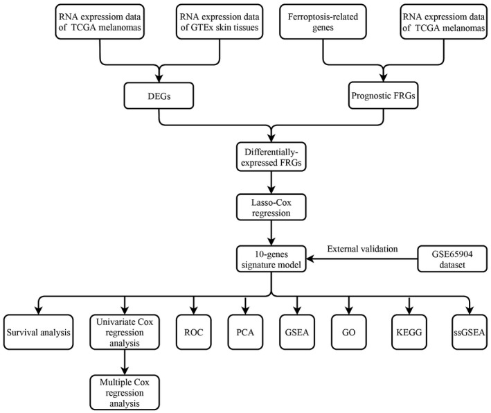
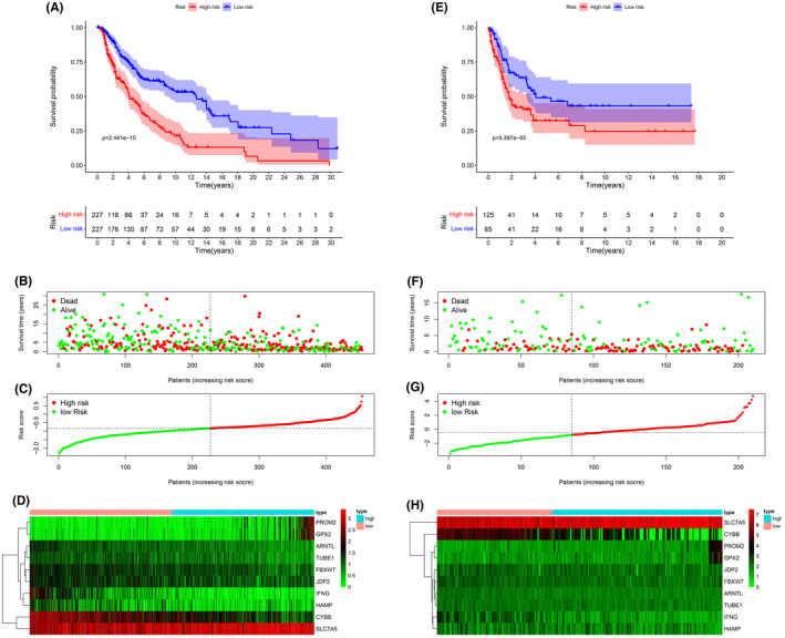
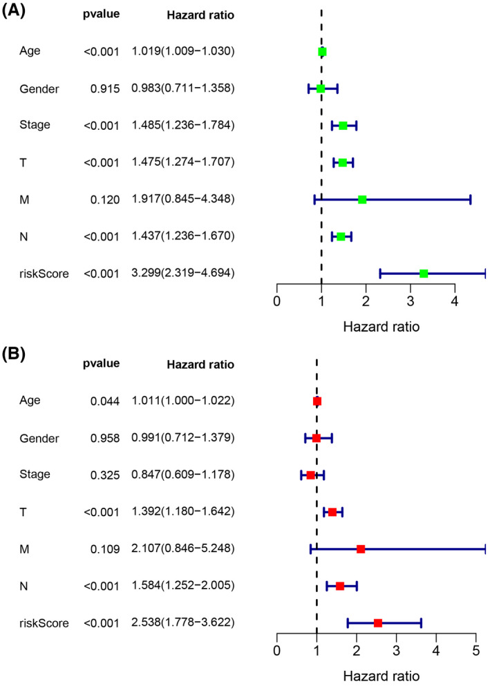
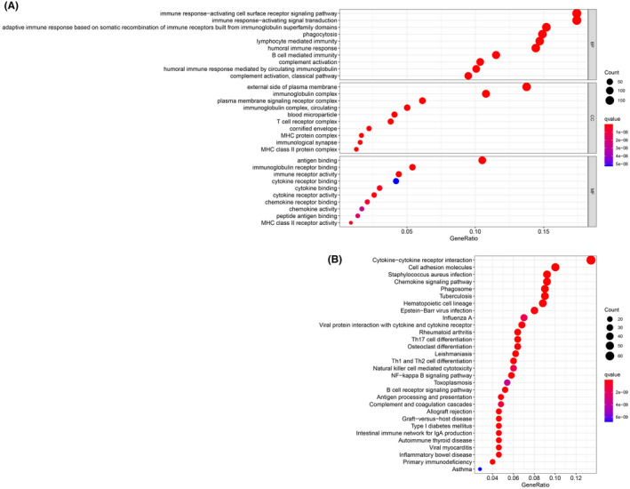
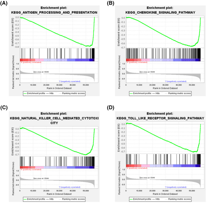
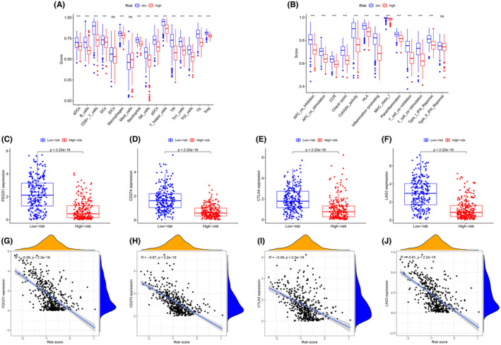
Methods: RNA-sequencing data and clinical information of melanoma patients were extracted from TCGA, GEO, and GTEx. Univariate, multivariate, and LASSO regression analyses were conducted to identify the gene signature. A 10 FRG signature was an independent and strong predictor of survival. The predictive performance was assessed using ROC curve. The functions of this gene signature were assessed by GO and KEGG analysis. The statuses of low-risk and high-risk groups according to the gene signature were compared by GSEA. In addition, we investigated the possible relationship of FRGs with immunotherapy efficacy.

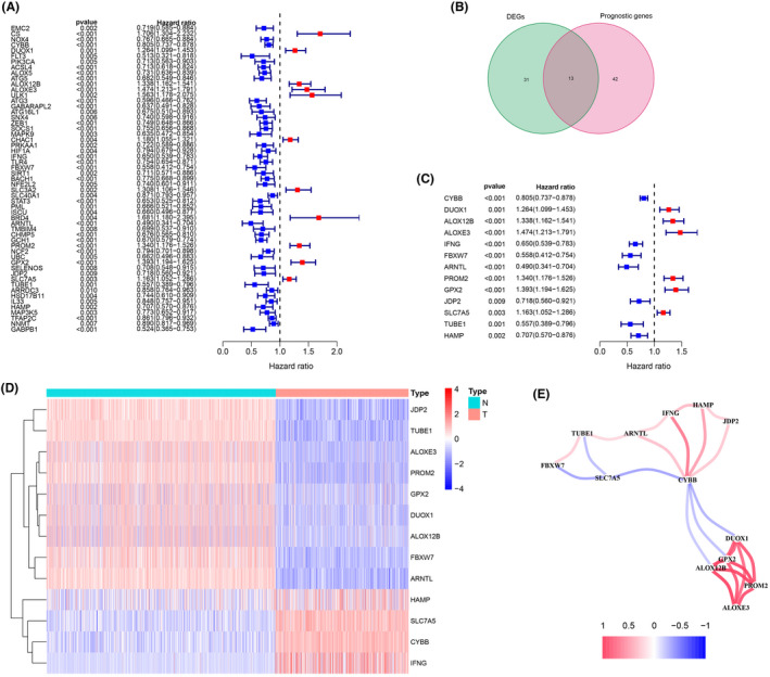
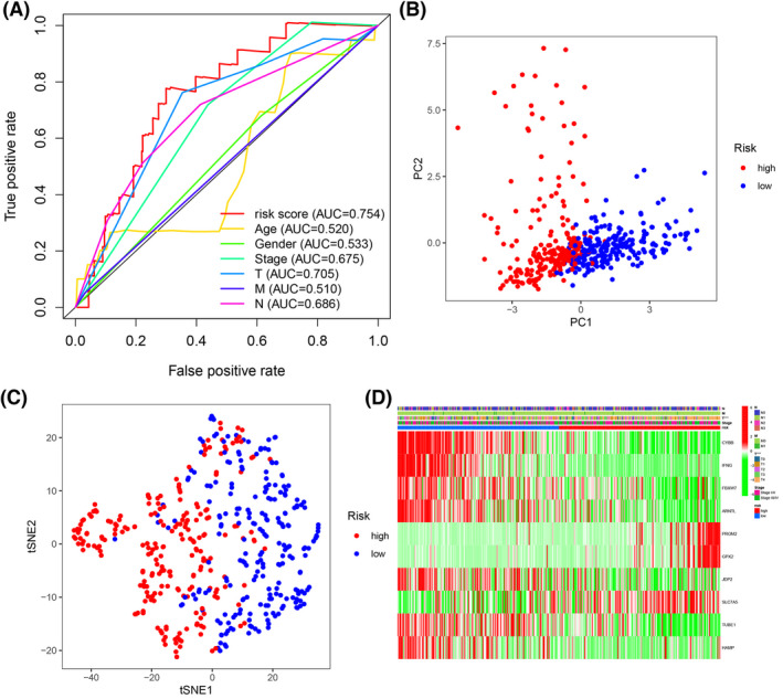
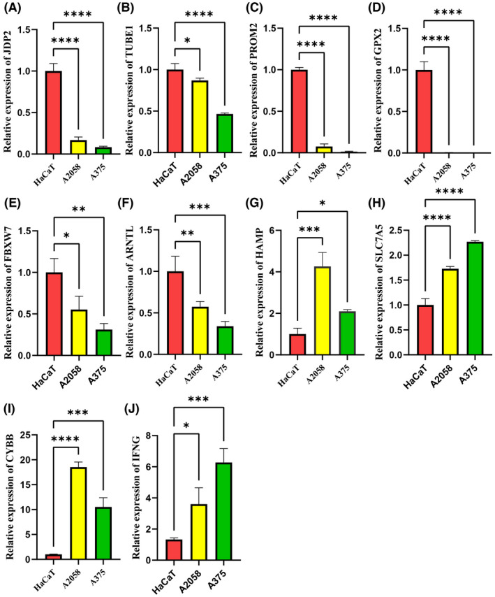
Results: A prognostic signature with 10 FRGs (CYBB, IFNG, FBXW7, ARNTL, PROM2, GPX2, JDP2, SLC7A5, TUBE1, and HAMP) was identified by Cox regression analysis. This signature had a higher prediction efficiency than clinicopathological features (AUC = 0.70). The enrichment analyses of DEGs indicated that ferroptosis-related immune pathways were largely enriched. Furthermore, GSEA showed that ferroptosis was associated with immunosuppression in the high-risk group. Finally, immune checkpoints such as PDCD-1 (PD-1), CTLA4, CD274 (PD-L1), and LAG3 were also differential expression in two risk groups.

Conclusions: The 10 FRGs signature were a strong predictor of OS in SKCM and could be used to predict therapeutic targets for melanoma.

Keywords: ferroptosis; gene signature; melanoma; overall survival; tumor immunity.


Conflict of interest statement

The authors declare no conflict of interest in this work.

Figures

FIGURE 1
FIGURE 1
The process flow of this study
FIGURE 2
FIGURE 2
Identification of the prognostic ferroptosis‐related DEGs in the TCGA. (A) Forest plot of the prognostic ferroptosis‐related gene (FRGs) of skin cutaneous melanoma in the TCGA. (B) Venn diagram of the overlapped genes between DEGs and FRGs. (C) Forest plots showing the 13 overlapping genes of the prognostic ferroptosis‐related DEGs. (D) A heat map of the 13 prognostic ferroptosis‐related DEGs, four genes of which were upregulated, and the remaining nine genes of which were downregulated in tumor tissue. (E) The correlation network of 13 prognostic ferroptosis‐related DEGs. The correlation coefficients are represented by different colors
FIGURE 3
FIGURE 3
Kaplan–Meier curves for overall survival in the high‐risk and low‐risk groups. (A) The training dataset, (E) validation dataset. The survival status, risk score distribution, and risk genes expression in the datasets. (B, C, D) Training dataset, (F, G, H) validation dataset. (green and red lines/dots represent low and high risk, respectively)
FIGURE 4
FIGURE 4
(A) The AUC for risk score and clinical features according to the ROC curves in the training dataset. Clinical feature: Age, gender, stage, and T, N, M stage. (B) PCA plot of the training dataset. (C) The t‐SNE analysis of the training dataset. (D) The differential expression of ferroptosis‐related genes in different clinical features. ***p < 0.001
FIGURE 5
FIGURE 5
Forest plot of (A) the univariate and (B) multivariate Cox regression analysis showing that the age, T stage, N stage, and risk score were independent prognostic predictors
FIGURE 6
FIGURE 6
GO and KEGG enrichment analysis of DEGs. (A) GO enrichment analysis of the DEGs. (B) KEGG enrichment analysis of the DEGs
FIGURE 7
FIGURE 7
Analysis of enriched pathways. KEGG analysis (A–D) of Gene Set Enrichment Analysis in the low‐risk groups in skin cutaneous melanoma
FIGURE 8
FIGURE 8
Comparison of the immune cell subpopulations and related functions between the different risk groups: (A) Scores of the 16 immune cells and (B) Scores of the 13 immune‐related functions. (C–F) The comparison of the expression levels of PDCD1 (PD‐1), CD274 (PD‐L1), CTLA‐4, and LAG‐3 between high‐risk and low‐risk groups. (G–J) Significant negative association between the risk score and ICB receptors PDCD‐1 (R = −0.59, p < 0.001, H), CD247 (R = −0.67, p < 0.001), CTLA4 (R = −0.49, p < 0.001), and LAG3 (R = −0.61, p < 0.001). *p < 0.05, **p < 0.01, ***p < 0.001, ns, no significance
FIGURE 9
FIGURE 9
The differential expression of ferroptosis‐related genes was detected by qPCR. (A–F) Compared with the HaCaT cell line, the mRNA of JDP2, TUBE1, PROM2, GPX2, FBXW7, and ARNTL were significantly lower in the A2058 and A375 cell lines, (G–J) while the mRNA of HAMP, SLC7A5, CYBB, and IFNG were significantly higher in the A2058 and A375 cell lines. *p < 0.05, **p < 0.01, ***p < 0.001, ****p < 0.0001

Similar articles

Cited by

References

    1. Siegel RL, Miller KD, Fuchs HE, Jemal A. Cancer Statistics, 2021. CA Cancer J Clin. 2021;71(1):7‐33. doi:10.3322/caac.21654 - DOI - PubMed
    1. Bomar L, Senithilnathan A, Ahn C. Systemic therapies for advanced melanoma. Dermatol Clin. 2019;37(4):409‐423. doi:10.1016/j.det.2019.05.001 - DOI - PubMed
    1. Leonardi GC, Candido S, Falzone L, Spandidos DA, Libra M. Cutaneous melanoma and the immunotherapy revolution (review). Int J Oncol. 2020;57(3):609‐618. doi:10.3892/ijo.2020.5088 - DOI - PMC - PubMed
    1. Villani A, Scalvenzi M, Fabbrocini G, Ocampo‐Candiani J, Ocampo‐Garza SS. Looking into a better future: novel therapies for metastatic melanoma. Dermatol Ther (Heidelb). 2021;11(3):751‐767. doi:10.1007/s13555-021-00525-9 - DOI - PMC - PubMed
    1. Guerrisi A, Loi E, Ungania S, et al. Novel cancer therapies for advanced cutaneous melanoma: the added value of radiomics in the decision making process‐a systematic review. Cancer Med. 2020;9(5):1603‐1612. doi:10.1002/cam4.2709 - DOI - PMC - PubMed

MeSH terms