Review
. 2023 Aug;29(4):445-460.
doi: 10.1177/10738584221082620. Epub 2022 Apr 2.

Sculpting Astrocyte Diversity through Circuits and Transcription

Affiliations
Review

Sculpting Astrocyte Diversity through Circuits and Transcription

Yi-Ting Cheng et al. Neuroscientist. 2023 Aug.

Abstract

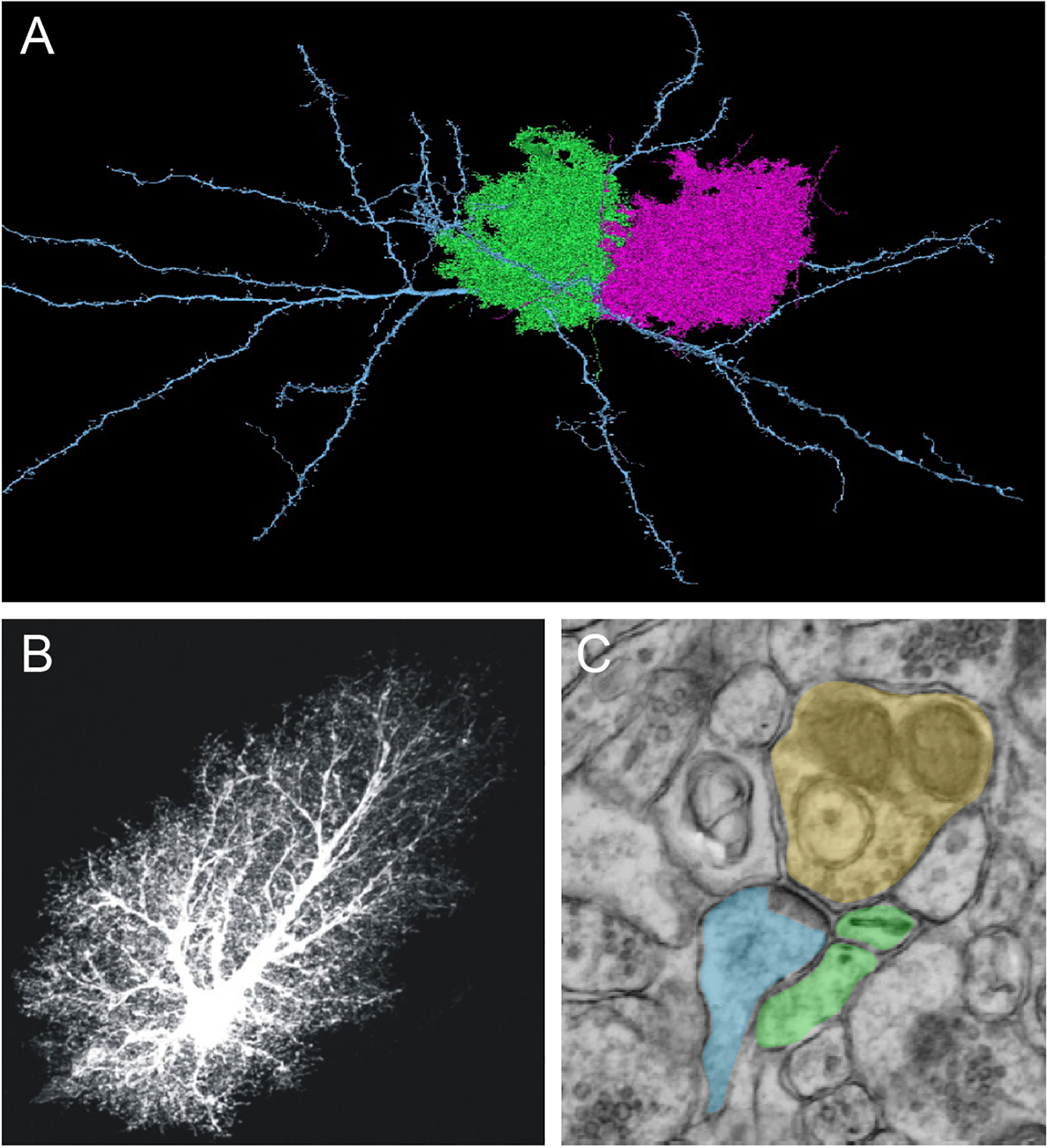
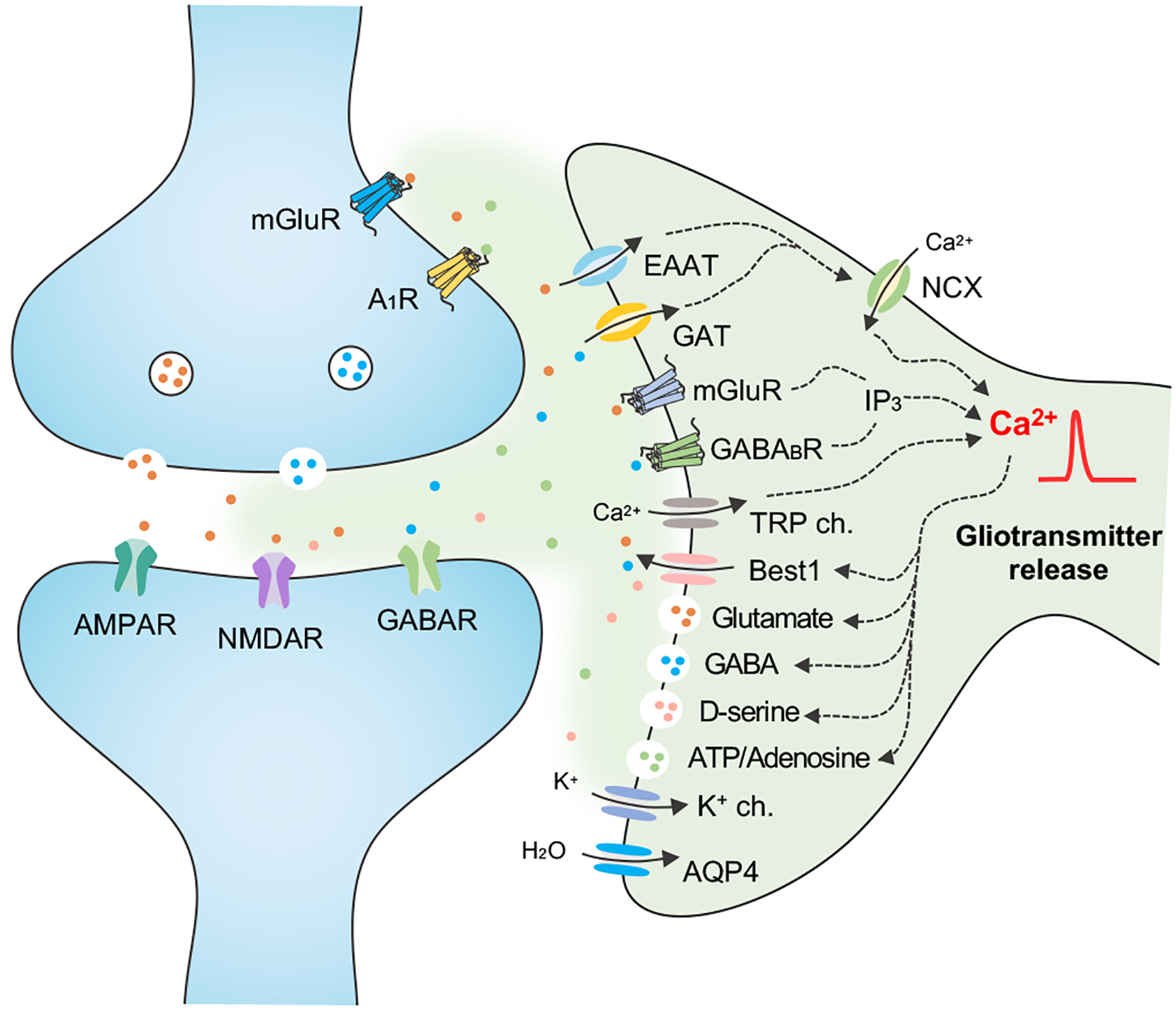
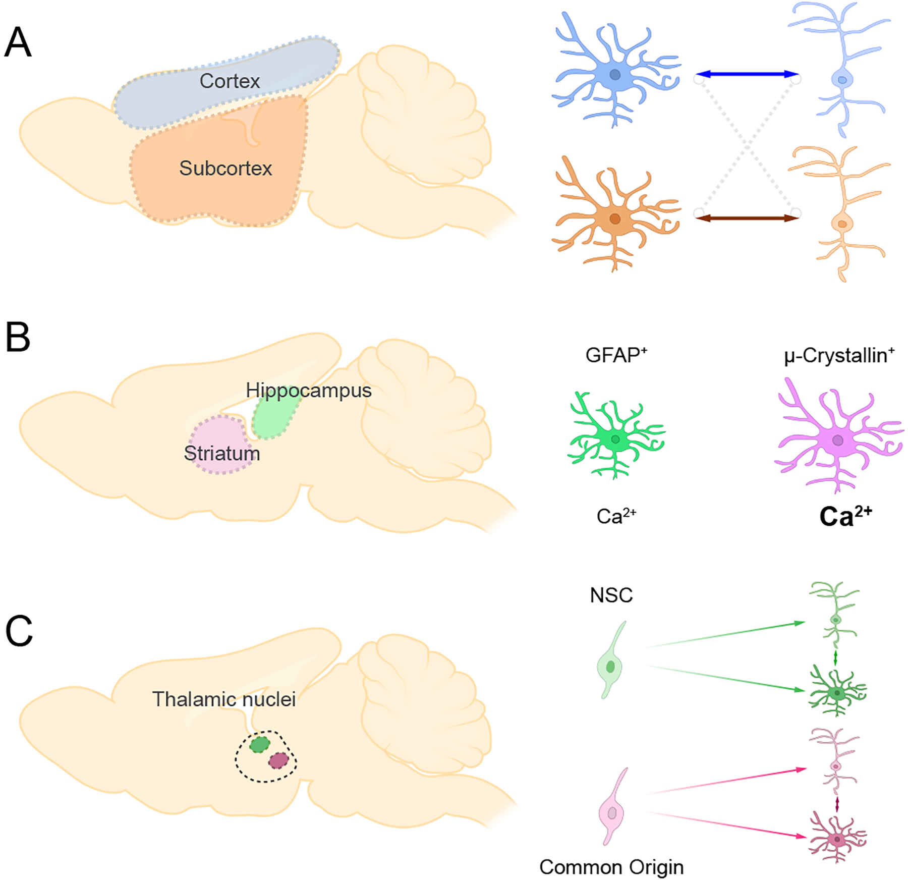
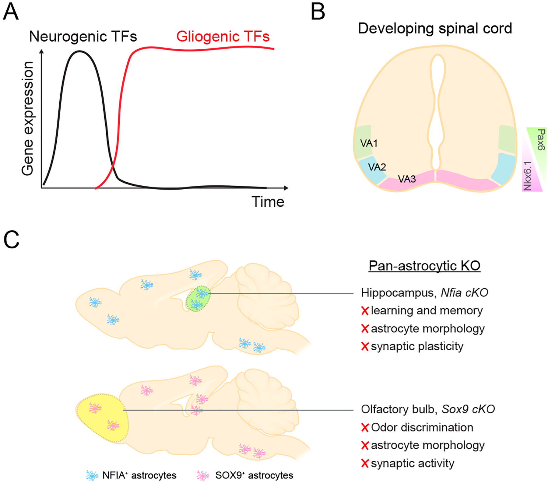
Astrocytes are the most abundant glial cell in the central nervous system and occupy a wide range of roles that are essential for brain function. Over the past few years, evidence has emerged that astrocytes exhibit cellular and molecular heterogeneity, raising the possibility that subsets of astrocytes are functionally distinct and that transcriptional mechanisms are involved in encoding this prospective diversity. In this review, we focus on three emerging areas of astrocyte biology: region-specific circuit regulation, molecular diversity, and transcriptional regulation. This review highlights our nascent understanding of how molecular diversity is converted to functional diversity of astrocytes through the lens of brain region-specific circuits. We articulate our understanding of how transcriptional mechanisms regulate this diversity and key areas that need further exploration to achieve the overarching goal of a functional taxonomy of astrocytes in the brain.

Keywords: astrocyte; brain circuits; cellular diversity; synapse; transcription factors.


Figures

Figure 1.
Figure 1.. Astrocyte Exhibit Complex Morphology and Proximity to Neurons
A) High resolution, serial EM reconstruction of astrocytes in layer 2/3 of mouse visual cortex. Image generated as part of the IARPA MICrONS project.(Consortium and others 2021) B) A prototypical astrocyte from the mouse hippocampus, loaded with fluorescent dye (Lucifer yellow). The images are from the Cell Centered Database, , at the National Center for Microscopy and Imaging Research (http://ccdb.ucsd.edu/index.shtm) and have Accession #1066, 1063; also shown in ref (Khakh and Sofroniew 2015). Note the complex morphology, featuring thousands of processes and leaflets that are peripheral to the soma. C) Electron microscopy image of an astrocyte process in close proximity to a neuron. Presynaptic (yellow), postsynaptic (blue), and astrocyte leaflet (green).
Figure 2.
Figure 2.. Astrocyte-Neuron Communication Regulates Synaptic Activity
Astrocytes maintain synaptic function through the uptake of ions and neuroactive molecules via channels, transporters, and receptors. This can elicit increases in Ca2+ activity through direct or indirect pathways. In response to Ca2+ activity astrocytes release neuroactive compounds, called ‘gliotransmitters’ such as glutamate, GABA, D-serine and ATP/adenosine to pre- and post- synaptic neurons. EAAT, excitatory amino acid (glutamate) transporter; GAT, GABA transporter; NCX, Na+-Ca2+ exchanger; mGluR, metabotropic glutamate receptor; IP3, inositol trisphosphate; TRP ch, transient receptor potential channel; Best1, bestrophin1 channel permeable to glutamate/GABA/D-serine; AQP4, aquaporin-4.
Figure 3.
Figure 3.. Astrocyte Regulation of Circuits and Behaviors Across Brain Regions.
Sagittal brain section demonstrating various brain regions and their associated behavioral function, summarized molecular mechanism, and the molecular/genetic tools used in functional studies. OB, olfactory bulb; ST, striatum; HP, hippocampus; AG, amygdala; SP, spinal cord.
Figure 4.
Figure 4.. Regional Astrocyte Diversity
A) Astrocytes preferentially support neurons from matched brain regions. Cortical astrocytes support cortical neurons, while subcortical astrocytes support subcortical neurons B) Striatal and hippocampal astrocytes exhibit distinct molecular and functional properties. C) Astrocytes and neurons that share common origin are clustered in the same thalamic nucleus
Figure 5.
Figure 5.. Local Astrocyte Diversity
Features of local astrocyte diversity in the brain. Local astrocyte subpopulations within a given brain region are heterogeneous at the molecular and functional levels. Astrocytes in the same region also demonstrate differences that are related to adjacent neurons, such as excitatory (green) or inhibitory (pink) neurons. Cortical astrocytes exhibit diverse morphological properties and gene expression gradients based on cortical layering.
Figure 6.
Figure 6.. Transcriptional Regulation of Astrocyte Function
A) Expression dynamics of transcription factors that regulate the generation of neuronal and glial fates in the developing and adult CNS. B) Patterned expression of development transcription factors Pax6 and Nkx6.1 in subsets of astrocytes in the developing spinal cord. C) Summary of region specific transcriptional dependencies for NFIA and Sox9. NFIA and Sox9 are universally expressed in astrocytes throughout the brain, but when knocked out specifically impact astrocyte function and associated circuits in a region specific manner.

References

    1. Adamsky A, Kol A, Kreisel T, Doron A, Ozeri-Engelhard N, Melcer T and others. 2018. Astrocytic Activation Generates De Novo Neuronal Potentiation and Memory Enhancement. Cell 174(1):59–71 e14. - PubMed
    1. Akdemir ES, Woo J, Bosquez Huerta NA, Lozzi B, Groves AK, Harmanci AS and others. 2021. Lunatic Fringe-GFP Marks Lamina-Specific Astrocytes That Regulate Sensory Processing. J Neurosci. - PMC - PubMed
    1. Allen NJ. 2014. Astrocyte regulation of synaptic behavior. Annu Rev Cell Dev Biol 30:439–63. - PubMed
    1. Araque A, Carmignoto G, Haydon PG, Oliet SH, Robitaille R, Volterra A. 2014. Gliotransmitters travel in time and space. Neuron 81(4):728–39. - PMC - PubMed
    1. Arnett HA, Fancy SP, Alberta JA, Zhao C, Plant SR, Kaing S and others. 2004. bHLH transcription factor Olig1 is required to repair demyelinated lesions in the CNS. Science 306(5704):2111–5. - PubMed

Publication types