Review
. 2022 Jul;11(13):e2102530.
doi: 10.1002/adhm.202102530. Epub 2022 May 4.

Intervertebral Disc Degeneration: Biomaterials and Tissue Engineering Strategies toward Precision Medicine

Affiliations
Review

Intervertebral Disc Degeneration: Biomaterials and Tissue Engineering Strategies toward Precision Medicine

Isma Liza Mohd Isa et al. Adv Healthc Mater. 2022 Jul.

Abstract

Intervertebral disc degeneration is a common cause of discogenic low back pain resulting in significant disability. Current conservative or surgical intervention treatments do not reverse the underlying disc degeneration or regenerate the disc. Biomaterial-based tissue engineering strategies exhibit the potential to regenerate the disc due to their capacity to modulate local tissue responses, maintain the disc phenotype, attain biochemical homeostasis, promote anatomical tissue repair, and provide functional mechanical support. Despite preliminary positive results in preclinical models, these approaches have limited success in clinical trials as they fail to address discogenic pain. This review gives insights into the understanding of intervertebral disc pathology, the emerging concept of precision medicine, and the rationale of personalized biomaterial-based tissue engineering tailored to the severity of the disease targeting early, mild, or severe degeneration, thereby enhancing the efficacy of the treatment for disc regeneration and ultimately to alleviate discogenic pain. Further research is required to assess the relationship between disc degeneration and lower back pain for developing future clinically relevant therapeutic interventions targeted towards the subgroup of degenerative disc disease patients.

Keywords: biomaterials; discogenic low back pain; intervertebral discs; precision medicine; tissue engineering.


Conflict of interest statement

The authors declare no conflict of interest.

Figures

Figure 1
Figure 1
Schematic summarizes the phases of intervertebral disc degeneration associated discogenic pain. A multifactorial condition initiates an imbalance of ECM metabolism in the early phase of degeneration by increasing degradative enzymes such as ADAMTS and MMPs to promote ECM degradation and reduce ECM synthesis, including aggrecan and collagen. Alteration of ECM biochemical compositions induce the production of proinflammatory mediators includes IL‐1β TNF, IFN and IL‐6. In phase II, inflammatory insult results in further ECM degradation and loss of cell density. Further, proinflammatory cytokines stimulate AF and NP cells and infiltration of immune cells to release growth factors such as VEGF and neurotrophins such as NGF and BDNF to promote neovascularization and nerve in‐growth in the IVD. Phase III is characterized by continuous neurogenic insults to induce pronociceptive molecules such as neuropeptides to be released in IVD and spinal level, and activation of pronociceptor to sensitize and develop pain. Advanced structural breakdown results in annular tears and mechanical instability, contributing to disc herniation.
Figure 2
Figure 2
Precision medicine approach in IVD tissue engineering and regenerative therapy targeting early to severe IVD degeneration. Molecular profiling in subgroup of degenerative disc disease patients using genomic or proteomic approaches for patient stratification at early, mild and severe stages. Understanding patient‐specific pathogenesis IVD degeneration underlying discogenic low back pain will lead to identifying therapeutic targets and the design of biomaterials incorporating with targeted therapeutic molecules or cells tailored to disease severity. The decision of treatment plans through biomaterial injection alone at an early stage or the combination of biomaterials and therapeutic molecules or cells via the minimally invasive injection or surgical implantation at an advanced stage improves the treatment efficacy. The schematic was created with BioRender.com.
Figure 3
Figure 3
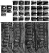
Classification system for IVD degeneration based on MRI pathological features. a) Pfirrmann grading system for the assessment of lumbar disc degeneration. Reproduced with permission.[ 40 ] Copyright 2001, Lippincott Williams & Wilkins, Inc. A) Grade I: The structure of the disc is homogeneous, with a bright hyperintense white signal intensity and a normal disc height. B) Grade II: The structure of the disc is inhomogeneous, with a hyperintense white signal. The distinction between nucleus and annulus is clear, and the disc height is normal, with or without horizontal gray bands. C) Grade III: The structure of the disc is inhomogeneous, with intermediate gray signal intensity. The distinction between nucleus and annulus is unclear, and the disc height is normal or slightly decreased. D) Grade IV: The structure of the disc is inhomogeneous, with a hypointense dark gray signal intensity. The distinction between nucleus and annulus is lost, and the disc height is normal or moderately decreased. E) Grade V: The structure of the disc is homogeneous, with hypointense black signal intensity. The distinction between nucleus and annulus is lost, and the disc space is collapsed. Grading is performed on T2‐weighted midsagittal (repetition time 5000 msec/echo time 130 msec) fast spin‐echo images. b) Modified Pfirrmann grading system for lumbar disc degeneration. Image reference panel shows increasing severity of disc degeneration. Reproduced with permission.[ 41 ] Copyright 2007, Lippincott Williams & Wilkins, Inc. c) Modic changes. Reproduced with permission.[ 43 ] Modic type I change: hyperintense on T2WI (A arrow), hypointense on T1WI (B arrow) at inferior endplate of L4. Modic type II change: hyperintense on T2WI (C upper arrows), hyperintense on T1WI (D upper arrows) at superior endplate of L3. Modic type III change: hypointense on T2WI (C inferior arrows), hypointense on T1WI (D inferior arrow) at superior endplate of L4. Permission of figures adaptation from.[ 40 , 41 , 43 ]
Figure 4
Figure 4
Schematic inflammatory pain processing and inhibitory target at peripheral and spinal levels through local injection IVD treatment to produce an antinociceptive effect. a) At peripheral tissue, disc degeneration causes the release of inflammatory and neurogenic mediators (IL‐1, IL‐6, TNF, IFN, NGF, and PGE2) by resident cells, including AF and NP cells, macrophages, and mast cells. The inflammatory signaling molecules activate nociceptors by binding to cell surface receptors or ion channels such as voltage‐gated sodium channels, transient receptor potential cation channel (TRP) channels, receptor tyrosine kinases (RTK), G protein coupled receptors, acid‐sensitive ion channels (ASIC) on first‐order neurons, thus initiating membrane depolarization for transmitting action potentials to the spinal cord. b) Action potentials are transmitted along nociceptive fibers in the DRG to the presynaptic neurons in the dorsal horn. Prolonged noxious stimulation activates C and Aδ fibers to release excitatory neurotransmitters (glutamate, substance P, CGRP, and BDNF) onto synapses in lamina I of the dorsal horn to mediate central sensitization through NMDA glutamate receptors or TrkB receptor. This neurotransmission can increase intracellular calcium to activate calcium‐dependent signaling pathways and second messengers (c‐Fos, MAPK, and protein kinase), increasing the excitability of the postsynaptic neurons to transmit pain to the brain. c) The axons of second‐order neurons decussate at the spinal cord and anterolaterally project to the thalamic nuclei and brainstem. The third‐order neurons project to several cortical regions encodes for sensory‐discriminative aspects of pain. Overall, the inhibition of nociceptive transduction at peripheral tissue will suppress persistent noxious stimulation at the spinal level to inhibit excitatory glutamatergic and peptidergic neurotransmissions up to supra‐spinal level, thereby producing an antinociceptive effect. The schematic was created with BioRender.com.

References

    1. Vos T., Lim S. S., Abbafati C., Abbas K. M., Abbasi M., Abbasifard M., Abbasi‐Kangevari M., Abbastabar H., Abd‐Allah F., Abdelalim A., Abdollahi M., Abdollahpour I., Abolhassani H., Aboyans V., Abrams E. M., Abreu L. G., Abrigo M. R. M., Abu‐Raddad L. J., Abushouk A. I., Acebedo A., Ackerman I. N., Adabi M., Adamu A. A., Adebayo O. M., Adekanmbi V., Adelson J. D., Adetokunboh O. O., Adham D., Afshari M., Afshin A., et al., Lancet 2020, 396, 1204. - PubMed
    1. Raftery M. N., Sarma K., Murphy A. W., De L. a., Harpe D., Normand C., McGuire B. E., Pain 2011, 152, 1096. - PubMed
    1. Fullen B., Hurley D. A., Power C., Canavan D., O'Keeffe D., Ir. J. Med. Sci. 2006, 175, 68. - PubMed
    1. Hall J. A., Jowett S., Lewis M., Oppong R., Konstantinou K., Pain 2021, 162, 702. - PubMed
    1. Belitskaya‐Levy I., David Clark J., Shih M. C., Bair M. J., J. Pain Res. 2021, 14, 161. - PMC - PubMed

Publication types

MeSH terms

Substances

LinkOut - more resources