. 2022 Apr 4;79(4):222.
doi: 10.1007/s00018-022-04255-9.

Aβ/tau oligomer interplay at human synapses supports shifting therapeutic targets for Alzheimer's disease

Affiliations

Aβ/tau oligomer interplay at human synapses supports shifting therapeutic targets for Alzheimer's disease

Michela Marcatti et al. Cell Mol Life Sci. .

Abstract

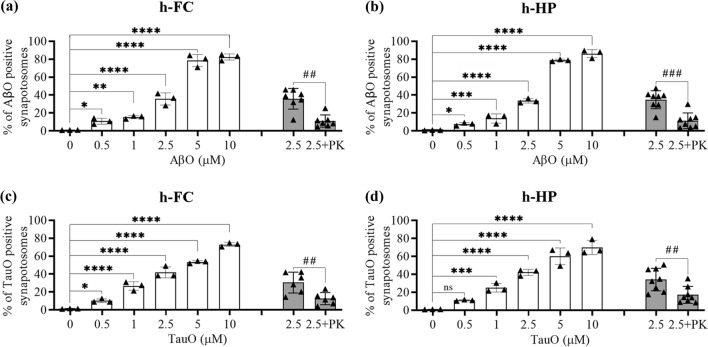
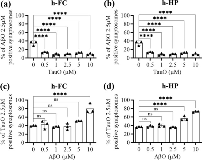
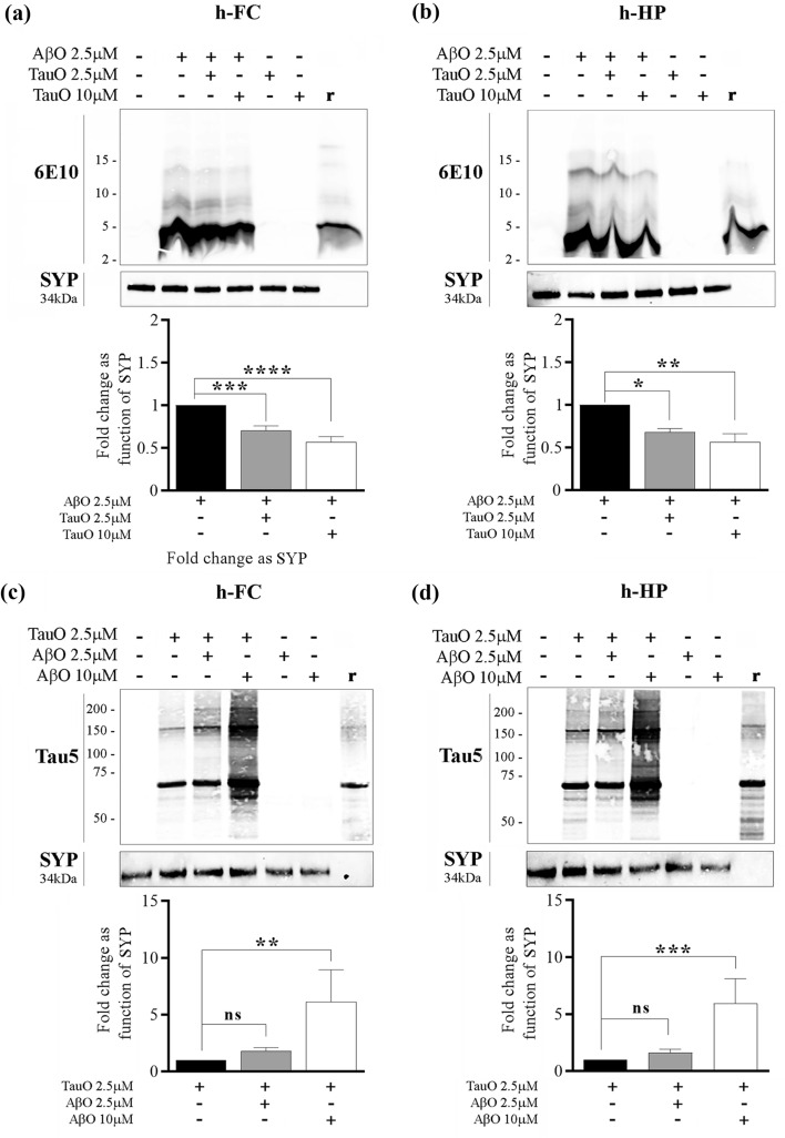
Background: Alzheimer's disease (AD) is characterized by progressive cognitive decline due to accumulating synaptic insults by toxic oligomers of amyloid beta (AβO) and tau (TauO). There is growing consensus that preventing these oligomers from interacting with synapses might be an effective approach to treat AD. However, recent clinical trial failures suggest low effectiveness of targeting Aβ in late-stage AD. Researchers have redirected their attention toward TauO as the levels of this species increase later in disease pathogenesis. Here we show that AβO and TauO differentially target synapses and affect each other's binding dynamics.

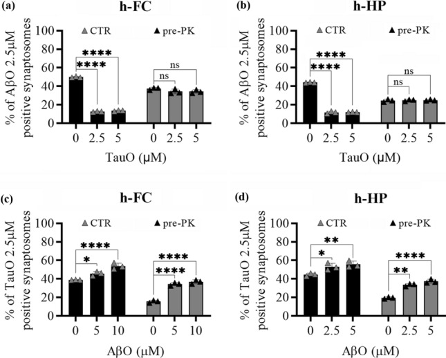
Methods: Binding of labeled, pre-formed Aβ and tau oligomers onto synaptosomes isolated from the hippocampus and frontal cortex of mouse and postmortem cognitively intact elderly human brains was evaluated using flow-cytometry and western blot analyses. Binding of labeled, pre-formed Aβ and tau oligomers onto mouse primary neurons was assessed using immunofluorescence assay. The synaptic dysfunction was measured by fluorescence analysis of single-synapse long-term potentiation (FASS-LTP) assay.

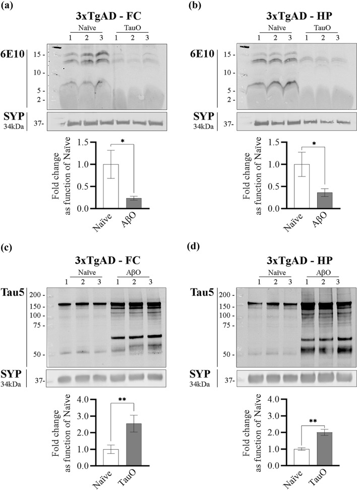
Results: We demonstrated that higher TauO concentrations effectively outcompete AβO and become the prevailing synaptic-associated species. Conversely, high concentrations of AβO facilitate synaptic TauO recruitment. Immunofluorescence analyses of mouse primary cortical neurons confirmed differential synaptic binding dynamics of AβO and TauO. Moreover, in vivo experiments using old 3xTgAD mice ICV injected with either AβO or TauO fully supported these findings. Consistent with these observations, FASS-LTP analyses demonstrated that TauO-induced suppression of chemical LTP was exacerbated by AβO. Finally, predigestion with proteinase K abolished the ability of TauO to compete off AβO without affecting the ability of high AβO levels to increase synaptic TauO recruitment. Thus, unlike AβO, TauO effects on synaptosomes are hampered by the absence of protein substrate in the membrane.

Conclusions: These results introduce the concept that TauO become the main synaptotoxic species at late AD, thus supporting the hypothesis that TauO may be the most effective therapeutic target for clinically manifest AD.

Keywords: Amyloid; Dementia; Synaptic binding; Synaptosomes; Tau.


Conflict of interest statement

The authors declare that they have no competing interests.

Figures

Fig. 1
Fig. 1
Characterization of dose dependent AβO and TauO binding to human synapses. a White columns—AβO binding to a pool of synaptosomes isolated from human FC samples. Data represent the mean ± SD; *P = 0.05, **P = 0.0075, and ****P < 0.0001 compared with synaptosomes not challenged with AβO (biological replicates n = 5; independent experiments n = 3; one-way ANOVA plus Dunnett’s multiple comparisons); gray columns—proteinase K (PK) enzymatic digestion of human FC synaptosomes post-challenge with AβO 2.5 µM. Data represent the mean ± SD; **P = 0.0023 compared with control (biological replicates n = 5; independent experiments n = 7; paired t-test, two-tailed). b White columns—AβO binding to a pool of synaptosomes isolated from human HP samples. Data represent the mean ± SD; *P = 0.05, ***P = 0.0010, and ****P < 0.0001 compared with synaptosomes not challenged with AβO (biological replicates n = 6; independent experiments n = 3; one-way ANOVA plus Dunnett’s multiple comparisons); gray columns—proteinase K (PK) enzymatic digestion of human HP synaptosomes post-challenge with AβO 2.5 µM. Data represent the mean ± SD; ***P = 0.0002 compared with control (biological replicates n = 6; independent experiments n = 8; paired t-test, two-tailed). c White columns—TauO binding to a pool of synaptosomes isolated from human FC samples. Data represent the mean ± SD; *P = 0.0329, and ****P < 0.0001 compared with synaptosomes not challenged with TauO (biological replicates n = 5; independent experiments n = 3; one-way ANOVA plus Dunnett’s multiple comparisons); gray columns—proteinase K (PK) enzymatic digestion of human FC synaptosomes post-challenge with TauO 2.5 µM. Data represent the mean ± SD; **P = 0.0086 compared with control (biological replicates n = 5; independent experiments n = 6; paired t-test, two-tailed); d White columns—TauO binding to a pool of synaptosomes isolated from human HP samples. Data represent the mean ± SD; ***P = 0.0009, and ****P < 0.0001 compared with synaptosomes not challenged with TauO (biological replicates n = 6; independent experiments n = 3; one-way ANOVA plus Dunnett’s multiple comparisons); gray columns—proteinase K (PK) enzymatic digestion of human HP synaptosomes post-challenge with TauO 2.5 µM. Data represent the mean ± SD; **P = 0.0081 compared with control (biological replicates n = 6; independent experiments n = 8; paired t-test, two-tailed)
Fig. 2
Fig. 2
TauO reduces AβO binding to human FC and HP synaptosomes, while AβO increases TauO binding. Effect of increasing TauO concentrations (0.5–1–2.5–5–10 µM) on AβO binding to synaptosomes isolated from human a FC and b HP samples. Effect of increasing AβO concentrations (0.5–1–2.5–5–10 µM) on TauO binding to synaptosomes isolated from human c FC and d HP samples. Data represent the mean ± SD; ***P = 0.002, and ****P < 0.0001 compared with synaptosomes not challenged with AβO and TauO (biological replicates n = 5–6; independent experiments n = 3; ordinary one-way ANOVA plus Dunnett’s multiple comparison test)
Fig. 3
Fig. 3
Western blot analyses confirming changes in AβO/TauO synaptic binding in the presence of each other. a, b Representative western blots (upper panels) and relative densitometric analyses (lower panels) of oligomeric species (selected area of quantification from about 8 kDa to about 16 kDa) to evaluate the percentages of AβO 2.5 µM bound to synaptosomes isolated from human a FC and b HP in the presence of TauO 2.5 and 10 µM. Data represent the mean ± SD; *P = 0.0109, **P = 0.0015, ***P = 0.0002, and ****P < 0.0001 compared with control synaptosomes (challenged with AβO alone) (biological replicates n = 5–6; independent experiments n = 5; one-way ANOVA plus Dunnett’s multiple comparison test); c, d Representative western blots (upper panels) and relative densitometric analyses (lower panels) of oligomeric species (selected area of quantification from about 60 kDa to up) to evaluate the percentages of TauO 2.5 µM bound to synaptosomes isolated from human c FC and d HP in the presence of AβO 2.5 and 10 µM. Data represent the mean ± SD; **P = 0.0034 (c), **P = 0.0007 (d), compared with control synaptosomes (challenged with TauO alone) (biological replicates n = 5–6; independent experiments n = 5; one-way ANOVA plus Dunnett’s multiple comparison test)
Fig. 4
Fig. 4
AβO/TauO synaptic binding in mice brain slices treated ex vivo with AβO plus TauO. Mice brain slices were treated with increasing concentrations of AβO and TauO, then synaptosomes were isolated and subjected to flow cytometric analysis to evaluate the dose-dependent binding percentages of a AβO and b TauO; Data represent the mean ± SD; **P = 0.0077 and ****P < 0.0001 compared with synaptosomes derived from untreated mouse brain slices (biological replicates n = 14–30; independent experiments n = 5; ordinary one-way ANOVA plus Dunnett’s multiple comparisons); mice brain slices were treated with a combination of AβO 2.5 µM and TauO 1 µM before the synaptosomes were isolated for flow cytometric analyses to evaluate the binding percentage of c AβO and d TauO. Data represent the mean ± SD; ****P < 0.0001 compared with brain slices challenged with AβO or TauO alone (biological replicates n = 10; independent experiments n = 2; paired t-test, two-tailed)
Fig. 5
Fig. 5
Immunofluorescence analyses of AβO and TauO binding dynamics in mouse primary neuron synapses. a Representative confocal images of neuronal projections (30-µm long) from mouse primary cortical neurons treated with AβO 2.5 µM (blue), TauO 2.5 µM (green) and their combination (blue + green) and stained for MAP2 (gray) and PSD95 (red); original magnification 63 × , scale bar 2 µm. b Representative pixel maps of AβO/PSD95 and TauO/PSD95 showing colocalizations in gray (white arrowheads) of neuronal projections showed in a . c Quantitative analysis of PSD95 puncta within 30-µm length of neuronal projections randomly selected from mouse primary cortical neurons treated with AβO 2.5 µM (blue), TauO 2.5 µM (green), and their combination (blue + green) as compared with untreated cells (red). Data represent the mean ± SD, ****P < 0.0001 compared with control cells (neuronal projections analyzed n = 18; independent experiments n = 3; ordinary one-way ANOVA plus Dunnett’s multiple comparisons). d Quantitative analysis of synaptic AβO within 30-µm lengths of neuronal projections randomly selected from mouse primary cortical neurons treated with AβO 2.5 µM (blue) and the combination of AβO 2.5 µM and TauO 2.5 µM (blue + green); data represent the mean ± SD, ***P = 0.0004 (neuronal projections analyzed n = 18; independent experiments n = 3; unpaired t-test, two-tailed). e Quantitative analysis of synaptic TauO within 30-µm lengths of neuronal projections randomly selected from mouse primary cortical neurons treated with TauO 2.5 µM (green) and the combination of AβO 2.5 µM and TauO 2.5 µM (blue + green); Data represent the mean ± SD, *P = 0.0128 (neuronal projections analyzed n = 18; independent experiments n = 3; unpaired t-test, two-tailed)
Fig. 6
Fig. 6
In vivo treatment of 3xTgAD mice with AβO and TauO. a, b Representative western blots (upper panels) and relative densitometric analyses (lower panels) of oligomeric species (selected area of quantification from about 8 kDa to about 16 kDa) to evaluate the levels of AβO bound to synaptosomes from a FC and b HP of 3xTgAD mice ICV with TauO 0.55 µM; data represent the mean ± SD; *P = 0.0147 (a) and *P = 0.0186 (b) as compared with FC and HP synaptosomes isolated from 3xTgAD naïve mice (biological replicates n = 3/group; independent experiments n = 3; unpaired t-test, two-tailed); c, d Representative western blots (upper panels) and relative densitometric analyses (lower panels) of oligomeric species (selected area of quantification from about 60 kDa to up) to evaluate the levels of TauO bound to synaptosomes from c FC and d HP of 3xTgAD mice ICV with AβO 0.55 µM. Data represent the mean ± SD; **P = 0.009 (c) and **P = 0.001 as compared with FC and HP synaptosomes isolated from 3xTgAD naïve mice (biological replicates n = 3/group; independent experiments n = 7; unpaired t test, two-tailed)
Fig. 7
Fig. 7
Effects of PK pret-reatment of human FC and HP synaptosomes on AβO/TauO binding dynamics. The surface protein component of human synaptosomes was digested with 1 mg/ml PK before the synaptosomes were challenged with AβO and/or TauO, and the resulting binding was detected by flow cytometric analysis. CTR, synaptosomes not PK pre-treated, pre-PK, synaptosomes PK pre-treated. Binding dynamics of AβO 2.5 µM to human a FC and b HP synaptosomes in the presence of increasing concentrations of TauO (2.5 and 5 µM); Data represent the mean ± SD; ****P < 0.0001 compared with control synaptosomes (challenged with AβO and TauO alone) (biological replicates n = 5–6; independent experiments n = 3; two-way ANOVA plus Tukey’s multiple comparison test). Binding dynamics of TauO 2.5 µM to human c FC and d HP synaptosomes in the presence of increasing concentrations of AβO (5 and 10 µM). Data represent the mean ± SD; *P = 0.05, **P = 0.0083 (d—CTR samples) and **P = 0.0015 (d—PK samples), and ****P < 0.0001 compared with control synaptosomes (challenged with AβO and TauO alone) (biological replicates n = 5–6; independent experiments n = 3; two-way ANOVA plus Tukey’s multiple comparison test)
Fig. 8
Fig. 8
Impact of AβO and TauO on physiological synaptic transmission of human synapses. FASS-LTP identifies potentiated synapses by tracking GluA1 and Nrx1ß surface expression in size-gated synaptosomes. Percentages of GluA1+Nrx1ß+ size-gated synaptosomes isolated from human a FC and b HP post-challenged with AβO and/or TauO 0.5 µM. Data represent the mean ± SEM; *P = 0.05, **P = 0.0088 (b TauO vs CTR) and **P = 0.0065 (b AβO + TauO vs CTR) and ****P < 0.0001 compared with the basal levels (biological replicates n = 5–6; independent experiments n = 3; ordinary one-way ANOVA plus Dunnett’s multiple comparison test)

References

    1. Fabiani C, Antollini SS. Alzheimer’s disease as a membrane disorder: Spatial cross-talk among beta-amyloid peptides, nicotinic acetylcholine receptors and lipid rafts. Front Cell Neurosci. 2019;13(July):1–28. - PMC - PubMed
    1. Tiwari S, Venkata A, Kaushik A, Adriana Y, Nair M. Alzheimer’s disease diagnostics and therapeutics market. Int J Nanomedicine. 2019;2019(14):5541–5554. doi: 10.2147/IJN.S200490. - DOI - PMC - PubMed
    1. Spires-Jones TL, Hyman BT. The intersection of amyloid beta and tau at synapses in Alzheimer’s disease. Neuron. 2014;82(4):756–771. doi: 10.1016/j.neuron.2014.05.004. - DOI - PMC - PubMed
    1. Ashraf G, Greig N, Khan T, Hassan I, Tabrez S, Shakil S, et al. Protein misfolding and aggregation in Alzheimer’s disease and type 2 diabetes mellitus. CNS Neurol Disord - Drug Targets. 2014;13(7):1280–1293. doi: 10.2174/1871527313666140917095514. - DOI - PMC - PubMed
    1. John A, Reddy PH. Synaptic basis of Alzheimer’s disease: focus on synaptic amyloid beta, P-tau and mitochondria. Ageing Res Rev. 2021;65(July 2020):101208. doi: 10.1016/j.arr.2020.101208. - DOI - PMC - PubMed

Substances