Review
. 2022 Apr 4;12(1):14.
doi: 10.1186/s12348-022-00292-4.

Choroidal involvement in systemic vasculitis: a systematic review

Affiliations
Review

Choroidal involvement in systemic vasculitis: a systematic review

Pınar Çakar Özdal et al. J Ophthalmic Inflamm Infect. .

Abstract

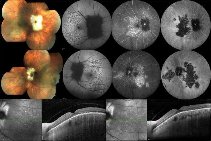
Systemic vasculitides are a large group of heterogeneous diseases characterized by inflammatory destruction of blood vessels targeting diverse organs and tissues including the eye. As the most vascularized layer of the eye, the choroid is expected to be affected in multiple systemic rheumatologic diseases with vascular involvement. While there are plenty of studies investigating retinal vascular involvement, choroidal vascular involvement in systemic vasculitides has not been investigated in isolation. However, choroidal manifestations including thickness changes, choroidal vasculitis and ischemia may be the earliest diagnostic features of systemic vasculitic diseases. Thus, multimodal imaging of the choroid may help early detection of choroidal involvement which may also have prognostic implications in these life-threatening diseases. This article aimed to review involvement of the choroid in systemic vasculitic diseases.

Keywords: Choroidal involvement; Inflammation; Ischemia; Multimodal imaging; Systemic vasculitis.


Conflict of interest statement

Pınar Çakar Özdal and Ilknur Tugal-Tutkun have both received speaker and consultant honoraria from AbbVie, Turkey.

Figures

Fig. 1
Fig. 1
Fundus photograph of the right eye of a patient with hypertensive retinopathy showing cotton wool spots, serous detachment of the macula, and numerous deep pale yellow Elschnig spots surrounding the posterior pole
Fig. 2
Fig. 2
Montage color fundus photograph of the right eye of a patient with hypertensive retinopathy showing severe papilledema with exudation and pigmented Elschnig spots with atrophic halo in the peripheral retina (Courtesy of Prof. Mehmet Yasin Teke, Department of Ophthalmology, University of Health Sciences, Ulucanlar Eye Education and Research Hospital, Ankara, Turkey)
Fig. 3
Fig. 3
Color fundus photographs of a patient with hypertensive disease and renal insufficiency showing peripheral wedge-shaped triangular areas of pigmentary changes in both eyes indicating sequelae of the Amalric sign of choroidal infarction. (Courtesy of Prof. Gürsel Yilmaz, Baskent University Faculty of Medicine, Department of Ophthalmology, Ankara, Turkey)
Fig. 4
Fig. 4
Fluorescein angiography and optical coherence tomography imaging of the left eye of a 20 year-old woman with Takayasu arteritis. Fluorescein angiography shows delayed patchy filling of the chororoid at 14 s (top left), 40 s (top middle), and 3 min (top right), pinpoint leaks at the level of retinal pigment epithelium seen at 3 min (top right) becoming more evident at 6 min (bottom left). Temporal peripheral frame at 6 min shows choroidal nonperfusion and focal leak of peripheral retinal vessels (bottom middle). SD-OCT scan of the macula shows serous retinal detachment (bottom right)
Fig. 5
Fig. 5
Dual fluorescein-indocyanine green angiography and optical coherence tomography imaging of a 36 year-old woman with systemic lupus erythematosus unresponsive to aggressive immunomodulatory treatment. Early phase fluorescein angiography (FA) shows extensive retinal pigment epithelial changes with window defects in the right (top row, first frame) and the left eye (top row, third frame); and late phase FA shows hyperfluorescence of the optic disc and peripapillary vasculature as well as subretinal staining/pooling (arrows) in the right eye (middle row, first frame) and staining of the optic disc and vessel walls in the left eye (middle row, third frame). Early phase indocyanine green angiography (ICGA) shows dilated and hyperfluorescent choroidal vessels in both the right (top row, second frame) and the left eye (top row, fourth frame); and late phase ICGA shows more numerous and larger hyperfluorescent areas than revealed by FA in the right eye (middle row, second frame) and also scattered hyperfluorescent spots in the left eye (middle row, fourth frame). SD-OCT shows extensive damage to the outer retina with chronic cystoid changes, subretinal hyperreflective material, and retinal pigment epithelium loss and irregularities in both the right (bottom row, left) and the left eye (bottom row, right)
Fig. 6
Fig. 6
Color fundus photography, dual fluorescein-indocyanine green angiography and optical coherence tomography imaging of a 63 year-old woman who presented with photopsias and loss of vision after tapering oral corticosteroid given in combination with azathioprine for the treatment of granulomatosis with polyangiitis diagnosed 2 months prior. Montage color fundus photographs of the right (top row, first frame) and the left (middle row first frame) eye show myelinated nerve fibers obscuring the optic disc margins, deep creamy-white serpiginoid choroiditis involving the posterior pole in both eyes and inferonasal to the disc in the left eye. Fundus autofluorescence imaging shows a granular pattern of hyper- and hypoautofluorescence of the lesions in the right (top row, second frame) and the left eye (middle row, second frame). Dual fluorescein-indocyanine green angiography frames at 8 min show window defects and staining on fluorescein angiography and diffuse hypofluorescence of the lesions on indocyanine green angiography in the right (top row, third and fourth frames, respectively) and the left eye (middle row, third and fourth frames, respectively). EDI-OCT of the left eye shows loss of outer retinal layers, RPE irregularities, thickening of the choroid, and hyperreflective dots in the choroid in the involved area (bottom row, left). After intravenous pulse methylprednisolone 1 g for 3 days, EDI-OCT shows partial improvement of RPE irregularity and reduced thickening of the choroid (bottom row, right). Visual acuity increased from counting fingers in both eyes to 20/40 in the right and 20/32 in the left eye. Active and latent tuberculosis had already been ruled out and the patient subsequently received cyclophosphamide therapy

References

    1. Tugal-Tutkun I. Systemic vasculitis and the eye. Curr Opin Rheumatol. 2017;29(1):24–32. doi: 10.1097/BOR.0000000000000345. - DOI - PubMed
    1. Aghayev A, Steigner ML. Systemic vasculitides and the role of multitechnique imaging in the diagnosis. Clin Radiol. 2021;76(7):488–501. doi: 10.1016/j.crad.2021.03.002. - DOI - PubMed
    1. Jennette JC, Falk RJ, Bacon PA, Basu N, Cid MC, Ferrario F, Flores-Suarez LF, Gross WL, Guillevin L, Hagen EC, Hoffman GS, Jayne DR, Kallenberg CG, Lamprecht P, Langford CA, Luqmani RA, Mahr AD, Matteson EL, Merkel PA, Ozen S, Pusey CD, Rasmussen N, Rees AJ, Scott DG, Specks U, Stone JH, Takahashi K, Watts RA. 2012 revised international Chapel Hill consensus conference nomenclature of Vasculitides. Arthritis Rheum. 2013;65(1):1–11. doi: 10.1002/art.37715. - DOI - PubMed
    1. Watkins AS, Kempen JH, Choi D, Liesegang TL, Pujari SS, Newcomb C, Nussenblatt RB, Rosenbaum JT, Thorne JE, Foster CS, Jabs DA, Levy-Clarke GA, Suhler EB, Smith JR. Ocular disease in patients with ANCA-positive vasculitis. J Ocul Biol Dis Infor. 2009;3(1):12–19. doi: 10.1007/s12177-009-9044-4. - DOI - PMC - PubMed
    1. Rothschild PR, Pagnoux C, Seror R, Brézin AP, Delair E, Guillevin L. Ophthalmologic manifestations of systemic necrotizing vasculitides at diagnosis: a retrospective study of 1286 patients and review of the literature. Semin Arthritis Rheum. 2013;42(5):507–514. doi: 10.1016/j.semarthrit.2012.08.003. - DOI - PubMed