. 2022 May 24;16(5):7233-7241.
doi: 10.1021/acsnano.1c10703. Epub 2022 Apr 4.

Bottom-Up Assembly of Synthetic Cells with a DNA Cytoskeleton

Affiliations

Bottom-Up Assembly of Synthetic Cells with a DNA Cytoskeleton

Kevin Jahnke et al. ACS Nano. .

Abstract

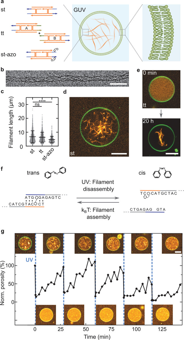
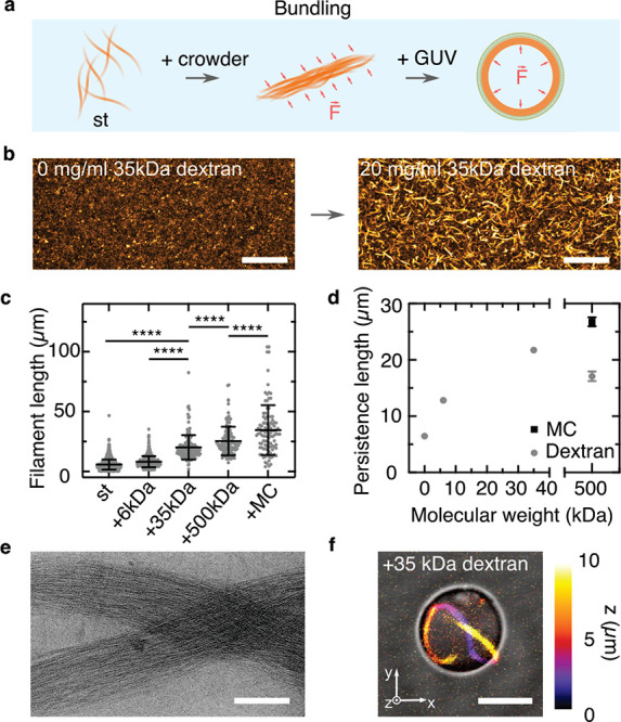
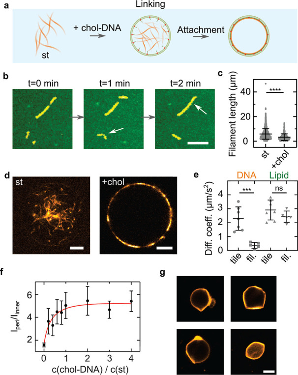
Cytoskeletal elements, like actin and myosin, have been reconstituted inside lipid vesicles toward the vision to reconstruct cells from the bottom up. Here, we realize the de novo assembly of entirely artificial DNA-based cytoskeletons with programmed multifunctionality inside synthetic cells. Giant unilamellar lipid vesicles (GUVs) serve as cell-like compartments, in which the DNA cytoskeletons are repeatedly and reversibly assembled and disassembled with light using the cis-trans isomerization of an azobenzene moiety positioned in the DNA tiles. Importantly, we induced ordered bundling of hundreds of DNA filaments into more rigid structures with molecular crowders. We quantify and tune the persistence length of the bundled filaments to achieve the formation of ring-like cortical structures inside GUVs, resembling actin rings that form during cell division. Additionally, we show that DNA filaments can be programmably linked to the compartment periphery using cholesterol-tagged DNA as a linker. The linker concentration determines the degree of the cortex-like network formation, and we demonstrate that the DNA cortex-like network can deform GUVs from within. All in all, this showcases the potential of DNA nanotechnology to mimic the diverse functions of a cytoskeleton in synthetic cells.

Keywords: DNA nanotechnology; DNA nanotube; azobenzene; bottom-up synthetic biology; giant unilamellar vesicles; synthetic cell.


Conflict of interest statement

The authors declare no competing financial interest.

Figures

Figure 1
Figure 1
DNA cytoskeletons can be assembled reversibly inside GUVs as lipid-bilayer-enclosed synthetic cell models. (a) Schematic representation of a GUV containing a DNA cytoskeleton composed of DNA filaments. DNA cytoskeletons were assembled from a single tile (st) with sticky overhangs, two tiles (tt) with orthogonal complementarity, or single tiles modified internally with two azobenzene moieties (st-azo). The asterisk indicates the position of a single-stranded overhang modified with a fluorophore. (b) Cryo-electron micrograph of an st DNA filament. Scale bar: 50 nm. (c) DNA filament length (n > 1000 filaments, mean ± SD). The st and tt filaments have the same length (p = 0.16), and st-azo filaments are shorter (p ≤ 0.001). (d) Confocal image of an st DNA cytoskeleton (orange, labeled with Cy3, λex = 561 nm) inside a GUV (green, 69% DOPC, 30% DOPG, 1% Atto488-DOPE, λex = 488 nm). Scale bar: 10 μm. (e) Confocal images of tt cytoskeletons prior to (0 h) and after assembly (20 h) inside a GUV. Scale bar: 10 μm. (f) Schematic representation of the st overhang modified with azobenzene (st-azo) for reversible cytoskeleton assembly with UV light. (g) Light-mediated reversible assembly of 500 nM st-azo cytoskeletons inside a GUV. The porosity measures the degree of filament polymerization over time. Time points of UV illumination (15 s) are indicated (blue dashed line). Insets depict confocal images of the same GUV at the respective time points. Scale bar: 10 μm.
Figure 2
Figure 2
DNA filament bundling leads to the formation of ring-like structures within GUVs. (a) Schematic representation of the bundling of DNA filaments caused by the addition of a molecular crowder and the subsequent formation of a cortex-like network inside GUVs. (b) Confocal z-projection of st DNA filaments (orange, labeled with Cy3, λex = 561 nm) in the absence and presence of 20 mg/mL 35 kDa dextran. Scale bar: 50 μm. (c) Length distribution of DNA filaments in the absence of bundling agents (st, n = 1896) and st DNA filaments in the presence of 20 mg/mL 6 kDa dextran (n = 510), 35 kDa dextran (n = 180), 500 kDa dextran (n = 129), and 500 kDa methylcellulose (MC, n = 104). All conditions are significantly different in length (p ≤ 0.001). (d) Persistence length of DNA filaments over molecular weight of the crowder (n = 11–15, mean ± SD). (e) Cryo-electron micrograph of bundled st filaments in the presence of 20 mg/mL dextran (MW = 35 kDa). Scale bar: 200 nm. (f) Overlay of color-coded confocal z-projection and bright-field image of 50 nM st filaments in the presence of 20 mg/mL 35 kDa dextran inside a GUV. Scale bar: 5 μm.
Figure 3
Figure 3
Deformation of GUVs from within by a membrane-linked DNA cortex-like network. (a) Schematic illustration of the linkage of DNA filaments to the GUV membrane with cholesterol-tagged DNA. (b) Confocal images of cholesterol-linked DNA filaments (st-chol, Cy3, λex = 561 nm) on a supported lipid bilayer (SLB, green, Atto488-DOPE, λex = 488 nm). The st-chol filaments diffuse and grow on the SLB. Scale bar: 10 μm. (c) Length distribution of st DNA filaments on glass (n = 1896) and st-chol-DNA filaments on SLBs (n = 429). The st-chol filaments are significantly shorter (p ≤ 0.001, mean ± SD). (d) Confocal images of 500 nM st (left) and st-chol filaments inside GUVs. For st-chol-containing GUVs, SUVs were incubated with 2 μM chol-link DNA for 2 min. Scale bar: 5 μm. (e) Diffusion coefficients of DNA filaments on SLBs determined by FRAP. Disassembled st-chol filaments (tile) exhibit 6-fold increased diffusion speeds compared to polymerized st-chol filaments (fil., p = 0.0007). The diffusion of the lipids of the SLB is not influenced by the polymerization state of the DNA filaments (n = 5–7, mean ± SD). (f) Fluorescence ratio Iperi/Iinner of 500 nM st-chol filaments inside GUVs at varying chol-DNA to st ratios (n = 10–18 analyzed GUVs). (g) Confocal images of deformed GUVs containing 1 μM st-chol filaments at an osmolarity ratio of cout/cin = 600 mOsm/300 mOsm = 2. Scale bar: 5 μm. (h) Circularity of deflated (cout/cin = 600 mOsm/300 mOsm = 2) and undeflated (cout/cin = 300 mOsm/300 mOsm = 1) GUVs containing 1 μM st-chol filaments (n = 4 and n = 7, respectively, mean ± SD, p = 0.008).

References

    1. Litschel T.; Kelley C. F.; Holz D.; Adeli Koudehi M.; Vogel S. K.; Burbaum L.; Mizuno N.; Vavylonis D.; Schwille P. Reconstitution of Contractile Actomyosin Rings in Vesicles. Nat. Commun. 2021, 12, 2254.10.1038/s41467-021-22422-7. - DOI - PMC - PubMed
    1. Maan R.; Loiseau E.; Bausch A. R. Adhesion of Active Cytoskeletal Vesicles. Biophys. J. 2018, 115, 2395–2402. 10.1016/j.bpj.2018.10.013. - DOI - PMC - PubMed
    1. Bashirzadeh Y.; Redford S. A.; Lorpaiboon C.; Groaz A.; Moghimianavval H.; Litschel T.; Schwille P.; Hocky G. M.; Dinner A. R.; Liu A. P. Actin Crosslinker Competition and Sorting Drive Emergent GUV Size-Dependent Actin Network Architecture. Communications Biology 2021, 4, 1136.10.1038/s42003-021-02653-6. - DOI - PMC - PubMed
    1. Lee K. Y.; Park S.-J.; Lee K. A.; Kim S.-H.; Kim H.; Meroz Y.; Mahadevan L.; Jung K.-H.; Ahn T. K.; Parker K. K.; Shin K. Photosynthetic Artificial Organelles Sustain and Control ATP-Dependent Reactions in a Protocellular System. Nat. Biotechnol. 2018, 36, 530–535. 10.1038/nbt.4140. - DOI - PubMed
    1. Vutukuri H. R.; Hoore M.; Abaurrea-Velasco C.; van Buren L.; Dutto A.; Auth T.; Fedosov D. A.; Gompper G.; Vermant J. Active Particles Induce Large Shape Deformations in Giant Lipid Vesicles. Nature 2020, 586, 52–56. 10.1038/s41586-020-2730-x. - DOI - PubMed

Publication types