. 2022 Apr 27;10(2):e0236121.
doi: 10.1128/spectrum.02361-21. Epub 2022 Apr 4.

Potential Role of the Host-Derived Cell-Wall Binding Domain of Endolysin CD16/50L as a Molecular Anchor in Preservation of Uninfected Clostridioides difficile for New Rounds of Phage Infection

Affiliations

Potential Role of the Host-Derived Cell-Wall Binding Domain of Endolysin CD16/50L as a Molecular Anchor in Preservation of Uninfected Clostridioides difficile for New Rounds of Phage Infection

Wichuda Phothichaisri et al. Microbiol Spectr. .

Abstract

Endolysin is a phage-encoded cell-wall hydrolase which degrades the peptidoglycan layer of the bacterial cell wall. The enzyme is often expressed at the late stage of the phage lytic cycle and is required for progeny escape. Endolysins of bacteriophage that infect Gram-positive bacteria often comprises two domains: a peptidoglycan hydrolase and a cell-wall binding domain (CBD). Although the catalytic domain of endolysin is relatively well-studied, the precise role of CBD is ambiguous and remains controversial. Here, we focus on the function of endolysin CBD from a recently isolated Clostridioides difficile phage. We found that the CBD is not required for lytic activity, which is strongly prevented by the surface layer of C. difficile. Intriguingly, hidden Markov model analysis suggested that the endolysin CBD is likely derived from the CWB2 motif of C. difficile cell-wall proteins but possesses a higher binding affinity to bacterial cell-wall polysaccharides. Moreover, the CBD forms a homodimer, formation of which is necessary for interaction with the surface saccharides. Importantly, endolysin diffusion and sequential cytolytic assays showed that CBD of endolysin is required for the enzyme to be anchored to post-lytic cell-wall remnants, suggesting its physiological roles in limiting diffusion of the enzyme, preserving neighboring host cells, and thereby enabling the phage progeny to initiate new rounds of infection. Taken together, this study provides an insight into regulation of endolysin through CBD and can potentially be applied for endolysin treatment against C. difficile infection. IMPORTANCE Endolysin is a peptidoglycan hydrolase encoded in a phage genome. The enzyme is attractive due to its potential use as antibacterial treatment. To utilize endolysin for the therapeutic propose, understanding of the fundamental role of endolysin becomes important. Here, we investigate the function of cell-wall binding domain (CBD) of an endolysin from a C. difficile phage. The domain is homologous to a cell-wall associating module of bacterial cell-wall proteins, likely acquired during phage-host coevolution. The interaction of CBD to bacterial cell walls reduces enzyme diffusion and thereby limits cell lysis of the neighboring bacteria. Our findings indicate that the endolysin is trapped to the cell-wall residuals through CBD and might serve as an advantage for phage replication. Thus, employing a CBD-less endolysin might be a feasible strategy for using endolysin for the treatment of C. difficile infection.

Keywords: Clostridioides difficile; bacteria-phage coevolution; cell-wall binding domain; endolysin; peptidoglycan; surface layer.


Conflict of interest statement

The authors declare no conflict of interest.

Figures

FIG 1
FIG 1
The endolysin CD16/50L is a modular cell-wall hydrolase. (A) Schematic representation of domain structure of endolysin CD16/50L and N-terminally hexahistidine (6xHis)-tagged CD16/50L variants used in this study. CD16/50L is composed of the N-terminal enzymatically active domain (EAD) and the C-terminal cell-wall binding domain (CBD). Numbers indicate amino acid positions in the full-length protein. (B) The EAD of CD16/50L confers a peptidoglycan hydrolase activity. Recombinant protein variants of CD16/50L were expressed in E. coli. Crude lysate (L) and purified proteins (P) were resolved by SDS-PAGE followed by Coomassie blue staining or zymogram analysis using C. difficile peptidoglycan as substrate. Asterisk indicates a false positive band of a highly positively charged protein (see also Fig. S1C; [42]). (C) The full-length and EAD of CD16/50L are able to lyse intact cells of C. difficile. Cytolytic activity of endolysin was assessed by using turbidity reduction assay. Exponentially growing cells were harvested and resuspended in 50 mM Tris-HCl (pH 7.4) in an absence or presence of 2.5 μM purified CD16/50L variants and incubated at 37°C in anaerobic condition for 180 min. The decrement of OD600 over time was monitored and plotted. Mean ± SD are shown (n = 3).
FIG 2
FIG 2
The CBD of CD16/50L interacts with surface polysaccharide of C. difficile. (A) The CBD of CD16/50L binds to cells lacking the surface layer (S-layer) more effectively than the intact cells. Binding of the CBD to whole bacterial cells was investigated using a cell-based pulldown assay. Purified 6xHis-CBD was mixed with buffer, untreated C. difficile, or cells treated with acidic glycine buffer. After incubation and centrifugation, fractions of cell pellet (P) and supernatant (S) were separated in SDS-PAGE, followed by Coomassie blue stain. The high molecular weight (HMW), low molecular weight (LMW) subunits of surface-layer protein (SLP) and the purified 6xHis-CBD are indicated by arrowheads. (B) The CBD of CD16/50L binds to purified peptidoglycan and polysaccharide (PG-PS) complex. Analysis similar to Fig. 2A, but purified PG-PS complex was used. (C) The CD16/50L CBD localizes to the periphery of C. difficile cells deficient in functional S-layer. Affinity purified mCherry-fusion CBD was incubated with exponentially growing wild-type (R20291) or S-layer-deficient (FM2.5) C. difficile cells at 37°C for 30 min. Fluorescence microscopy shows the localization of CBD on the periphery of FM2.5 cells but not that of the R20291. The scale bar represents 5 μm. (D) The CBD of CD16/50L interacts with the secondary polysaccharide of C. difficile cell wall. Purified PG-PS complex, PG, and PS were spotted onto nitrocellulose membrane. Far Western blotting was performed by incubated the membrane with (right panel) or without (left panel) purified 6xHis-CBD in TBS-T containing 2% bovine serum albumin (BSA). Bound CBD was detected by anti-His antibodies followed by HRP-conjugated secondary antibodies and chemiluminescence detection. Spotted 6xHis-CBD and BSA serve as positive and negative controls of far Western blotting assay.
FIG 3
FIG 3
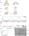
The CBD of CD16/50L forms a homodimer. (A) Modeled structures of CD16/50L’s CBD homodimer predicted by SWISS-MODEL. Ribbon diagram displays the head-on dimer (left) and the side-by-side dimer (right). The interface residues are labeled and shown as a stick model. (B) Scheme of reporter constructs used in a split complementary assay (left panel). Expression of each reporter construct is driven by the xylose-inducible promoter. The full-length luciferase reporter BitLucopt serves as positive control. Negative controls are the CBD fused with only the large N-terminus (LgBit) or the small C-terminus (SmBit) of BitLucopt. To observe an in vivo dimerization, wild-type (WT) or mutant (mut) variants of CBD were fused with each fragment of the reporter. If dimerization occurred, bright luminescent signal could be observed (right panel). Locations of ribosome-binding site (RBS) and linker are shown. (C) CD16/50L’s CBD most likely forms a homodimer in vivo. The split complementary luciferase assay using the reporter constructs shown in Fig. 3B was performed. Exponentially growing C. difficile cells harboring each reporter construct were induced with 4% (wt/vol) xylose for 2 h. Furimazine substrate was added and incubated for 5 min. The luminescent signal was then detected and plotted. CBD WT fusions exhibited a strong luciferase activity, while CBD(W257A) and CBD(Y202A, W257A) mutant variants significantly reduced the signal. Mean ± SD are shown (n = 3) (***P < 0.001). (D) The CBD of CD16/50L forms a homodimer in vitro and the W257 residue is crucial the dimerization. Size exclusion chromatography (SEC; Superdex 75 10/300 GL) analysis of purified 6xHis-CBD protein variants was performed and elution profiles plotted. Elution peaks of protein standards of indicated molecular weight are indicated as black rectangles at the top. (E) SDS-PAGE analysis of the CBD protein variants fractionated by SEC (Fig. 3D). Proteins were separated on SDS-PAGE and detected by Coomassie blue stain. (F) CBD dimerization is most likely crucial for the interaction with the PG-PS complex. Similar to Fig. 2B, but purified CBD mutant variants were analyzed. IN, material before pull-down; S, supernatant; W, wash fraction; P, pellet fraction. (G) Relative binding quantified from Fig. 3F. Protein levels relative to the CBD WT were calculated and plotted. Mean ± SD are shown (n = 3) (*P < 0.05, **P < 0.01, NS, not significant).
FIG 4
FIG 4
The CBD of CD16/50L is evolutionarily homologous to CWB2 domain of C. difficile cell-wall proteins and binds to PG-PS complex more tightly than the host counterpart. (A) Ribbon diagrams represent the structure of CWB2 (for cell wall binding 2) domain of Cwp8 (PDB: 5J6Q, gold) and CD16/50L’s CBD (similar to Fig. 3A, pink). The top panel shows an oligomer while the bottom panel indicates a monomer. The N- and C-terminus are indicated. (B) Structural superimposition of CWB2 monomer (gold) and the CD16/50L CBD (pink) suggests a remote structural homology. Two views of the structures are related by a 90-degree rotation. (C) Comparison of RMSD of Cα atoms between Cwp8 CWB2:Cwp6 CWB2 (blue) and Cwp8 CWB2:CD16/50L CBD (orange) is shown. The Cwp6 CWB2 serves as a control. The amino acid sequence and secondary structure of the Cwp8 CWB2 are shown. An alpha helix (red), a beta sheet (green), and a loop (gray) are indicated. (D) Scheme of Cwp8 and N-terminally 6xHis-tagged Cwp8 CWB2. The Cwp8 consists of the N-terminal domain and a triangular trimer of CWB2 domain. The number at the start and end of each protein/domain indicates the amino acid position. (E) The CD16/50L CBD binds to PG-PS complex more tightly than the bacterial CWB2 domain. Similar to Fig. 2B but PG-PS binding activity of CBD and CWB2 was compared. An equal volume of 5 μM purified CD16/50L CBD or CWB2 was incubated with indicated amount of PG-PS complex at 37°C for 20 min. After centrifugation and wash, pellets were resuspended in one tenth (×10) of the original volume. Then an equal volume of input (IN), supernatant (S), and pellet (P) were subjected to SDS-PAGE, followed by Coomassie blue stain. The 6xHis-CWB2 and 6xHis-CBD position are marked as arrowhead. The asterisk denotes a degraded fragment of the purified protein. One representative experiment from three biological replicates is shown. (F) Protein band intensity shown in Fig. 4E was quantified, percentage of PG-PS bound protein over the input calculated and plotted. Mean ± SD are shown (n = 3) (*P < 0.05).
FIG 5
FIG 5
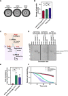
The CBD of CD16/50L anchors the endolysin to bacterial post-lytic remnants and prevents a successive round of cytolysis. (A) The CBD of CD16/50L and its dimerization decrease protein diffusion across a layer of soft agar containing heat-inactivated C. difficile cell debris. A 7-mm diameter hole was created and filled with 100 μM purified CD16/50L protein variants. After incubation at 37°C overnight, diffusion of proteins was assessed by the formation of a lysis halo. One representative experiment from three biological replicates is shown. (B) The diameter of the lytic halo from Fig. 5A was measured and plotted. A dash line indicates the diameter of each hole. Mean ± SD are shown (n = 3) (*P < 0.05, **P < 0.01). (C) A diagram depicting a sequential cytolytic assay. The experiment comprises two steps of bacterial cell lysis. Firstly, exponentially growing cells are harvested and resuspended in buffer containing 5 μM purified CD16/50L variants. The mixtures are then incubated at 37°C in anaerobic conditions for 3 h to let endolysin-mediated cytolysis occur. A soluble and insoluble fraction are separated by centrifugation and analyzed by SDS-PAGE followed by Coomassie blue stain. Subsequently, unanchored proteins of soluble fraction are mixed with fresh growing cells, reduction of OD600 over time is followed and plotted. (D) The CBD of CD16/50L and its dimerization facilitate the anchoring of the enzyme to cell remnants after endolysin-mediated cytolysis. SDS-PAGE analysis of the CD16/50L protein variants fractionated by centrifugation after the first cytolysis (Fig. 5C). Proteins were separated on SDS-PAGE and detected by Coomassie blue stain. (E) Ratio of soluble and insoluble fraction was quantified from Fig. 5D and plotted. Mean ± SD are shown (n = 3) (*P < 0.05, **P < 0.01). (F) Unanchored CD16/50L is able to perform a successive cytolysis of C. difficile cells in vitro. Similar to Fig. 1C, but post-centrifuged supernatants after the first cytolysis were analyzed. Mean ± SD are shown (n = 3).

Similar articles

Cited by

References

    1. Centers for Disease Control and Prevention. 2019. Antibiotic resistance threats in the United States, 2019. Centers for Disease Control and Prevention.
    1. Finn E, Andersson FL, Madin-Warburton M. 2021. Burden of Clostridioides difficile infection (CDI) - a systematic review of the epidemiology of primary and recurrent CDI. BMC Infect Dis 21:456. doi:10.1186/s12879-021-06147-y. - DOI - PMC - PubMed
    1. Vedantam G, Clark A, Chu M, McQuade R, Mallozzi M, Viswanathan VK. 2012. Clostridium difficile infection: toxins and non-toxin virulence factors, and their contributions to disease establishment and host response. Gut Microbes 3:121–134. doi:10.4161/gmic.19399. - DOI - PMC - PubMed
    1. Francis MB, Allen CA, Shrestha R, Sorg JA. 2013. Bile acid recognition by the Clostridium difficile Germinant Receptor, CspC, is important for establishing infection. PLoS Pathog 9:e1003356. doi:10.1371/journal.ppat.1003356. - DOI - PMC - PubMed
    1. Paredes-Sabja D, Shen A, Sorg JA. 2014. Clostridium difficile spore biology: sporulation, germination, and spore structural proteins. Trends Microbiol 22:406–416. doi:10.1016/j.tim.2014.04.003. - DOI - PMC - PubMed

Publication types

MeSH terms

LinkOut - more resources