. 2022 May 2;154(5):e202112912.
doi: 10.1085/jgp.202112912. Epub 2022 Apr 4.

The action of Con-ikot-ikot toxin on single AMPA-type glutamate receptors

Affiliations

The action of Con-ikot-ikot toxin on single AMPA-type glutamate receptors

Jelena Baranovic et al. J Gen Physiol. .

Abstract

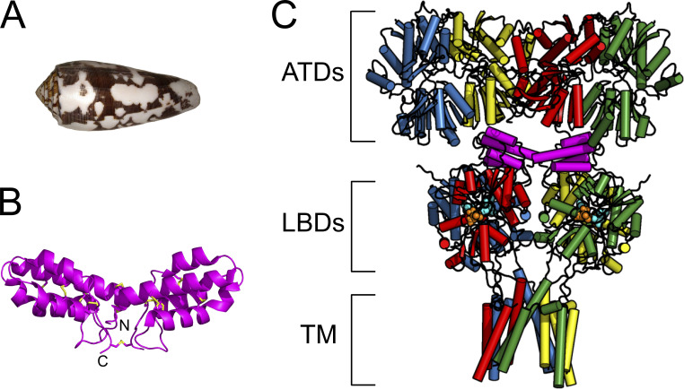
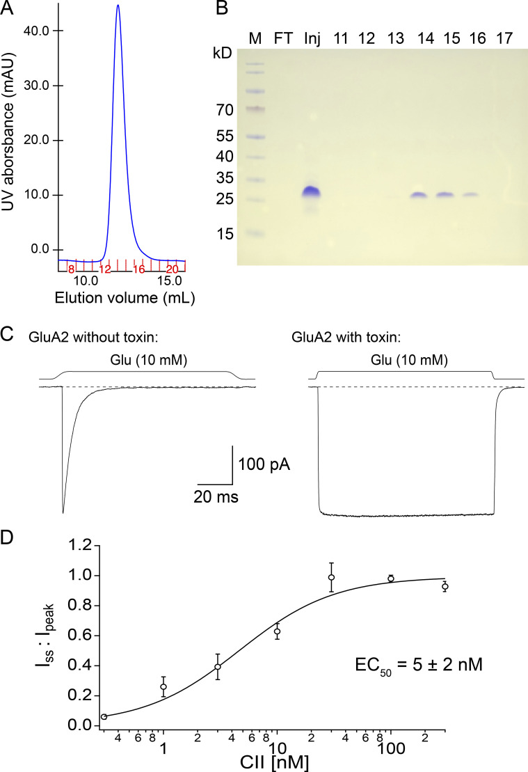
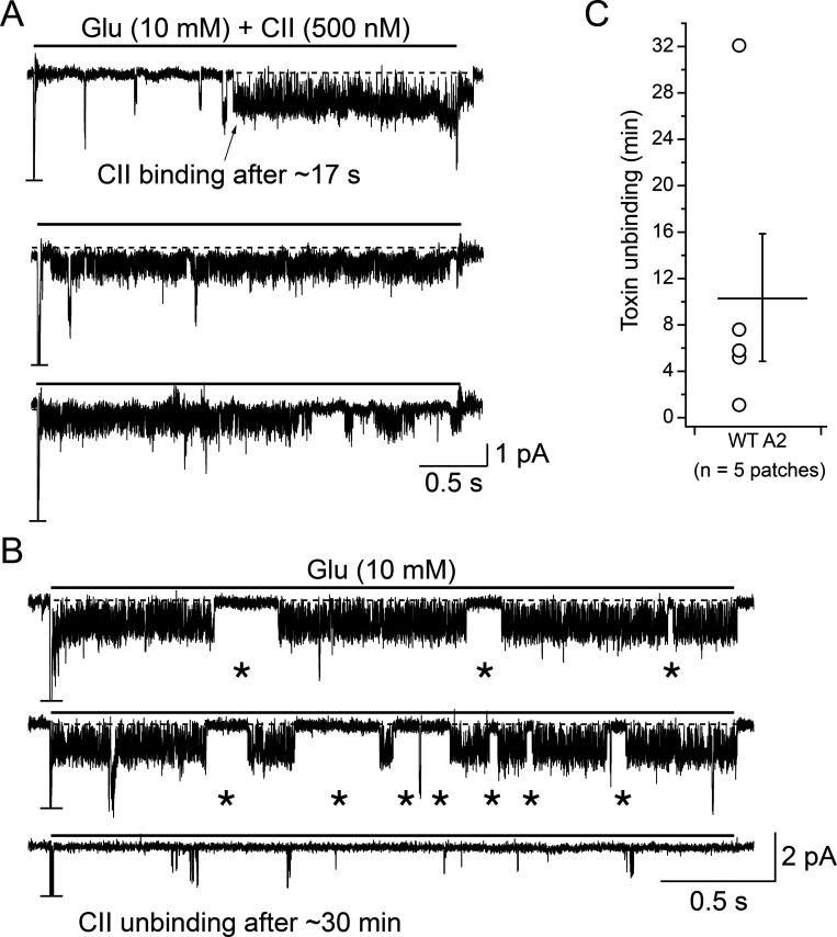
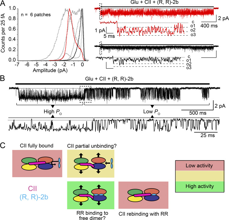
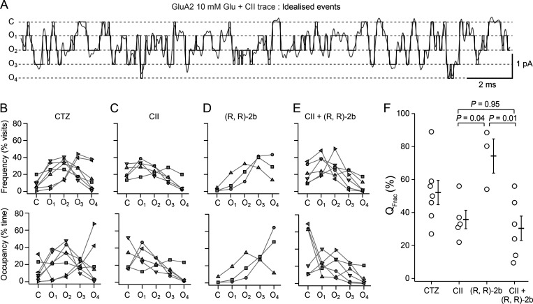
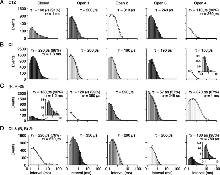
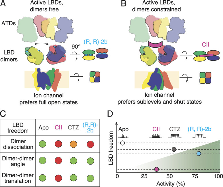
Conotoxins are a large group of naturally occurring toxic peptides produced by the predatory sea snails of the genus Conus. Many of these toxins target ion channels, often with high specificity and affinity. As such, they have proven to be invaluable for basic research, as well as acting as leads for therapeutic strategies. Con-ikot-ikot is the only conotoxin so far identified that targets AMPA-type glutamate receptors, the main mediators of excitatory neurotransmission in the vertebrate brain. Here, we describe how the toxin modifies the activity of AMPA receptors at the single-channel level. The toxin binds to the AMPA receptor with EC50 of 5 nM, and once bound takes minutes to wash out. As shown previously, it effectively blocks desensitization of AMPA receptors; however, compared to other desensitization blockers, it is a poor stabilizer of the open channel because toxin-bound AMPA receptors undergo frequent brief closures. We propose that this is a direct consequence of the toxin's unique binding mode to the ligand-binding domains (LBDs). Unlike other blockers of desensitization, which stabilize individual dimers within an AMPA receptor tetramer, the toxin immobilizes all four LBDs of the tetramer. This result further emphasizes that quaternary reorganization of independent LBD dimers is essential for the full activity of AMPA receptors.


Figures

Figure 1.
Figure 1.
Structure of CII toxin. (A) Shell of a Conus striatus snail, the natural source of CII toxin. (urjsa, 2008 is marked with CC BY-SA 2.0). (B) Crystal structure of the toxin homodimer; each CII monomer is a four-helix bundle containing five disulfide bridges, connected to another monomer via additional three disulfide bridges (yellow; PDB accession no. 4U5H; Chen et al., 2014). (C) Crystal structure of the full-length AMPA receptor (subunit GluA2) in complex with a toxin (magenta), partial agonist kainate, and desensitization blocker (R, R)-2b (PDB accession no. 4U5D; Chen et al., 2014); each AMPA receptor subunit is colored differently with the domains indicated with square brackets as: ATDs, amino terminal domains; LBDs, ligand binding domains; and TM, transmembrane region. Red and blue subunits are forming one and green and yellow the other LBD dimer. The V-shaped toxin dimer (magenta) sits on top of LBDs.
Figure 2.
Figure 2.
CII toxin purification and characterization. (A) Size-exclusion chromatogram (SEC) of purified CII toxin with fraction numbers indicated in red. (B) Coomassie-stained non-reducing SDS-PAGE gel of the SEC fractions. Wells marked 11–17 are SEC fractions, Inj is the concentrated sample injected onto the SEC column, and FT is the flow-through after concentrating the sample and before loading it onto the SEC column (to check for any protein loss). Fractions 14–16 contain the purified CII dimer. (C) The activity of the purified toxin was checked with outside-out patches: left trace shows the current produced by GluA2 AMPA receptors in the presence of glutamate (10 mM) without CII bound and right trace is from a different patch with CII toxin bound (after overnight incubation in 100 nM toxin). The dashed line indicates baseline and square trace above solution exchange in the respective patch. Voltage was approximately −40 mV in both recordings. (D) CII toxin dose-response curve for AMPA receptors (0.3–300 nM CII, n = 3–8 patches/toxin concentration). Toxin effect was measured as desensitization block, i.e., ratio of steady-state current over peak current for each patch (one for complete desensitization block). Fit by Hill equation gave EC50 of 5 ± 2 nM and Hill slope of 1 ± 0.3 (95% confidence interval).
Figure 3.
Figure 3.
Binding and unbinding of toxin to individual AMPA receptors. (A) Current trace showing the binding of the toxin to one AMPA receptor in a patch containing multiple receptors, as indicated by the truncated peak current response at the start of glutamate application. Arrow indicates toxin binding event after about 17 s. A horizontal black line above the current trace indicates a jump from perfusing external solution (free of glutamate and toxin) into a perfusing solution containing glutamate (Glu) and the toxin (CII) for 3 s. Jumps were repeated as long as the patch was stable. (B) To detect toxin unbinding, patches with bound toxin were perfused in toxin-free solutions (3 s jumps into 10 mM Glu as described in A). In this example, long bursts of activity characteristic for toxin-bound AMPA receptors (top two traces) were replaced by short bursts of activity interspersed by long closures (bottom trace), after about 30 min. In both, A and B, the dashed line indicates the baseline. Voltage was clamped at −80 mV for both traces. Asterisks mark longer shut epochs (>5 ms) during the toxin-bound phase. (C) After binding of the toxin to one AMPA receptor in a patch, it took on average 10.4 ± 5.5 min (n = 5) to observe unbinding in toxin-free solutions, with regular 3 s jumps into glutamate.
Figure 4.
Figure 4.
Comparison of CII toxin with AMPA receptor desensitization blockers CTZ and (R, R)-2b. (A) All-point-amplitude histograms of single-channel recordings of GluA2 wild type receptors perfused in glutamate (10 mM) and positive modulator cyclothiazide (CTZ, 100 μM), nCTZ = 7 (∼56 s of recording in total). A histogram was made for each patch, using only the parts of the recording where the patch was perfused in glutamate (Glu) and CTZ. Histograms were normalized to the maximum value. Representative single-channel currents from two color-coded patches are drawn next to the corresponding histograms. Dashed box indicates the part of the trace shown at greater magnification below. Dotted lines are baseline (closed level [c] in zoom), and dashed lines are open levels in each patch (O1–O4). All openings are downwards, traces were obtained at approximately −80 mV and low pass filtered at 1 kHz for presentation. (B and C) Same as in A, but with different modulators: (R, R)-2b (10 μM; nRR = 4; ∼81 s of recording in total), CII toxin (100–500 nM; nCII = 7; ∼77 s of recording in total).
Figure 5.
Figure 5.
AMPA receptor activity in CII toxin and (R, R)-2b. (A) As in Fig. 4, a normalized histogram was made for each patch. Representative single-channel currents from two color-coded patches are drawn on the same scale. The red trace had comparatively high activity, whereas the black record shows much less activity. Dashed box indicates the part of the trace shown at greater magnification below (again with common scale). Dotted lines are baseline (closed level [c] in zoom), and dashed lines are open levels in each patch (O1–O3, O4 not reached). All openings are downwards, traces were obtained at approximately −80 mV and low pass filtered at 1 kHz for presentation. Patches were taken from cells incubated in 100–500 nM CII and exposed to 10 μM (R, R)-2b; nRR+CII = 6, ∼72 sec of recording in total. (B) Example of a mode switch from high Popen activity (similar to (R, R)-2b alone and red trace from A) to low Popen activity (similar to CII alone and black trace from A) during a 3 s recording in the presence of 10 mM glutamate, 10 μM (R, R)-2b, and saturating CII. (C) Diagram indicating how binding and unbinding of CII toxin (magenta) and (R, R)-2b (cyan) might lead to different activity levels of an AMPA receptor (red sections, low activity; yellow sections, intermediate activity; green sections, high activity). LBDs of AMPA receptors (top view) are indicated by four differently coloured ellipses. Thick, black arrows indicate freedom of movement for LBDs.
Figure 6.
Figure 6.
Occupancy of closed and open levels for GluA2 bound by modulators of desensitization. (A) An example of the idealization from ASCAM (black), overlaid on the current trace (grey; low-pass filtered at 1 kHz for presentation) obtained in glutamate (Glu, 10 mM) and toxin (CII, 500 nM). Dashed lines indicate the four open conductance levels (O1–O4) and the closed level, C. (B–E) CTZ (100 μM; B), toxin alone (100–500 nM; C), (R, R)-2b (10 μM; D) and toxin + (R, R)-2b conditions (E). The frequency of visits to each level (upper row) and occupancy of each level as a fraction of the total time (bottom row) are shown for each patch. Different symbols in each condition represent different patches. (F) Fractional activity (QFrac, %) was determined as the ratio of the transferred charge to the charge that would be transferred if the channel were open continuously to the maximum open amplitude; CTZ: 53 ± 7% (n = 7), CII: 36 ± 6% (n = 5), (R, R)-2b: 74 ± 10% (n = 3), and toxin and (R, R)-2b: 30 ± 6% (n = 6). Horizontal bars are mean values and bars represent standard error of the mean. Probabilities of no difference shown are from Tukey’s HSD two-tailed test.
Figure 7.
Figure 7.
Dwell times of AMPA receptors in the presence of desensitization modulators. (A–D) Dwell time histograms were generated for the closed and each of the open levels (1–4), for CTZ (100 µM; A), CII toxin (100–500 nM; B), (R, R)-2b (10 μM; C), and toxin and (R, R)-2b (D) conditions. Shown are histograms from one representative patch for each condition. Each histogram was plotted on a log-square root scale and fit with one or two exponential components (grey curves). Mean time constants and amplitudes for each condition are given in Table 2.
Figure 8.
Figure 8.
Conformational freedom of ligand binding domain layer dictates activity of AMPA receptors. (A) AMPA receptor subunits are color-coded, with green and yellow forming one LBD dimer, and blue and red another. AMPA receptors freely desensitize as long as the dimers are allowed to disassociate, resulting in very low activity of wild type receptors. Fixing the two monomers within each dimer, by CTZ or (R, R)-2b (cyan, two bars indicating its double-headed structure), blocks AMPA receptor desensitization and increases activity. (B) Preventing lateral movements and/or rotations of the LBD dimers, through binding of CII toxin (magenta) lowers the activity of the ion channel. (C) Summary of degrees of freedom of the LBD layer–green meaning high, red meaning low, and orange meaning intermediate. (R, R)-2b has lower EC50 when binding to LBD dimers than CTZ. (D) Fictive single-channel currents indicate increasing activity over four conditions, including apo from previous work (Carbone and Plested, 2012). (R, R)-2b supports the highest activity (QFrac; Fig. 6) because it holds LBD dimers tightly together whilst allowing their free rotation and lateral translation to the optimal position(s).

References

    1. Baranovic, J., and Plested A. J.. 2018. Auxiliary subunits keep AMPA receptors compact during activation and desensitization. Elife. 7:e40548. 10.7554/eLife.40548 - DOI - PMC - PubMed
    1. Baranovic, J., Chebli M., Salazar H., Carbone A.L., Faelber K., Lau A.Y., Daumke O., and Plested A.J.R.. 2016. Dynamics of the ligand binding domain layer during AMPA receptor activation. Biophysj. 110:896–911. 10.1016/j.bpj.2015.12.033 - DOI - PMC - PubMed
    1. Carbone, A.L., and Plested A.J.R.. 2012. Coupled control of desensitization and gating by the ligand binding domain of glutamate receptors. Neuron. 74:845–857. 10.1016/j.neuron.2012.04.020 - DOI - PubMed
    1. Chen, L., Durr K.L., and Gouaux E.. 2014. X-ray structures of AMPA receptor-cone snail toxin complexes illuminate activation mechanism. Science. 345:1021–1026. 10.1126/science.1258409 - DOI - PMC - PubMed
    1. Christie, L.A., Russell T.A., Xu J., Wood L., Shepherd G.M.G., and Contractor A.. 2010. AMPA receptor desensitization mutation results in severe developmental phenotypes and early postnatal lethality. Proc. Natl. Acad. Sci. USA. 107:9412–9417. 10.1073/pnas.0908206107 - DOI - PMC - PubMed

Publication types

MeSH terms