. 2022 Jun 1;71(6):1246-1260.
doi: 10.2337/db21-0681.

DOC2b Enhances β-Cell Function via a Novel Tyrosine Phosphorylation-Dependent Mechanism

Affiliations

DOC2b Enhances β-Cell Function via a Novel Tyrosine Phosphorylation-Dependent Mechanism

Diti Chatterjee Bhowmick et al. Diabetes. .

Abstract

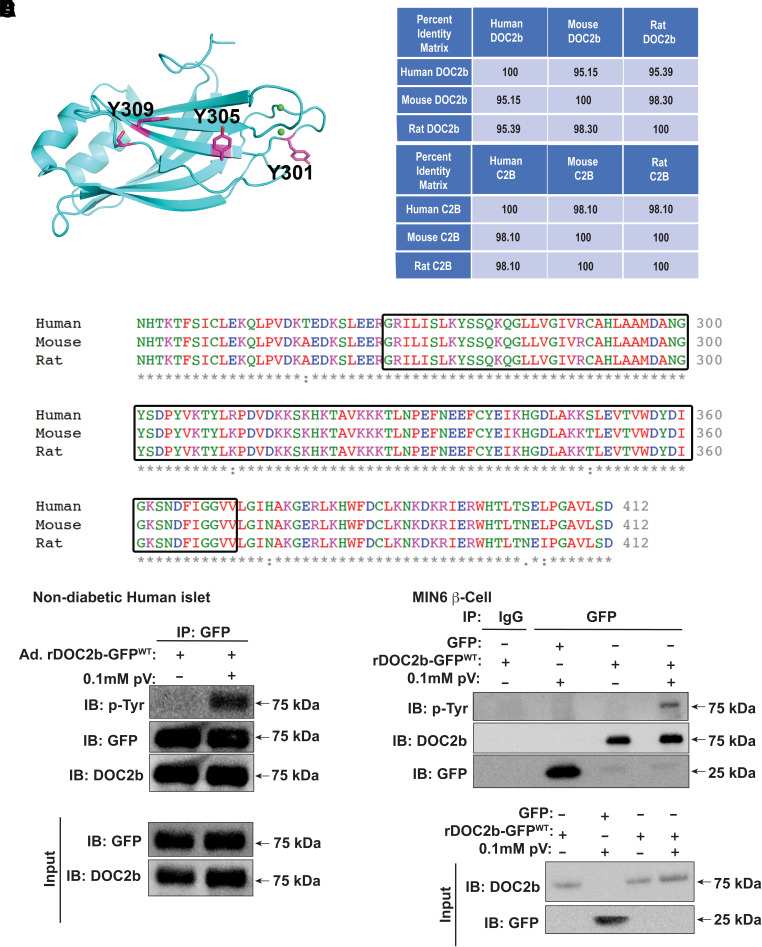
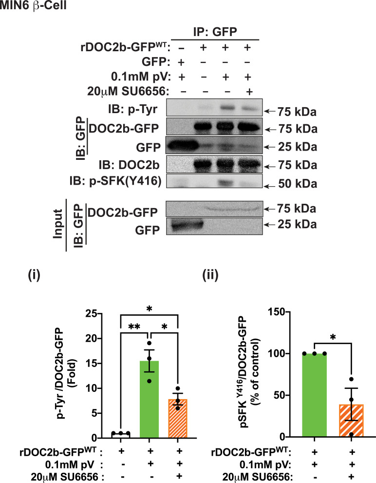
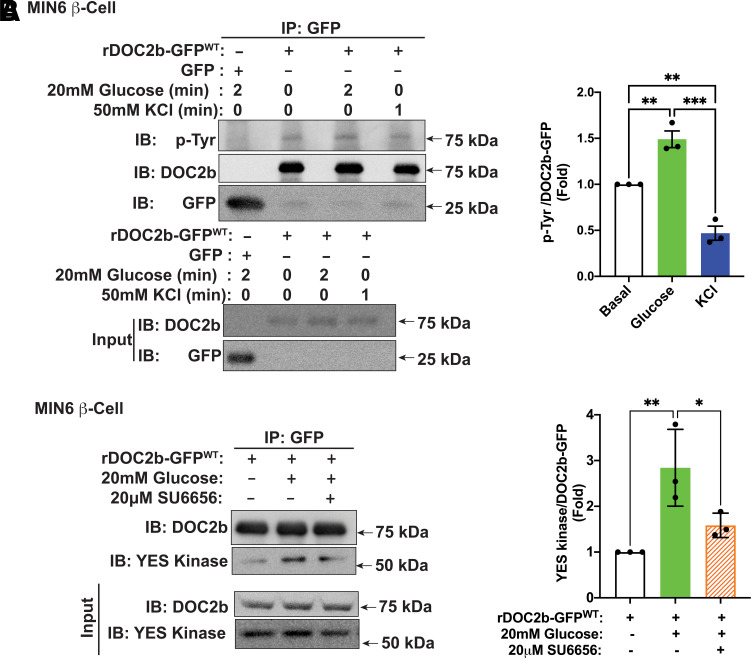
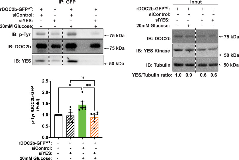
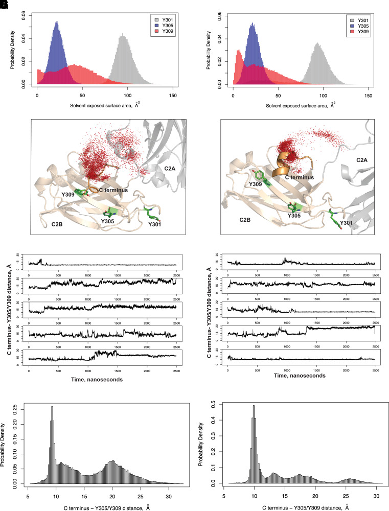
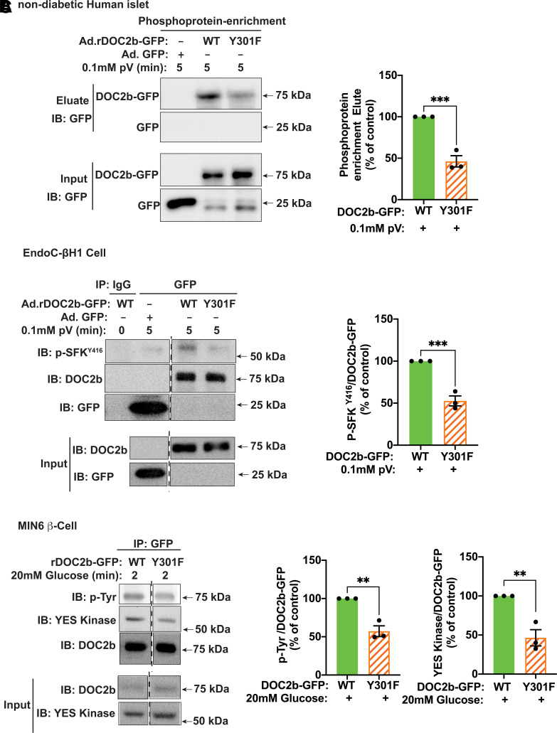
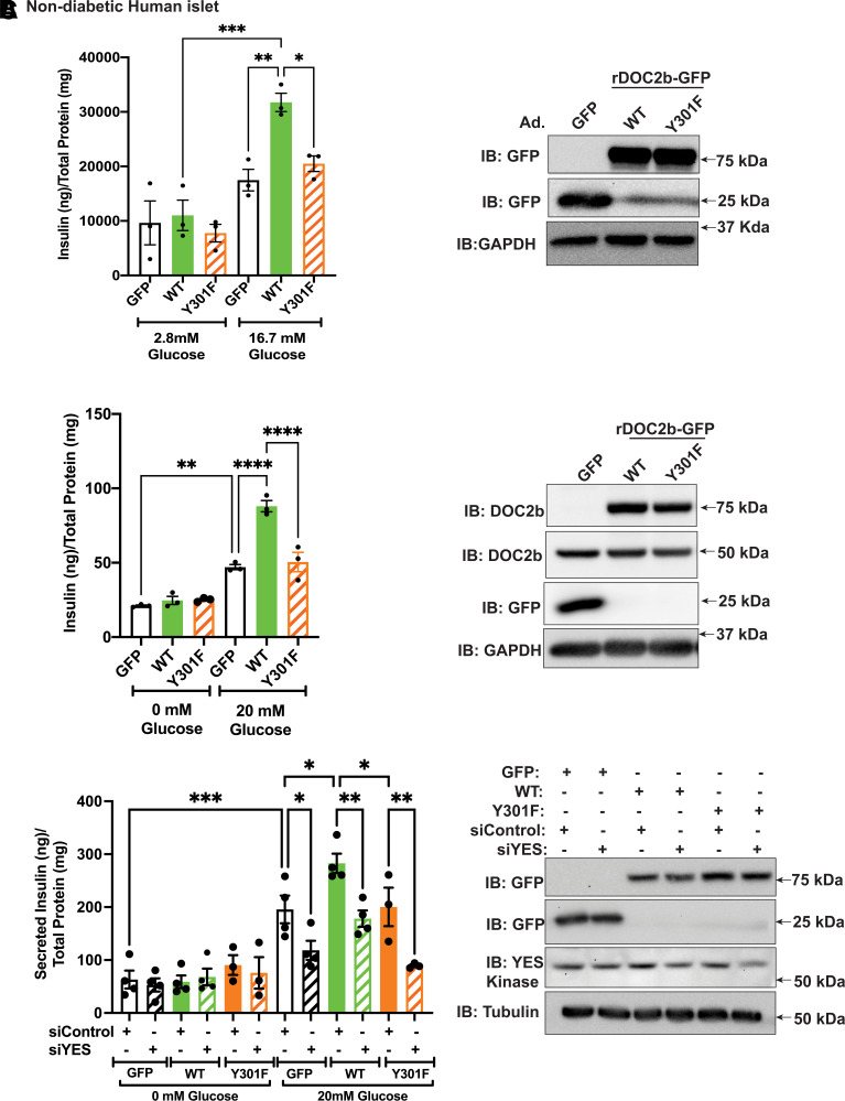
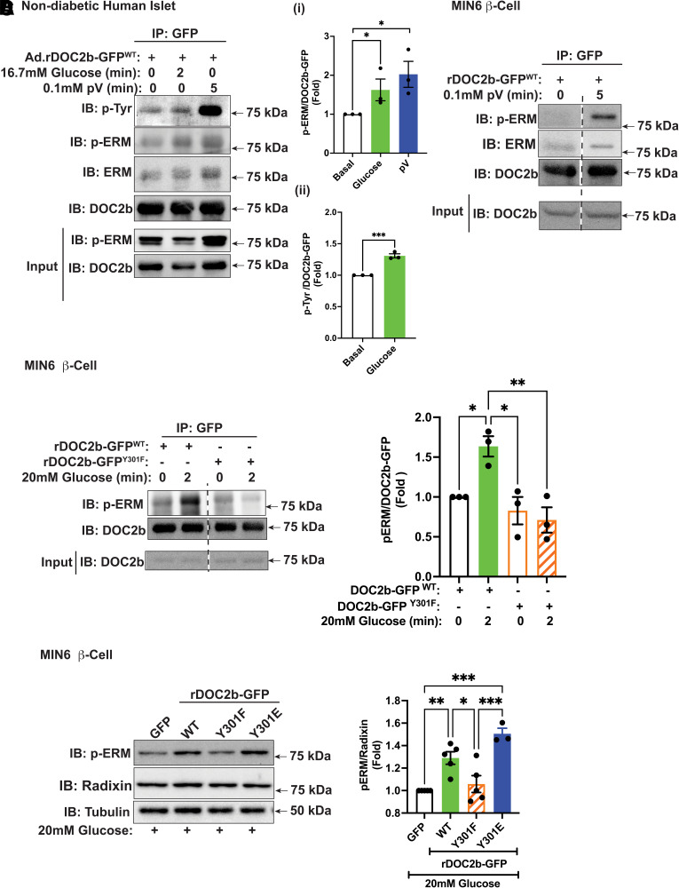
Double C2 domain Β (DOC2b) protein is required for glucose-stimulated insulin secretion (GSIS) in β-cells, the underlying mechanism of which remains unresolved. Our biochemical analysis using primary human islets and human and rodent clonal β-cells revealed that DOC2b is tyrosine phosphorylated within 2 min of glucose stimulation, and Src family kinase member YES is required for this process. Biochemical and functional analysis using DOC2bY301 mutants revealed the requirement of Y301 phosphorylation for the interaction of DOC2b with YES kinase and increased content of VAMP2, a protein on insulin secretory granules, at the plasma membrane (PM), concomitant with DOC2b-mediated enhancement of GSIS in β-cells. Coimmunoprecipitation studies demonstrated an increased association of DOC2b with ERM family proteins in β-cells following glucose stimulation or pervanadate treatment. Y301 phosphorylation-competent DOC2b was required to increase ERM protein activation, and ERM protein knockdown impaired DOC2b-mediated boosting of GSIS, suggesting that tyrosine-phosphorylated DOC2b regulates GSIS via ERM-mediated granule localization to the PM. Taken together, these results demonstrate the glucose-induced posttranslational modification of DOC2b in β-cells, pinpointing the kinase, site of action, and downstream signaling events and revealing a regulatory role of YES kinase at various steps in GSIS. This work will enhance the development of novel therapeutic strategies to restore glucose homeostasis in diabetes.


Figures

Figure 1
Figure 1
DOC2b is tyrosine phosphorylated in the β-cell. A: Predicted structure of rat DOC2b showing the C2B domain with the three putative tyrosine phosphorylation sites. B: Percent identity matrix for human, mouse, and rat full-length DOC2b (top panel) and C2B domain (bottom panel). C: Multiple sequence alignment of human, rat, and mouse DOC2b using Clustal Omega multiple sequence alignment tool (EMBL-European Bioinformatics Institute). Boxes highlight the conserved amino acid residues in the C2B domain. D: DOC2b tyrosine phosphorylation level in Ad.DOC2b-GFPWT-transduced nondiabetic human islets after pV treatment (0.1 mmol/L, 5 min) followed by IP and immunoblot (IB) analysis. E: The DOC2b tyrosine phosphorylation level in the GFP/DOC2b-GFPWT-transfected MIN6 β-cells after pV (0.1 mmol/L, 5 min) treatment followed by IP and IB analysis. Data are representative of three independent experiments.
Figure 2
Figure 2
SFK activity is required for the tyrosine phosphorylation of the DOC2b in MIN6 β-cells. Top panel: Representative Western blot images of MIN6 β-cells transfected with either GFP or rDOC2b-GFPWT plasmids and treated with 0.1 mmol/L pV (5 min) with or without 20 μmol/L SU6656 pretreatment (2 h) followed by IP and immunoblot (IB) analysis. Bottom panel: Quantification of the IBs pTyr/DOC2b-GFP (i) and p-SFKY416/DOC2b-GFP (ii) interactions from three independent experiments using different passages of cells. Data are shown as mean ± SEM. *P < 0.05; **P < 0.002.
Figure 3
Figure 3
Glucose stimulation augments DOC2b tyrosine phosphorylation and interaction with YES kinase in MIN6 β-cells. A, left: Representative Western blot images of DOC2b-GFPWT–transfected MIN6 β-cells treated with either 20 mmol/L glucose for 2 min or 50 mmol/L KCl for 1 min followed by IP and immunoblot (IB). A, right: Quantification of the IBs from three independent experiments. B, left: Representative Western blot images of MIN6 β-cells transfected with rDOC2b-GFPWT plasmid and stimulated with 20 mmol/L glucose (2 min) with or without 20 μmol/L SU6656 pretreatment (2 h) followed by IP and IB analysis. B, right: Quantification of the IBs from three independent experiments. Data are shown as mean ± SEM. *P < 0.05; **P < 0.002; ***P < 0.0002.
Figure 4
Figure 4
YES kinase knockdown ablates tyrosine phosphorylation of DOC2b in MIN6 β-cells. Representative Western blot images of MIN6 β-cells transfected with rDOC2b-GFPWT plasmid along with control (siControl) or siYES oligonucleotides. After a 48-h incubation, the cells were preincubated in modified Krebs-Ringer bicarbonate buffer for 2 h and stimulated with 20 mmol/L glucose (2 min) followed by IP and immunoblot (IB) analysis. Vertical dashed lines denote splicing of lanes from within the same gel exposure. YES/tubulin ratios demonstrate ∼40% siYES-induced knockdown efficiency. Bar graph quantification of the IBs from six independent experiments. Data are shown as mean ± SEM. *P < 0.05; **P < 0.002.
Figure 5
Figure 5
Y301 has a higher solvent-exposed surface area compared with Y305 and Y309 in both rat and human DOC2b. Rat (A) and human (B) DOC2b MD simulations showing the probability distributions of the solvent-exposed surface areas of Y301, Y305, and Y309. Representative conformations showing the C termini and the C2B domains of rat (C) and human (D) DOC2b from the highest populated clusters from MD. The C terminus positions from the individual MD frames are shown as red dots. MD simulation data showing rat (E) and human (F) DOC2b distances from the C terminus to Y305/Y309 with time (n = 5 independent MD simulations). The probability distribution of the distance between the rat (G) and human (H) DOC2b C terminus and Y305/Y309 is shown.
Figure 6
Figure 6
Y301F mutation reduces the tyrosine phosphorylation of DOC2b and blocks its interaction with activated SFK and YES in the β-cell. A, left: Representative Western blot images of nondiabetic human islets transduced with Ad.GFP, Ad.rDOC2b-GFPWT, or Ad.rDOC2b-GFPY301F, incubated for 48 h, and treated with pV (0.1 mmol/L, 5 min) followed by phosphoprotein enrichment and immunoblot (IB) analysis. A, right: Quantification of the IBs from three independent experiments. B, left: Representative IB images of human EndoC-βH1 cells transduced with Ad.GFP, Ad.rDOC2b-GFPWT, or Ad.rDOC2b-GFPY301F and treated with or without 0.1 mmol/L pV for 5 min followed by IP and IB analysis. B, right: Quantification of the IBs from three independent experiments. C, left: Representative Western blot images of MIN6 β-cells transfected with either rDOC2b-GFPWT or rDOC2b-GFPY301F and stimulated with or without glucose for 2 min followed by IP and IB analysis. Vertical dashed lines denote splicing of lanes from within the same gel exposure. C, right: Quantification of the IBs from three independent experiments. Data are shown as mean ± SEM. **P < 0.002; ***P < 0.0002.
Figure 7
Figure 7
Y301 of DOC2b mediates enhanced GSIS in human islet and rodent β-cells. A, left: Static GSIS of nondiabetic human islet cells transduced with Ad.GFP/Ad.rDOC2b-GFPWT/Ad.rDOC2b-GFPY301F. Glucose stimulation was performed for total 2 h using 10 medium-sized (250–300-µm) islets per experimental group. A, right: Representative Western blot images of nondiabetic human islet cells transduced with Ad.GFP/Ad.rDOC2b-GFPWT/Ad.rDOC2b-GFPY301F. Insulin was quantified using human (catalog number HI-14K; Millipore) insulin radioimmunoassay. B, left: GSIS of MIN6 β-cells transfected with GFP, rDOC2b-GFPWT, or rDOC2b-GFPY301F. Glucose stimulation was performed for 30 min. Insulin was quantified using a Mouse Insulin ELISA (catalog number 80-INSMSH-E01; Alpco). B, right: Representative Western blot images of MIN6 β-cells transfected with GFP, rDOC2b-GFPWT, or rDOC2b-GFPY301F. C, left: GSIS of MIN6 β-cells transfected with siControl or siYES plus GFP, rDOC2b-GFPWT, or rDOC2b-GFPY301F. Glucose stimulation was performed for 30 min. Insulin was quantified using a Mouse Insulin ELISA (catalog number 80-INSMSH-E01). C, right: Representative Western blot images of transfected MIN6 β-cells. Western blot images are representative of three independent experiments. Anti-GAPDH/tubulin antibodies were used as a loading control. Data are shown as mean ± SEM. *P < 0.05; **P < 0.002; ***P < 0.0002; ****P < 0.0001.
Figure 8
Figure 8
Glucose stimulation and pV treatment augment the binding of DOC2b with p-ERM in β-cells. A: Representative Western blot images of nondiabetic human islets transduced with Ad.rDOC2b-GFPWT for 48 h and treated with or without 20 mmol/L glucose (2 min) or 0.1 mmol/L pV (5 min) followed by IP and immunoblot (IB) analysis. Ai: p-ERM/DOC2b ratio, quantified from three independent sets of experiments. Aii: pTyr/DOC2b ratio, quantified from three independent experiments. B: Representative Western blot images of MIN6 β-cells transfected with rDOC2b-GFPWT for 48 h and treated with or without 0.1 mmol/L pV (5 min), followed by IP and IB analysis. Images are representative of three independent experiments using independent passages of cells. C, left: Representative IB images of MIN6 β-cells transfected with rDOC2b-GFPWT or rDOC2b-GFPY301F for 48 h and treated with or without 20 mmol/L glucose (2 min) followed by IP and IB analysis. Bar graph quantification of three independent experiments shown at right. D, left: Representative images of MIN6 β-cells transfected with GFP or rDOC2b-GFPWT or rDOC2b-GFPY301F or rDOC2b-GFPY301E for 48 h and treated with 20 mmol/L glucose for 30 min followed by IB analysis. Bar graph quantification of the IBs from three independent experiments shown at right. Anti-tubulin antibody was used as a loading control. Data are shown as mean ± SEM. *P < 0.05; **P < 0.002; ***P < 0.0002.

Similar articles

Cited by

References

    1. Ashcroft FM, Harrison DE, Ashcroft SJ. Glucose induces closure of single potassium channels in isolated rat pancreatic beta-cells. Nature 1984;312:446–448 - PubMed
    1. Daniel S, Noda M, Straub SG, Sharp GW. Identification of the docked granule pool responsible for the first phase of glucose-stimulated insulin secretion. Diabetes 1999;48:1686–1690 - PubMed
    1. Rorsman P, Ashcroft FM, Trube G. Single Ca channel currents in mouse pancreatic B-cells. Pflugers Arch 1988;412:597–603 - PubMed
    1. Satin LS, Cook DL. Voltage-gated Ca2+ current in pancreatic B-cells. Pflugers Arch 1985;404:385–387 - PubMed
    1. Rorsman P, Braun M. Regulation of insulin secretion in human pancreatic islets. Annu Rev Physiol 2013;75:155–179 - PubMed

Publication types

MeSH terms