. 2022 Apr 12;119(15):e2119952119.
doi: 10.1073/pnas.2119952119. Epub 2022 Apr 4.

Cryo-EM structure of RNA-induced tau fibrils reveals a small C-terminal core that may nucleate fibril formation

Affiliations

Cryo-EM structure of RNA-induced tau fibrils reveals a small C-terminal core that may nucleate fibril formation

Romany Abskharon et al. Proc Natl Acad Sci U S A. .

Abstract

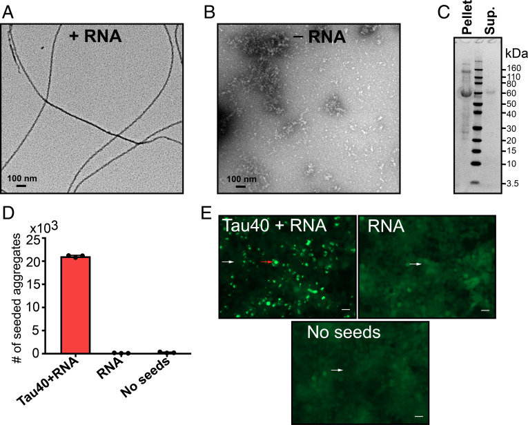
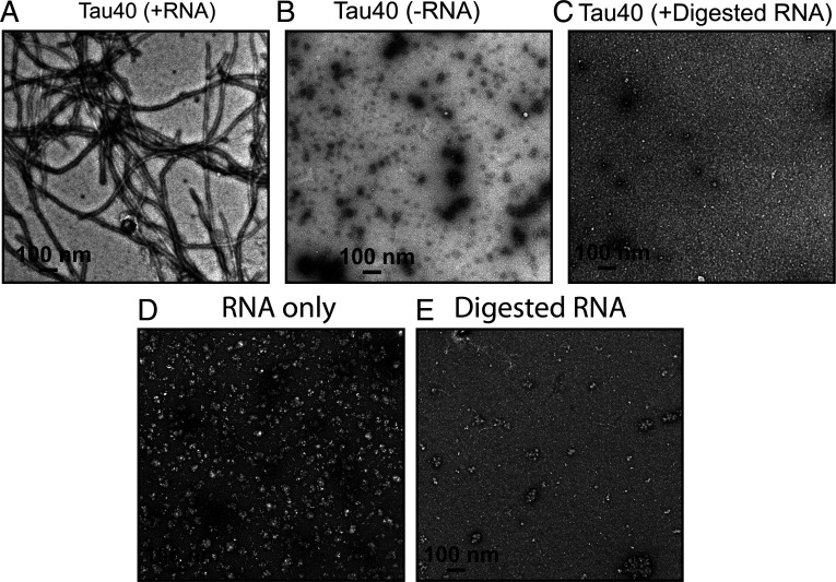
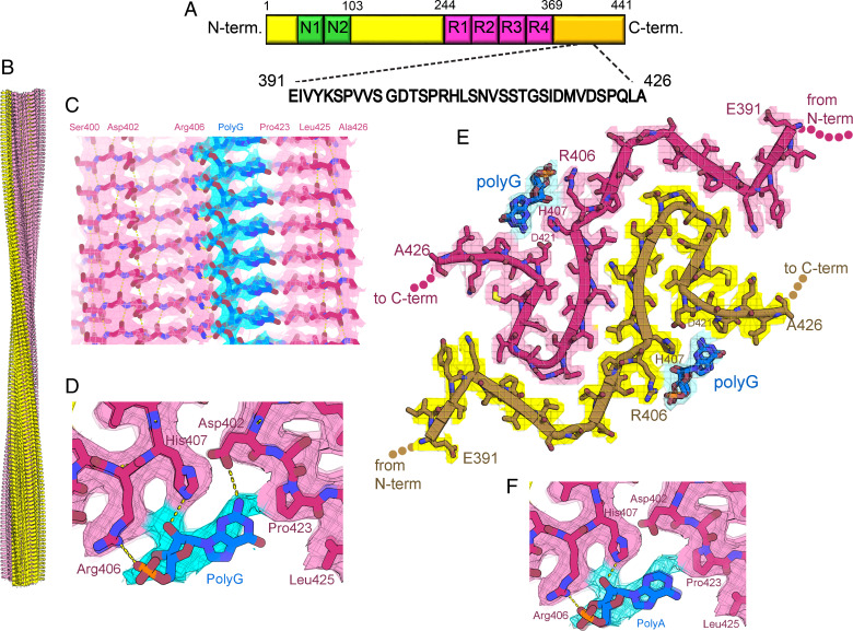
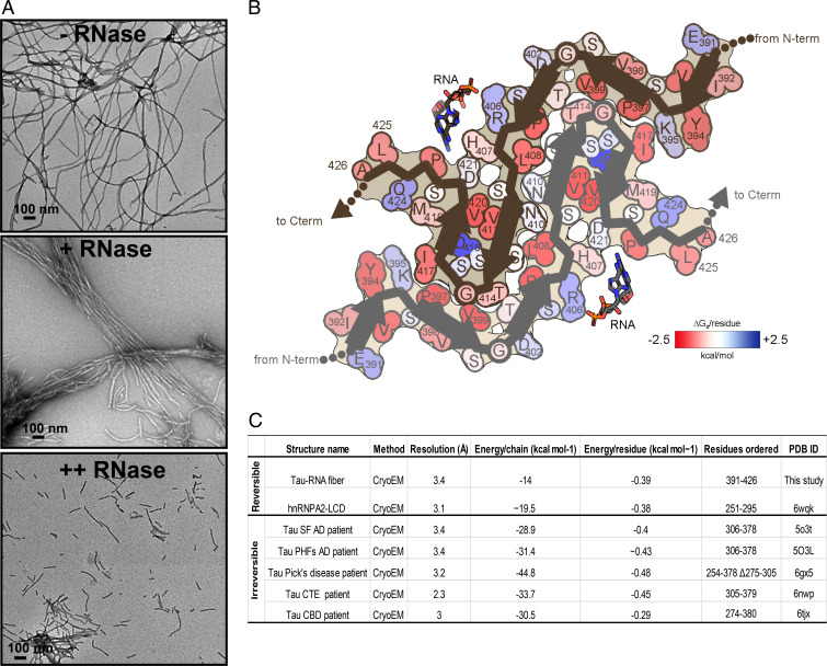
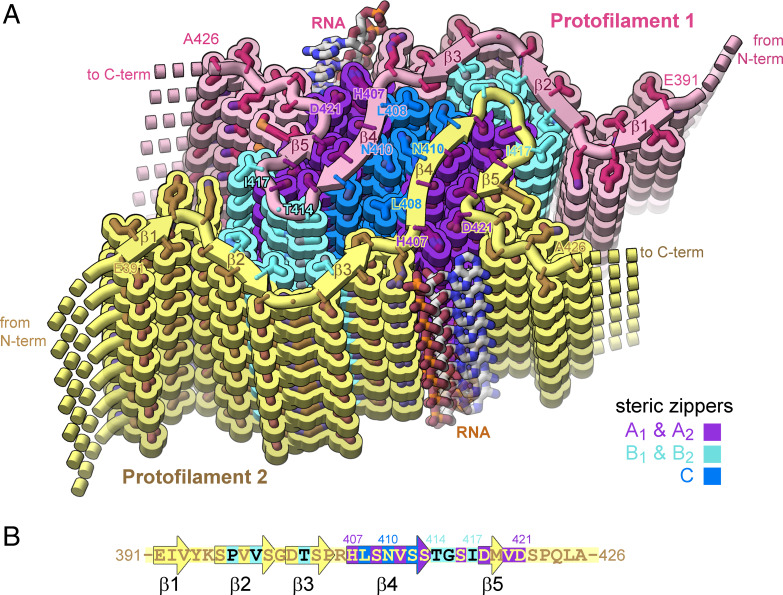
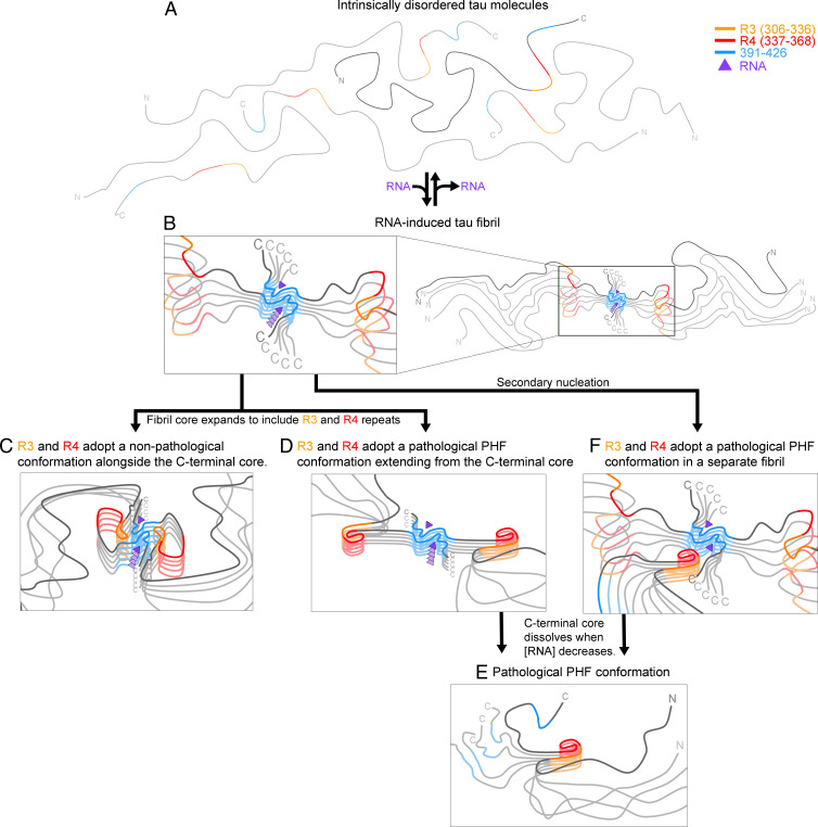
In neurodegenerative diseases including Alzheimer’s and amyotrophic lateral sclerosis, proteins that bind RNA are found in aggregated forms in autopsied brains. Evidence suggests that RNA aids nucleation of these pathological aggregates; however, the mechanism has not been investigated at the level of atomic structure. Here, we present the 3.4-Å resolution structure of fibrils of full-length recombinant tau protein in the presence of RNA, determined by electron cryomicroscopy (cryo-EM). The structure reveals the familiar in-register cross-β amyloid scaffold but with a small fibril core spanning residues Glu391 to Ala426, a region disordered in the fuzzy coat in all previously studied tau polymorphs. RNA is bound on the fibril surface to the positively charged residues Arg406 and His407 and runs parallel to the fibril axis. The fibrils dissolve when RNase is added, showing that RNA is necessary for fibril integrity. While this structure cannot exist simultaneously with the tau fibril structures extracted from patients’ brains, it could conceivably account for the nucleating effects of RNA cofactors followed by remodeling as fibrils mature.

Keywords: Alzheimer’s; RNA; amyloid; cryo-EM; tau.


Conflict of interest statement

Competing interest statement: D.S.E. is an advisor and equity shareholder in ADRx, Inc.

Figures

Fig. 1.
Fig. 1.
RNA induces formation of full-length tau amyloid fibrils. (A) Negative stain electron micrograph of tau40 fibrils grown in the presence of total RNA extracted from mouse liver. Tau fibrils were prepared by mixing 200 µL of 50 µM of monomeric tau40 in 20 mM ammonium acetate buffer pH7 with 400 µg/mL RNA and incubated with shaking for 2 d at 37 °C. (B) Electron micrograph of monomeric tau40 in the absence of RNA under the conditions of A. (C) SDS/PAGE analysis of tau-RNA fibrils after centrifugation at 159,000 × g and washing twice with RNA free water. (D) Quantification of the seeding activity of tau-RNA fibrils, measured in HEK293 biosensor cells expressing YFP-tagged tau-K18. (E) Representative images of aggregates produced by seeding in HEK293 biosensor cells. Cells seeded with tau-RNA fibrils (Left) and RNA only as a control (Right). The red arrow highlights a cell representative of those that contain aggregates. The white arrows highlight cells representative of those that contain no aggregates. (Scale bar, 25 µm.)
Fig. 2.
Fig. 2.
RNA enables in vitro fibril formation of tau. (A) Representative EM image of recombinant tau40 in the presence of RNA. (B) EM image of tau40 in the absence of RNA. (C) EM image of tau40 in the presence of predigested RNA. (D) Representative EM image of RNA only. (E) EM image of the digested RNA only. The digestion of RNA was performed by incubating the RNA with RNase at 4:1 ratio at 37 °C for 6 h. (Scale bar, 100 nm.)
Fig. 3.
Fig. 3.
Cryo-EM structure of full-length recombinant tau fibril bound to RNA. (A) Schematic representation of full-length tau (tau40, residues 1 to 441) including the two alternatively spliced N-terminal domains and four microtubule binding domains (R1 to R4). The sequence of the ordered core in our tau-RNA fibril structure is shown in black. (B) The cryo-EM reconstruction of our tau-RNA fibril, showing two identical protofilaments (pink and yellow) with left-handed twist. (C) Parallel, in register alignment of tau molecules (pink) and polyG RNA (cyan) running parallel to the fibril axis. (D) Close-up view of tau fibril-RNA model showing H-bonding between Arg406, His407, and Asp402 and residues and polyG RNA. (E) Atomic model and density map of one cross-sectional layer of the tau fibril core viewed down the fibril axis. (F) Close-up view of tau fibril–RNA interaction showing modeled H-bonding between Arg406, His407 residues, and polyA RNA.
Fig. 4.
Fig. 4.
Tau-RNA fibrils are reversible amyloid. (A) Influence of RNase on tau fibril stability. EM image of tau-RNA fibrils in the absence of RNase (Top). Tau fibrils begin to cluster and break down in the presence of RNase at 1:0.6 molar ratio (fibrils:RNase; Middle). Fibrils treated with higher molar ratio of RNase (1:3) break down into short fragments (Bottom). RNase was incubated with tau fibrils for 2 h at 37 °C in 20 mM ammonium acetate buffer, pH 7.0. (Scale bar, 100 nm.) (B) Solvation energy maps of tau-RNA fibrils ordered segment. Residues are colored according to their stabilization energies from unfavorable (blue; +2.5 kcal mol−1) to favorable (red; −2.5 kcal mol−1). (C) Comparison of the solvation energy values of the tau-RNA fiber structure with other amyloid structures.
Fig. 5.
Fig. 5.
Structure of tau-RNA fibril. (A) Cartoon of the two identical protofilaments (pink and yellow) of tau-RNA fibrils. Eight tau layers of each protofilament are shown. β-sheet surfaces are mated together in five steric zipper interfaces (A1, A2, B1, B2, and C, colored by letter). (B) The amino acid sequence of the ordered fibril core of tau-RNA cryo-EM structure. The RNA-tau fibril core is composed of residues Glu391 to Ala426 arranged on a cross-β scaffold. The steric zipper interfaces involve β-strands 2, 3, 4, and 5 (β1, Glu391 to Val393; β2, Ser396 to Ser400; β3, Asp402 to Pro405; β4, His407 to Ser413; and β5, Asp418-Val420) that stack in layers perpendicular to the fibril axis.
Fig. 6.
Fig. 6.
RNA-induced tau fibrils might be protective, or they might facilitate formation of pathogenic tau conformations. (A) When intrinsically disordered tau molecules become concentrated in the presence of RNA, (B) RNA induces the C-terminal segment (residues 391 to 426) to form an amyloid core. We anticipate four possible outcomes: loss of RNA causes the fibrils to dissolve, returning tau to the soluble state (A); (C) the amyloid-RNA core facilitates ordering of R3 and R4 which pack aside the C-terminal core in a conformation that protects tau from forming pathological conformations as suggested by Dregni et al. (33); (D) the amyloid-RNA core facilitates ordering of R3 and R4 in a pathological conformation, such as the C-shaped conformation of paired helical filaments (PHF) in AD, and then the C-terminal core disassembles, leaving (E) the PHF conformations; or (F) the RNA-amyloid core epitaxially nucleates a pathological tau conformation.

References

    1. Ulrich J., Alzheimer changes in nondemented patients younger than sixty-five: Possible early stages of Alzheimer’s disease and senile dementia of Alzheimer type. Ann. Neurol. 17, 273–277 (1985). - PubMed
    1. Apicco D. J., et al. , Reducing the RNA binding protein TIA1 protects against tau-mediated neurodegeneration in vivo. Nat. Neurosci. 21, 72–80 (2018). - PMC - PubMed
    1. Fichou Y., et al. , Cofactors are essential constituents of stable and seeding-active tau fibrils. Proc. Natl. Acad. Sci. U.S.A. 115, 13234–13239 (2018). - PMC - PubMed
    1. Sawaya M. R., Hughes M. P., Rodriguez J. A., Riek R., Eisenberg D. S., The expanding amyloid family: Structure, stability, function, and pathogenesis. Cell 184, 4857–4873 (2021). - PMC - PubMed
    1. Goedert M., et al. , Assembly of microtubule-associated protein tau into Alzheimer-like filaments induced by sulphated glycosaminoglycans. Nature 383, 550–553 (1996). - PubMed