. 2022 Apr 12;119(15):e2122682119.
doi: 10.1073/pnas.2122682119. Epub 2022 Apr 4.

GPCR large-amplitude dynamics by 19F-NMR of aprepitant bound to the neurokinin 1 receptor

Affiliations

GPCR large-amplitude dynamics by 19F-NMR of aprepitant bound to the neurokinin 1 receptor

Benxun Pan et al. Proc Natl Acad Sci U S A. .

Abstract

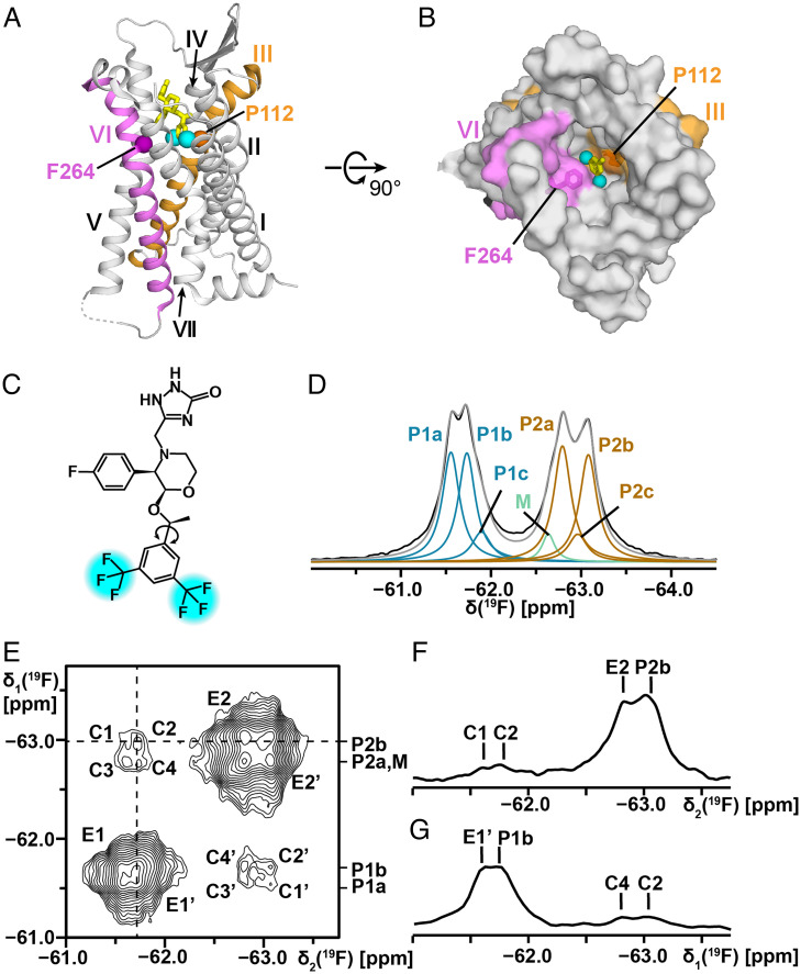
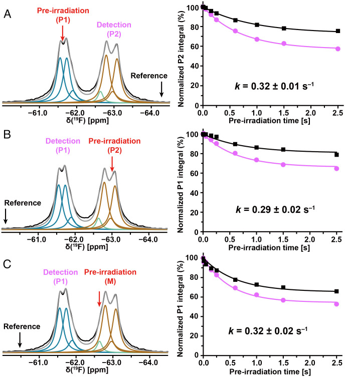
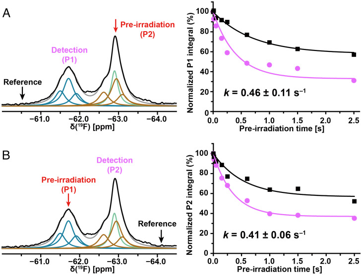
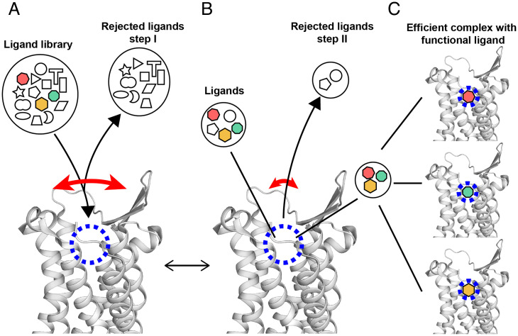
Comparisons of G protein-coupled receptor (GPCR) complexes with agonists and antagonists based on X-ray crystallography and cryo-electron microscopy structure determinations show differences in the width of the orthosteric ligand binding groove over the range from 0.3 to 2.9 Å. Here, we show that there are transient structure fluctuations with amplitudes up to at least 6 Å. The experiments were performed with the neurokinin 1 receptor (NK1R), a GPCR of class A that is involved in inflammation, pain, and cancer. We used 19F-NMR observation of aprepitant, which is an approved drug that targets NK1R for the treatment of chemotherapy-induced nausea and vomiting. Aprepitant includes a bis-trifluoromethyl-phenyl ring attached with a single bond to the core of the molecule; 19F-NMR revealed 180° flipping motions of this ring about this bond. In the picture emerging from the 19F-NMR data, the GPCR transmembrane helices undergo large-scale floating motions in the lipid bilayer. The functional implication is of extensive promiscuity of initial ligand binding, primarily determined by size and shape of the ligand, with subsequent selection by unique interactions between atom groups of the ligand and the GPCR within the binding groove. This second step ensures the wide range of different efficacies documented for GPCR-targeting drugs. The NK1R data also provide a rationale for the observation that diffracting GPCR crystals are obtained for complexes with only very few of the ligands from libraries of approved drugs and lead compounds that bind to the receptors.

Keywords: 2D [19F,19F]-EXSY; NMR saturation transfer; aromatic ring flips; nanodiscs; protein dynamics.


Conflict of interest statement

The authors declare no competing interest.

Figures

Fig. 1.
Fig. 1.
Chemical structure of the drug aprepitant, its location in the crystal structure of the complex with NK1R, and its 19F-NMR spectrum when bound to NK1R[2–335]. (A and B) Crystal structure of an NK1R complex with aprepitant (PDB ID code 6J20). (A) Side view in ribbon presentation. Aprepitant is shown in yellow stick presentation, with cyan spheres representing the trifluoromethyl groups. The TM helices are identified with Roman numerals. On the gray background the TM III and VI are highlighted in orange and purple, respectively. The locations of the α-carbon atoms of P1123.32 in TM III and F2646.51 in TM VI are represented by spheres. (B) View onto the extracellular surface in a space-filling atom presentation, same color code as in A. (C) Chemical structure of aprepitant. The two –CF3 groups are highlighted in cyan. The arrow indicates the single bond about which the bis-trifluoromethyl-phenyl ring undergoes 180° rotational flipping motions. (D) One-dimensional 19F-NMR spectrum of aprepitant bound to NK1R[2–335] in mixed micelles of LMNG and CHS at 298 K. The black line represents the experimental 19F-NMR signals of the two –CF3 groups. Lorentzian deconvolution revealed the following substates: P1a, P1b, and P1c (blue) represent one of the two –CF3 groups; M (green) represents micelle-associated “free” aprepitant; P2a, P2b, and P2c (brown) represent the second –CF3 group; the sum of P1, M, and P2 is shown in gray (SI Appendix, Table S1 for the relative integrals of the individual components). (E) Two-dimensional [19F,19F]-EXSY spectrum of NK1R[2–335] in LMNG/CHS micelles at 298 K, protein concentration ∼200 μM, mixing time = 600 ms. The cross-peaks C and C′ represent the exchange between P1 and P2, where exchange between pairs of substates is manifested by the fine structures of C and C′, with the components C1 to C4 and C1′ to C4′ (see text). The cross peaks E1, E1′, E2, and E2′ represent exchange among substates within the manifolds of P1 and P2. (F) One-dimensional cross-section along the dashed horizontal line in E. (G) One-dimensional cross-section along the dashed vertical line in E.
Fig. 2.
Fig. 2.
Large-amplitude structure fluctuations of NK1R[2–335] in mixed micelles of LMNG and CHS observed at 298 K by 19F saturation transfer between the two trifluoromethyl groups of bound aprepitant. (AC) In each experiment, the red arrow indicates the carrier position for the preirradiation, and the black arrow the position of the reference irradiation; the detection position is indicated with purple lettering. Lorentzian deconvolution of the 1D 19F-NMR spectra is used to identify a minimal number of overlapping signals that provide a quantitative fit of the experimental data. The plots on the right show the normalized integrals of the observed peak at different preirradiation times on-resonance and at the reference position. The Bloch–McConnell equation was used to fit the experimental data and derive the exchange rates.
Fig. 3.
Fig. 3.
Large-amplitude structure fluctuations of NK1R[2–335] in nanodiscs at 298 K detected by 19F-NMR observation of the bound drug aprepitant. The color code used for the Lorentzian deconvolution is presented the same as in Fig. 1D. A green line represents nanodiscs-associated “free” aprepitant. (A and B) 19F-NMR saturation transfer between the trifluoromethyl groups of aprepitant bound to NK1R[2–335]. Same presentation as in Fig. 2.
Fig. 4.
Fig. 4.
Hypothetical multistep selection of functional orthosteric ligands by GPCRs based on the NMR observation of transient large-amplitude structure fluctuations and observations with crystallization assays (39). A side-view of the extracellular part of the NK1R crystal structure (PDB ID code 6J20) is shown. The long curved red arrow indicates large transient openings of the ligand-binding groove. The short red arrow indicates a closed conformation of the GPCR, which is populated most of the time. The blue dashed circle indicates the location in the binding groove where ligands interact with specific amino acid residues of the GPCR. The black lines refer symbols to locations in the NK1R structure. Black arrowheads indicate flows of ligands leading to equilibrium at given concentrations of NK1R and ligands, solution conditions and temperature. (A) Initial screening of ligands by size and shape. (B) Selection of high-affinity ligands within the binding groove. (C) Different functional ligands are specifically bound to the receptor, yielding different functional states. Depending on the availability of such, ligands in the solution milieu (colored symbols in A) and their relative binding affinities, one of the ligands may be predominantly bound and imprint its efficacy on GPCR.

References

    1. Kramer M. S., et al. , Distinct mechanism for antidepressant activity by blockade of central substance P receptors. Science 281, 1640–1645 (1998). - PubMed
    1. Mantyh P. W., Substance P and the inflammatory and immune response. Ann. N. Y. Acad. Sci. 632, 263–271 (1991). - PubMed
    1. Harris J. A., et al. , Selective G protein signaling driven by substance P-neurokinin receptor dynamics. Nat. Chem. Biol. 18, 109–115 (2021). - PMC - PubMed
    1. Steinhoff M. S., von Mentzer B., Geppetti P., Pothoulakis C., Bunnett N. W., Tachykinins and their receptors: Contributions to physiological control and the mechanisms of disease. Physiol. Rev. 94, 265–301 (2014). - PMC - PubMed
    1. Hale J. J., et al. , Structural optimization affording 2-(R)-(1-(R)-3, 5-bis(trifluoromethyl)phenylethoxy)-3-(S)-(4-fluoro)phenyl-4- (3-oxo-1,2,4-triazol-5-yl)methylmorpholine, a potent, orally active, long-acting morpholine acetal human NK-1 receptor antagonist. J. Med. Chem. 41, 4607–4614 (1998). - PubMed

MeSH terms

LinkOut - more resources