Review
. 2022 Oct 1;102(4):1625-1667.
doi: 10.1152/physrev.00014.2021. Epub 2022 Apr 4.

Albumin uptake and processing by the proximal tubule: physiological, pathological, and therapeutic implications

Affiliations
Review

Albumin uptake and processing by the proximal tubule: physiological, pathological, and therapeutic implications

Bruce A Molitoris et al. Physiol Rev. .

Abstract

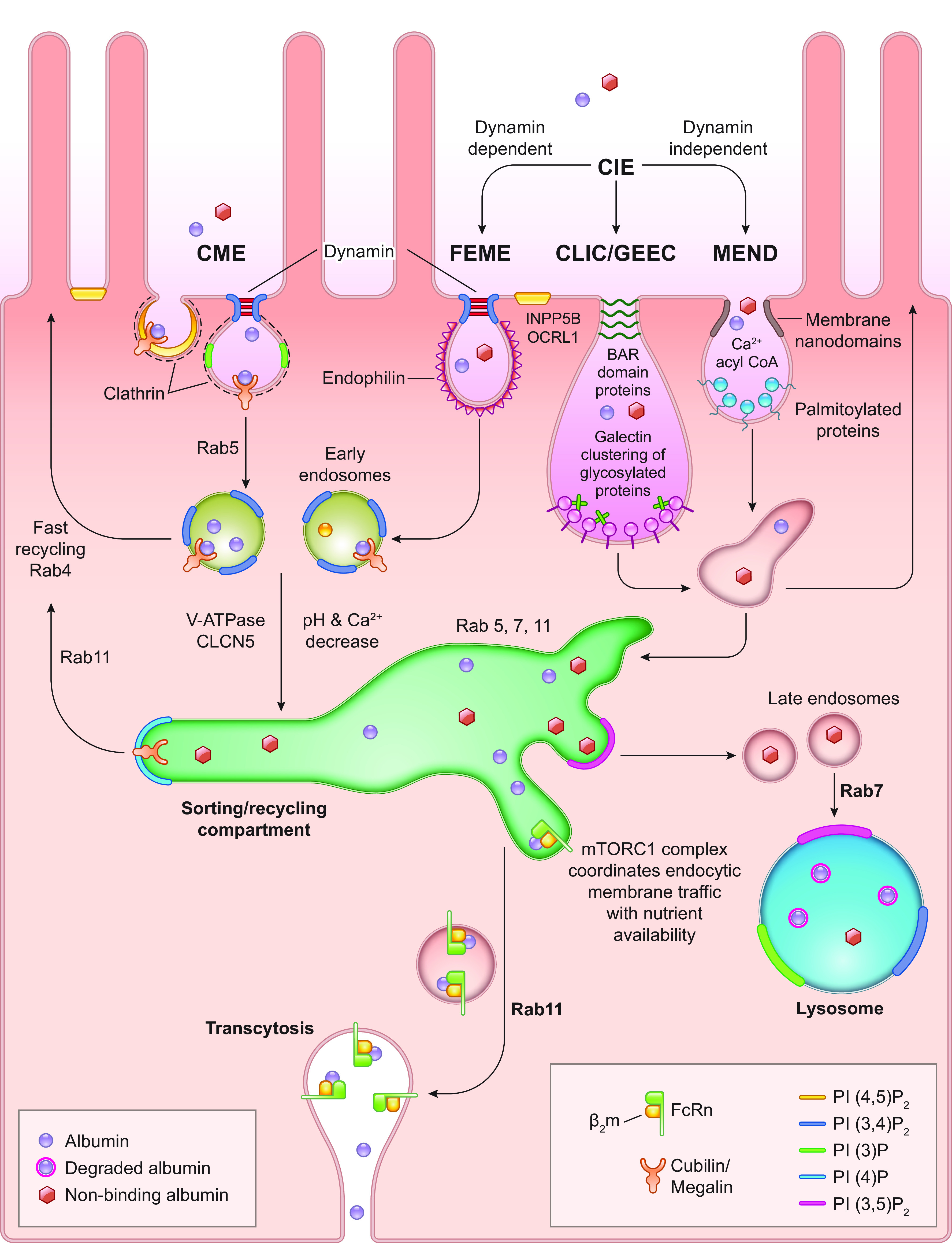
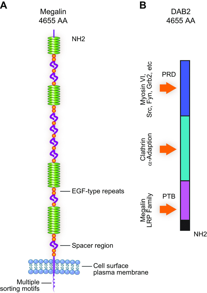
For nearly 50 years the proximal tubule (PT) has been known to reabsorb, process, and either catabolize or transcytose albumin from the glomerular filtrate. Innovative techniques and approaches have provided insights into these processes. Several genetic diseases, nonselective PT cell defects, chronic kidney disease (CKD), and acute PT injury lead to significant albuminuria, reaching nephrotic range. Albumin is also known to stimulate PT injury cascades. Thus, the mechanisms of albumin reabsorption, catabolism, and transcytosis are being reexamined with the use of techniques that allow for novel molecular and cellular discoveries. Megalin, a scavenger receptor, cubilin, amnionless, and Dab2 form a nonselective multireceptor complex that mediates albumin binding and uptake and directs proteins for lysosomal degradation after endocytosis. Albumin transcytosis is mediated by a pH-dependent binding affinity to the neonatal Fc receptor (FcRn) in the endosomal compartments. This reclamation pathway rescues albumin from urinary losses and cellular catabolism, extending its serum half-life. Albumin that has been altered by oxidation, glycation, or carbamylation or because of other bound ligands that do not bind to FcRn traffics to the lysosome. This molecular sorting mechanism reclaims physiological albumin and eliminates potentially toxic albumin. The clinical importance of PT albumin metabolism has also increased as albumin is now being used to bind therapeutic agents to extend their half-life and minimize filtration and kidney injury. The purpose of this review is to update and integrate evolving information regarding the reabsorption and processing of albumin by proximal tubule cells including discussion of genetic disorders and therapeutic considerations.

Keywords: FcRn; cubilin; drug delivery; endocytosis; megalin; transcytosis.


Conflict of interest statement

No conflicts of interest, financial or otherwise, are declared by the authors.

Figures

None
Graphical abstract
FIGURE 1.
FIGURE 1.
A: albumin uptake and processing by the proximal tubule. Albumin filtered across the glomerulus into Bowman’s space is reabsorbed after binding by the apical megalin-cubilin receptor complex. Both receptor-mediated endocytosis, via clathrin-coated vesicles, and fluid-phase endocytosis result in albumin reabsorption. After uptake, albumin can be transcytosed or undergo catabolism via lysosomal degradation. Albumin fragments in the urine result from lysosomal exocytosis of partially degraded albumin or peptide hydrolysis by apical membrane proteases. B: 25-μm 3-dimensional volume demonstrating Texas Red-labeled albumin endocytosed into proximal tubule cells, especially the S1 segment (S1). G, glomerular capillaries. Arrow indictes proximal tubule cells; bar = 20 μm. Figure modified from Ref. , with permission from the Journal of the American Society of Nephrology.
FIGURE 2.
FIGURE 2.
A–C: Sprague-Dawley rat kidney proximal tubules (PTs): segment S1 (A), S2 (B), and S3 (C) electron micrographs showing distinct differences in brush border microvilli, mitochondrial organization, and large vesicle/vacuoles between PT segments. L, lysosome; M, mitochondria; V, vacuole. Image from Ref. , with permission from Kidney International. D and E: Sprague-Dawley rat PT convoluted tubule at low (D; bar, 5 µm) and high (E; bar, 1 µm) magnification with helium ion microscopy. Note the bright and prominent brush border (BB) and the complex interdigitations of the lateral cellular membranes of PTs. Image from Ref. , with permission from PLoS One. F: scanning electron micrograph of a rabbit PT showing that lateral ridges (LR) begin below the apical microvilli (MV) and fan laterally. BM, basement membrane.
FIGURE 3.
FIGURE 3.
Initial filtration, binding, and internalization of fluorescent albumin by proximal tubule cells. An intravital 2-photon image of an S1 proximal tubule section is shown before an infusion of Texas Red-X-rat serum albumin (TR-RSA) in A. The adjacent tubule images (asterisks) are a continuation of the same S1 segment. The insets at bottom right in all panels show the S1 segment in a pseudocolor palette to better discern dimmer intensities not readily evident in black-and-white version. The micrograph in B was taken 15 s after the initial infusion. A portion of the glomerulus associated with the S1 segment is under the pseudocolor inset. The inset in B demonstrates early binding at the apical brush border membrane (arrowheads in inset). Early brush border binding progresses and eventually enriches in the subapical region of the S1 segment, appearing in C as a distinct band (arrows in inset). The end of the 100-s movie (D; Supplemental Movie 1) clearly shows small, distinct, early endocytic vesicles lining the subapical region, with a few appearing to have traversed well into the cytosol of the tubular epithelia. The individual time stamps are located at bottom left of all panels. In Supplemental Movie 1, the vascular intensity of albumin can be seen fluctuating in the earlier portion. This is due to the careful and protracted bolus infusion of TR-RSA in an effort to avoid saturation of fluorescence in the plasma. The bar located on right of D shows intensity equivalence between the black-and-white and pseudocolor display palettes. Bar, 20 µm. Data from Ref. .
FIGURE 4.
FIGURE 4.
Intracellular trafficking of albumin in proximal tubule cells. A high-resolution, 100-frame, 5-µm, 4-dimensional volume of Texas Red-X-albumin (TR-RSA) trafficking within rat renal proximal tubule cells shows vesicular and tubular-vesicular trafficking. Three micrographs from the data are shown in A–C, with respective time stamps from initial infusion of the fluorescent albumin. The data show small endocytic vesicles readily moving on the luminal side of the proximal tubule (lumen) shuttling around the apical region. These vesicles can also be seen moving toward the basolateral membrane, adjacent to the microvasculature showing rapidly flowing red blood cells. Arrowheads point to bright accumulations of the TR-RSA (with a dye-to-protein ratio of 1:1), showing distinct tubular-vesicular extensions projecting toward the basolateral membrane and appearing to merge with the interstitial space. Arrowheads point to regions where prominent extensions form and shuttle larger, brighter vesicles along these dimmer albumin-containing tracts. The often subtle fluorescence of these structures necessitated acquisition of these images with some degree of saturation in the brighter regions to allow for detection of the dimmer structures. The same structures can be seen in other cells throughout the proximal tubules shown here. Bar, 10 µm. Image from Ref. (Supplemental Movie 2), with permission from the Journal of the American Society of Nephrology.
FIGURE 5.
FIGURE 5.
Albumin reabsorption and trafficking by proximal tubule cells. Albumin reabsorbed by clathrin-mediated endocytosis (CME) or clathrin-independent endocytosis (CIE) undergoes endosomal acidification, resulting in dissociation of albumin from megalin-cubilin complexes for CME endosomes. Albumin binding to neonatal Fc receptor (FcRn) will occur as pH decreases, with a possible role for Ca2+ decrease. This transfer occurs in the dynamic sorting/recycling compartment. This exchange within the sorting compartment directs albumin either toward lysosomal degradation or to the transcytotic pathway. Both vesicular and tubular structures mediate albumin transcytosis to the basolateral membrane. Vesicle fusion with the basolateral membrane exposes its contents to the interstitial fluid, at elevated pH, resulting in dissociation of albumin from FcRn. FcRn undergoes recycling back to the sorting compartment. Reductions in albumin-FcRn binding within the endosomal compartment by albumin alterations such as oxidation, glycosylation, or carbamylation (nonbinding albumin) would reduce transcytosis of albumin. This provides an intracellular molecular sorting mechanism preserving physiological albumin and facilitating catabolism of albumin not binding to FcRn. It could also result in catabolism of albumin if concentrations exceed FcRn binding capacity. Note that multiple genetic mutations, knockouts, and specific manipulations to proteins involved in these intricate traffic and sorting pathways, i.e., Rabs, phosphatidylinositol (PI) kinase, phosphatases, V-ATPase, CLCN5, and mammalian target of rapamycin complex (mTORC)1 can lead to dysfunction. CLIC/GEEC, clathrin-independent carrier/glycosylphosphatidylinositol (GPI)-anchored protein-enriched early endosomal compartments; FEME, fast endophilin-mediated endocytosis; MEND, massive endocytosis; PI(3)P, phosphatidylinositol 3-phosphate; PI(4)P, phosphatidylinositol 4-phosphate; PI(3,4)P2, phosphatidylinositol 3,4-bisphosphate; PI(3,5)P2, phosphatidylinositol 3,4-bisphosphate; PI(4,5)P2, phosphatidylinositol 4,5-bisphosphate; β2m, β2-microglobulin.
FIGURE 6.
FIGURE 6.
Albumin’s structure, domains, and binding sites. Albumin domains are color coded, and fatty acid (FA) binding sites and physiologically relevant known drug sites are highlighted in Sudlow sites I (DIIA) and II (DIIIA) and other subdomains of albumin. The domains are color coded: red, IA; blue, IB; light brown, IIA; yellow, IIB; gray, IIIA; purple, IIIB. Data from Ref. .
FIGURE 7.
FIGURE 7.
Light sheet fluorescent microscopy and 3-dimensional image reconstruction image of a tissue-cleared mouse kidney at low (A; bar, 100 µm) and high (B; bar, 50 µm) magnification. Male C57BL/6 mice were injected with DyLight-649-tomato lectin (blue) and Alexa Fluor 555-albumin (yellow). Note that lectin labels glomeruli and filtered albumin is taken up by proximal tubules. Arrowhead in B represents the glomerulotubular junction. Figure from Ref. , with permission from Kidney360.
FIGURE 8.
FIGURE 8.
Cubilin and Amnionless domains and structural complex. A: Cubilin is a peripheral membrane protein containing an NH2-terminal stretch of 110 amino acids (AA), 8 epidermal growth factor (EGF)-type repeats, and 27 CUB domains (162). B: Amnionless is a transmembrane protein with a cytoplasmic domain of 75 amino acids containing 2 putative NPXY motifs followed by a transmembrane region (TM) and a cysteine-rich region that links to the NH2-terminal part of AMN that form 2 β-helix structures with hydrophobic cores. C: single-stem form of CUBAM with approximate dimensions of stem and crown regions followed by a representation of the double-stem form of CUBAM. Data from Ref. .
FIGURE 9.
FIGURE 9.
Megalin and DAB2 domains. A: Megalin is a large, 4,655-amino acid (AA), transmembrane protein with an extracellular domain that consists of 4 clusters of complement-type repeats, separated by 8 spacer regions containing YWTD motifs and 17 epidermal growth factor (EGF)-type repeats (162). Its cytoplasmic tail contains multiple sorting motifs including PDZ, PKC, SH3, and NPXY (188). B: DAB2 is composed of 3 principal domains. The NH2-terminal PTB domain binds to NPXY motifs, the middle domain interacts with clathrin and alpha-adaptin, and the COOH-terminal portion is a proline-rich domain (PRD) that can bind SH3-containing proteins such as Grb2, Fyn, and Src (189).
FIGURE 10.
FIGURE 10.
A: proximal tubules (PTs) determine the physiological state of the body by “sensing” urine and serum albumin levels. Proximal tubule cells (PTCs) can adjust uptake and secretion mechanisms to impact the physiological state both directly and indirectly. B: box plot showing quantification of Texas Red-X-rat serum albumin (TR-RSA) reabsorption by all surface PTs of control (n = 3 rats, 157 fields quantified) and albumin-overloaded (n = 8 rats, 176 fields quantified) rats. A significant reduction in albumin uptake (P < 0.01, KaleidaGraph, Student’s t test) was seen with albumin overloading. C: box plot showing the quantification of TR-RSA uptake in all surface PTs of control rats (n = 3 rats, 101 fields quantified) and rats treated with diphtheria toxin (DT) to increase glomerular albumin permeability (n = 3 rats, 106 fields quantified). There was a significant increase in PTC albumin uptake (P = 0.05, Student’s t test 1-tailed equal variance) when filtrate albumin concentration increased. Modified from Ref. , with permission from the Journal of the American Society of Nephrology.
FIGURE 11.
FIGURE 11.
A: vascular clearance of wild-type and modified rat serum albumins (RSAs) (6, 155). Fluorescently tagged (Texas Red-X or Oregon Green-X) RSA and one of the modified RSAs were injected simultaneously into the same rat, and blood was collected after injection at 15 min, 2 h, and 24 h. Each albumin was evaluated in 4 rats (male Sprague-Dawley rats, 180–220 g) that received both a control and a modified albumin. The 15 min collection time point was set to 100%, and the decrease in fluorescence followed at 2 h and 24 h. Note that modified albumins all had increased vascular clearance. GraphPad Prism was used to graph means ± SD for each 2 and 24 h time point (6, 155). cRSA, carbamylated RSA; MGO, methylglyoxal. B: albumin in kidney endothelial/interstitial regions (6). Quantification of albumins in kidney endothelial/interstitial regions showed significant increases in accumulation between RSA and albumin modified with potassium cyanate for 30 min, 2 h, or 4 h (cRSA2hr #P < 0.05 and RSA vs. cRSA4hr *P < 0.05) (6). Images from Refs. and , with permission from the American Physiological Society.
FIGURE 12.
FIGURE 12.
A: 3-dimensional structural view of human serum albumin (HSA)-neonatal Fc receptor (FcRn) complex (PDB ID 4N0F). HSA (DI, DII, and DIII), FcRn α-chain and β2-microglobulin (β2M) are shown in light blue and black, respectively (296). Albumin domains are colored as in FIGURE 5: red, IA; blue, IB; light brown, IIA; yellow, IIB; gray, IIIA; purple, IIIB. Green color highlights key amino acid changes and conditions that favor FcRn-albumin interaction (297, 298). Red color highlights amino acid mutations and conditions that destabilize FcRn-albumin interaction including carbamylation (*C), glycation (*G), oxidation (*O), and pH (116, 299, 300). The inset highlights 2 salt bridges critical for FcRn-HSA and β2M-HSA interactions that are affected by modifications (296). B: albumin structure (PDB ID 1E78) labeled for many of its various binding moieties and modifications including carbamylation, glycation, fatty acid (FA), oxidation, and Sudlow’s I and II sites known for binding different drugs, metabolites, and metal ions. The primary site of both glycation and carbamylation is K525 (blue), whereas R410 is a non-lysine glycated site and C34 is the oxidation site (, , , –303). Other sites are noted to emphasize the potential impact of modifications/associations on albumin’s many interactions. C: structure of the CUB5–8–IF–Cbl complex (PDB ID 3KQ4) (301) is presented in 2 different views. The concave interface (red) is required for the intrinsic factor (IF) interaction, which takes place mainly via Cub6 and Cub8 domains. The convex surface of Cub5–8 (blue) binds to albumin via interactions with mainly Cub7 and Cub8 residues. This is based on our cross linking mass spectrometry and docking studies (unpublished observations). Note that the albumin binding interface is distinct from IF binding site. The red balls designate Ca2+ binding sites.

References

    1. Benzing T, Salant D. Insights into glomerular filtration and albuminuria. N Engl J Med 384: 1437–1446, 2021. doi:10.1056/NEJMra1808786. - DOI - PubMed
    1. Dickson LE, Wagner MC, Sandoval RM, Molitoris BA. The proximal tubule and albuminuria: really!. J Am Soc Nephrol 25: 443–453, 2014. doi:10.1681/ASN.2013090950. - DOI - PMC - PubMed
    1. Wagner MC, Campos-Bilderback SB, Chowdhury M, Flores B, Lai X, Myslinski J, Pandit S, Sandoval RM, Wean SE, Wei Y, Satlin LM, Wiggins RC, Witzmann FA, Molitoris BA. Proximal tubules have the capacity to regulate uptake of albumin. J Am Soc Nephrol 27: 482–494, 2016. doi:10.1681/ASN.2014111107. - DOI - PMC - PubMed
    1. Sandoval RM, Wagner MC, Patel M, Campos-Bilderback SB, Rhodes GJ, Wang E, Wean SE, Clendenon SS, Molitoris BA. Multiple factors influence glomerular albumin permeability in rats. J Am Soc Nephrol 23: 447–457, 2012. doi:10.1681/ASN.2011070666. - DOI - PMC - PubMed
    1. Tenten V, Menzel S, Kunter U, Sicking EM, van Roeyen CR, Sanden SK, Kaldenbach M, Boor P, Fuss A, Uhlig S, Lanzmich R, Willemsen B, Dijkman H, Grepl M, Wild K, Kriz W, Smeets B, Floege J, Moeller MJ. Albumin is recycled from the primary urine by tubular transcytosis. J Am Soc Nephrol 24: 1966–1980, 2013. doi:10.1681/ASN.2013010018. - DOI - PMC - PubMed

Publication types

LinkOut - more resources