. 2022 Apr 4;12(1):40.
doi: 10.1186/s13578-022-00774-x.

Rebuilding hippocampus neural circuit with hADSC-derived neuron cells for treating ischemic stroke

Affiliations

Rebuilding hippocampus neural circuit with hADSC-derived neuron cells for treating ischemic stroke

Jian Wang et al. Cell Biosci. .

Abstract

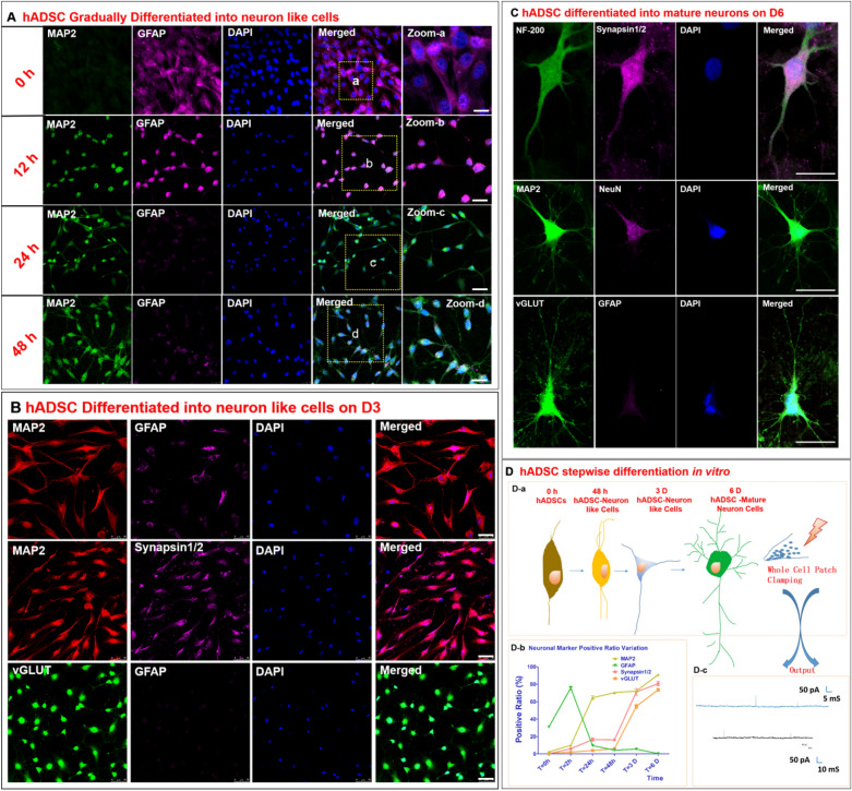
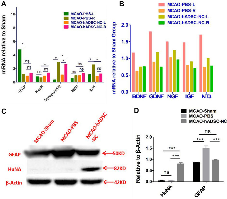
Background: Human adipose-derived stem cells (hADSCs) have been demonstrated to be a promising autologous stem cell source for treating various neuronal diseases. Our study indicated that hADSCs could be induced into neuron-like cells in a stepwise manner that are characterized by the positive expression of MAP2, SYNAPSIN 1/2, NF-200, and vGLUT and electrophysiological activity. We first primed hADSCs into neuron-like cells (hADSC-NCs) and then intracerebrally transplanted them into MCAO reperfusion mice to further explore their in vivo survival, migration, integration, fate commitment and involvement in neural circuit rebuilding.

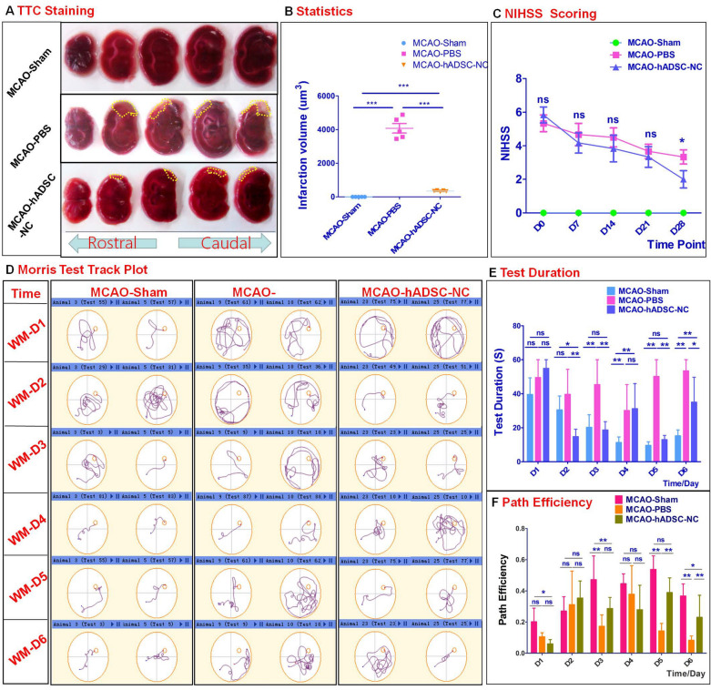
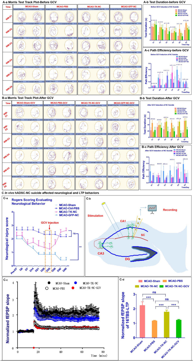
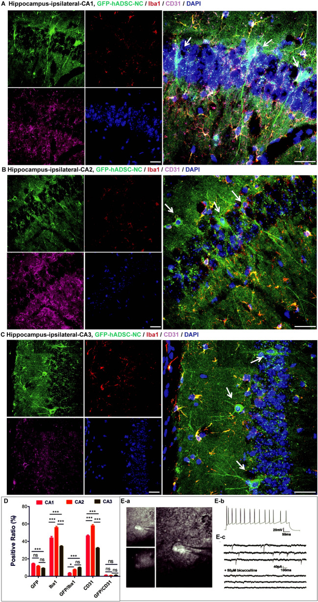
Results: The hADSC-NCs survived well and transformed into MAP2-positive, Iba1- or GFAP-negative cells in vivo while maintaining some proliferative ability, indicated by positive Ki67 staining after 4 weeks. hADSC-NCs could migrate to multiple brain regions, including the cortex, hippocampus, striatum, and hypothalamus, and further differentiate into mature neurons, as confirmed by action potential elicitation and postsynaptic currents. With the aid of a cell suicide system, hADSC-NCs were proven to have functionally integrated into the hippocampal memory circuit, where they contributed to spatial learning and memory rescue, as indicated by LTP improvement and subsequent GCV-induced relapse. In addition to infarction size shrinkage and movement improvement, MCAO-reperfused mice showed bidirectional immune modulation, including inhibition of the local proinflammatory factors IL-1α, IL-1β, IL-2, MIP-1β and promotion proinflammatory IP-10, MCP-1, and enhancement of the anti-inflammatory factors IL-15.

Conclusion: Overall, hADSC-NCs used as an intermediate autologous cell source for treating stroke can rebuild hippocampus neuronal circuits through cell replacement.

Keywords: Adipose-derived stem cells (ADSCs); Middle cerebral artery occlusion (MCAO); National Institute of Health Stroke Scale (NIHSS); Rogers scale system; hADSC-derived neuron-like cells (hADSC-NCs).


Conflict of interest statement

The authors declare no competing interests.

Figures

Fig. 1
Fig. 1
hADSCs are gradually induced to differentiate into neuron-like cells characterized by neuronal marker staining and action potentials. A Shows the variance in morphology and neuronal marker expression during stepwise induction from 0 to 48 h. B Shows that some functional neuronal markers could be detected and were distributed within the cell soma on day 3 (D3). C shows that hADSCs could be induced to differentiate into relatively mature neurons with complex neurites and neurotransmitters on Day 6 (D6). D Shows the cell morphological variance diagram (D-a), the change in the positive expression ratio over time for various neuronal markers (D-b) and the action potentials measured by whole-cell patch clamping on D6. n = 5, Scale bar = 50 μm in A-B, Scale bar = 25 μm in C
Fig. 2
Fig. 2
hADSC-NCs can decrease the MCAO mouse infarction volume and improve neurological scores, spatial learning and memory. A Shows TTC data to demonstrate the infarction volume variance among the groups. B shows the statistical analysis of the infarction volume. C Shows NIHSS data indicating the improvement in neurological behavior between the MCAO-PBS group and the MCAO-hADSC-NC group. D Compares the track plots from different groups at different time points. E shows the statistics for the test duration. F shows the path efficiency variance. n = 7. * indicates P < 0.05, ** indicates P < 0.001. *** indicates P < 0.0005
Fig. 3
Fig. 3
Tracking and identification of hADSC-NCs in MCAO mouse brains 4 weeks after transplantation. A Shows the working flowchart. Immunohistochemical staining data show that GFP and MAP2 double-positive hADSC-NC cells can be found in different areas of the mouse brain, including the cortex (B), hippocampus (C), striatum (D) and hypothalamus (E). Scale bar = 25 μm
Fig. 4
Fig. 4
hADSC-NC suicide leads to the relapse of spatial learning and memory. A Show that hADSC-NCs rescued the brain function of MCAO mice according to the Morris test, B shows hADSC-NC suicide can lead to spatial learning and memory relapse after i.p. injection of ganciclovir (GCV), a Shows the track plots of representative mice from each group, b shows the test duration in each group, c shows the path efficiency of each group. C shows that hADSC-NC suicide increases the Rogers scores and decreases long-term potentiation (LTP). C-a shows the neurological evaluation in each group, C-b shows the schematic of LTP recording, Cc shows the TBS-induced CA3-CA1 LTP in each group, C-d shows a summary of the data on the magnitude of LTP observed in C-c. n = 7. * indicates P < 0.05, ** indicates P < 0.001, *** indicates P < 0.0005
Fig. 5
Fig. 5
Characterization of hADSC-NCs in various MCAO mouse brain regions. A Shows the cell apoptosis marker cleaved Caspase 3. B Shows the negative expression of cleaved Caspase 3 in GFP-positive hADSC-NCs in the cortex. C Shows that the transplanted hADSC-NCs express cleaved Caspase 3, integrate well into the hippocampal tissue, and exhibit a healthy and complex neuron morphology. D, E Show similar cell survival, integration and neuron differentiation in the mouse striatum and hypothalamus. F Shows the statistical analysis of the hADSC-NC distribution, Caspase 3 positive percentage and GFP/Caspase 3 double-positive percentage. n = 5, Scale bar = 50 μm in BD, scale bar = 25 μm in E
Fig. 6
Fig. 6
hADSC-NC terminal differentiation and brain vasculature rebuilding in the MCAO mouse hippocampus. The minus hADSC-NCs express the microglial cell marker Iba1 and the blood vessel marker CD31 in 3 areas of the hippocampus: A for CA1, B for CA2 and C for CA3. D shows the statistical analysis of the hADSC-NC distribution, the Iba1-positive percentage and the CD31-positive percentage in CA1, CA2 and CA3. E shows the electrophysiological properties of GFP-positive hADSC-NCs in live hippocampal slices. a Representative image of GFP-positive hADSC-NC-derived neurons during patch-clamp recording. b Action potentials could be elicited by a current injection. c Spontaneous IPSCs were recorded in patched neurons and blocked by 50 μM bicuculine. n = 7/8, Scale bar = 25 μm
Fig. 7
Fig. 7
Molecular analysis to demonstrate the interaction between hADSC-NCs and brain host cells as well as the effects on neurotrophic factor secretion. A Show that hADSC-NCs affected the mRNA expression of the neuronal markers NeuN, synapsin 1/2, microglia marker Iba1 and astrocyte or neuronal stem cell marker GFAP. B Shows that hADSC-NCs scarcely changed the expression of neurotrophic factors at the mRNA level. C Shows that GFAP expression was inhibited by hADSC-NCs; HuNA could be detected in the mouse brain in the MCAO-hADSC-NC group, but HuNA could not be detected in the MCAO-Sham or the MCAO-PBS group by western blotting

References

    1. Abdanipour A, Tiraihi T, Delshad A. Trans-differentiation of the adipose tissue-derived stem cells into neuron-like cells expressing neurotrophins by selegiline. Iran Biomed J. 2011;15:113–121. - PMC - PubMed
    1. Albers GW, Diener HC, Grind M, Halperin JL, Horrow J, Olsson SB, Petersen P, Vahanian A, Frison L, Nevinson M, et al. Stroke prevention with the oral direct thrombin inhibitor ximelagatran compared with warfarin in patients with non-valvular atrial fibrillation (SPORTIF III): randomised controlled trial. Lancet. 2003;362:1691–1698. - PubMed
    1. Blecker D, Elashry MI, Heimann M, Wenisch S, Arnhold S. New insights into the neural differentiation potential of canine adipose tissue-derived mesenchymal stem cells. Anat Histol Embryol. 2017;46:304–315. - PubMed
    1. Burrow KL, Hoyland JA, Richardson SM. Human adipose-derived stem cells exhibit enhanced proliferative capacity and retain multipotency longer than donor-matched bone marrow mesenchymal stem cells during expansion in vitro. Stem Cells Int. 2017;2017(2017):2541275. - PMC - PubMed
    1. Chen AZ, Liu N, Huang H, Lin FF, Liu DS, Lin XH. Outgrowth of neuronal axons on adipose-derived stem cell transplanting for treatment of cerebral infarction in rats. Chin J Cell Mol Imm. 2011;27:868–871. - PubMed