Clinical Trial
. 2022 Apr;10(4):e004656.
doi: 10.1136/jitc-2022-004656.

Efficacy and safety of camrelizumab plus apatinib during the perioperative period in resectable hepatocellular carcinoma: a single-arm, open label, phase II clinical trial

Affiliations
Clinical Trial

Efficacy and safety of camrelizumab plus apatinib during the perioperative period in resectable hepatocellular carcinoma: a single-arm, open label, phase II clinical trial

Yongxiang Xia et al. J Immunother Cancer. 2022 Apr.

Abstract

Objective: This study aimed to assess the efficacy and safety of camrelizumab plus apatinib in patients with resectable hepatocellular carcinoma (HCC) as neoadjuvant therapy.

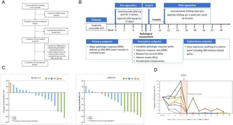
Methods: Initially, 20 patients with HCC were screened and 18 patients with resectable HCC were enrolled in this open-label, single-arm, phase II clinical trial. Patients received three cycles of neoadjuvant therapy including three doses of camrelizumab concurrent with apatinib for 21 days followed by surgery. Four to 8 weeks after surgery, patients received eight cycles of adjuvant therapy with camrelizumab in combination with apatinib. Major pathological reactions (MPR), complete pathological reactions (pCR), objective response rate (ORR), relapse-free survival (RFS), and adverse events (AE) were assessed. In addition, cancer tissue and plasma samples were collected before and after treatment, and genetic differences between responding and non-responding lesions were compared by tumor immune microenvironment (TIME) analysis, circulating tumor DNA (ctDNA) analysis and proteomics analysis.

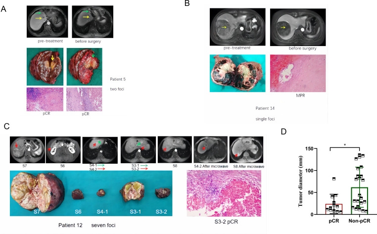
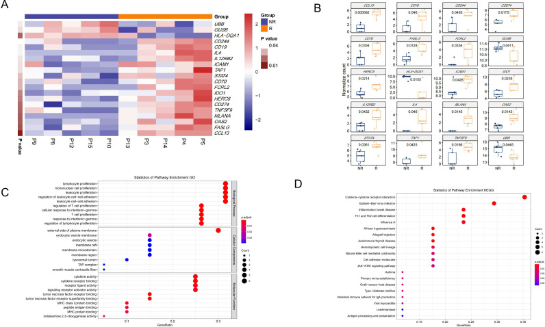
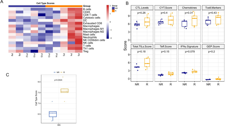
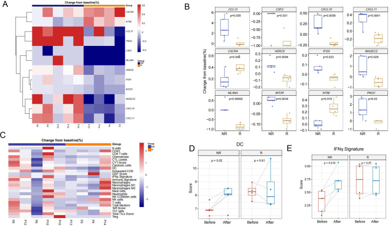
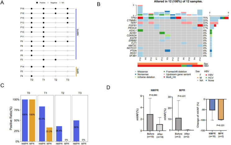
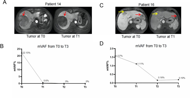
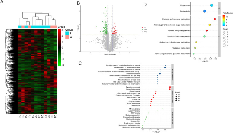
Results: In 18 patients with HCC who completed neoadjuvant therapy, 3 (16.7%) and 6 (33.3%) patients with HCC reached ORR based on Response Evaluation Criteria in Solid Tumors (RECIST) V.1.1 and modified RECIST criteria, respectively. Of the 17 patients with HCC who received surgical resection, 3 (17.6%) patients with HCC reported MPR and 1 (5.9%) patient with HCC achieved pCR. The 1-year RFS rate of the enrolled patients was 53.85% (95% CI: 24.77% to 75.99%). Grade 3/4 AEs were reported in 3 (16.7%) of the 18 patients, with the most common AEs being rash (11.1%), hypertension (5.6%), drug-induced liver damage (5.6%), and neutropenia (5.6%) in the preoperative phase. The 289 NanoString panel RNA sequencing showed that TIME cell infiltration especially dendritic cells (DCs) infiltration was better in responding tumors than in non-responding tumors. Our results of ctDNA revealed a higher positive rate (100%) among patients with HCC with stage IIb-IIIa disease. When comparing patients with pCR/MPR and non-MPR, we observed more mutations in patients who achieved pCR/MPR at baseline (6 mutations vs 2.5 mutations, p=0.025). Patients who were ctDNA positive after adjuvant therapy presented a trend of shorter RFS than those who were ctDNA negative. Proteomic analysis suggested that abnormal glucose metabolism in patients with multifocal HCC might be related to different sensitivity of treatment in different lesions.

Conclusion: Perioperative camrelizumab plus apatinib displays a promising efficacy and manageable toxicity in patients with resectable HCC. DCs infiltration might be a predictive marker of response to camrelizumab and apatinib as well as patients' recurrence. ctDNA as a compose biomarker can predict pathological response and relapse. Abnormal glucose metabolism in patients with multifocal HCC may be related to different sensitivity of treatment in different lesions.

Trial registration number: NCT04297202.

Keywords: antibodies, neoplasm; antineoplastic protocols; immunity; immunotherapy; liver neoplasms.


Conflict of interest statement

Competing interests: No, there are no competing interests.

Figures

Figure 1
Figure 1
Study design. (A) Flowchart of enrolled patients. (B) Flowchart of therapeutic regimen. (C) Treatment was evaluated for the enrolled patients based on RECIST V.1.1 criteria and mRECIST criteria. (D) Measuring of AFP level change during perioperative period in patients with surgical resectable HCC. HCC, hepatocellular carcinoma; mRECIST, modified RECIST; RECIST, Response Evaluation Criteria in Solid Tumors.
Figure 2
Figure 2
Response to treatment in typical cases. (A) Patient 5; male; 70 years old; stage IIb; total five lesions, maximum diameter 32.6 mm; after neoadjuvant therapy, RECIST V.1.1 and mRECIST evaluation were PR; surgical pathology only showed two large lesions and were both pCR. (B) Patient 14; female; 53 years old; stage IIIa; tumor diameter 108.1 mm; after neoadjuvant therapy, RECIST V.1.1 evaluation was stable disease, mRECIST evaluation was PR; surgical pathology was MPR. (C) Patient 12; male; 55 years old; stage IIb; seven tumor nodules; the largest of which was 80 mm in diameter; after neoadjuvant therapy, five tumors were resected, among which S3–2 lesion was treated with pCR, S4–2 and S8 lesions with microwave. DFS is 293 days. (D) The relationship between tumor diameter and pathological response. *P<0.05. Arrows represent lesions.RECIST, Response Evaluation Criteria in Solid Tumors; mRECIST, modified RECIST;PR, partial response; pCR, pathological reactions;MPR, major pathological reactions; DFS, Disease Free Survival.
Figure 3
Figure 3
Comparison of expression of immune-related genes between responding and non-responding lesions. (A–B) Expression of immune-related genes per pretreatment sample of response and non-response groups. In the heatmap (A), red indicates an increased gene expression and blue indicates a decreased gene expression. (C–D) GO (C) and Kyoto Encyclopedia of Genes and Genomes (D) pathway analysis of the pretreatment samples. The count represents the number of genes in each pathway and dot size corresponds to ‘count’. GO, gene ontology.
Figure 4
Figure 4
Comparison of cell type scores between responding and non-responding lesions. (A–B) Different cell type scores in per pretreatment sample of response and non-response groups. Red indicates an increased cell type scores and blue indicates a decreased cell type scores. (C) The cell type scores of DCs between response and non-response groups. CTL, cytotoxic T lymphocyte; CYT, cytolytic activity; DC, dendritic cell; GEP, gene expression profiling; IFN, interferon; NK, natural killer; Teff, T-effector; TIL, tumor infiltrating lymphocytes.
Figure 5
Figure 5
Pre-to-post-treatment changes of tumor immune microenvironment between responding and non-responding lesions. (A–B) Pre-to-post-treatment changes of immune-related genes expression according to response and non-response groups. In the heatmap (A), red indicates an increase and blue indicates a decrease. (C) Pre-to-post-treatment changes of cell type scores and immune signature scores according to response and non-response groups. Red indicates an increase and blue indicates a decrease. (D) Pre-to-post-treatment changes of IFN-γ signature scores between response and non-response groups. (E) Pre-to-post-treatment changes of DCs scores between response and non-response groups. CTL, cytotoxic T lymphocyte; CYT, cytolytic activity; DC, dendritic cell; GEP, gene expression profiling; IFN, interferon; NK, natural killer; Teff, T-effector; TIL, tumor infiltrating lymphocytes.
Figure 6
Figure 6
Detection of ctDNA and its association with pathologic response. (A) Plot of ctDNA status for group MPR and non-MPR at T0 (pre-neoadjuvant), T1 (post-neoadjuvant), T2 (post surgery) and T3 (post-adjuvant therapy). Non-MPR, group of patients did not achieve pathologic response of MPR/pCR. (B) Mutational landscape of 12 samples at baseline (T0). (C) Comparison of positive rates from T0 to T3 between group of MPR/pCR and non-MPR. (D) Maximum variant allele frequency (mVAF) in group of non-MPR, MPR/pCR before and after neoadjuvant therapy and comparison of the change of mVAF. ctDNA, circulating tumor DNA; MPR, major pathological reactions; pCR, pathological reactions.
Figure 7
Figure 7
Change of tumor lesion and mVAF of ctDNA in Patient 14 (A–B) and 16 (C–D). Arrows represent lesions. mVAF, maximum variant allele frequency.
Figure 8
Figure 8
The difference in proteomics between the responding and non-responding tumors. (A–B) Heatmap (A) and volcano map (B) of different proteins between responding and non-responding lesions. T indicates non-responding lesions and W indicates responding lesions. (C) Gene ontology analysis results of different proteins between responding and non-responding lesions. (D) Kyoto Encyclopedia of Genes and Genomes pathway analysis of different proteins between responding and non-responding lesions.

Similar articles

Cited by

References

    1. Sung H, Ferlay J, Siegel RL, et al. . Global cancer statistics 2020: GLOBOCAN estimates of incidence and mortality worldwide for 36 cancers in 185 countries. CA Cancer J Clin 2021;71:209–49. 10.3322/caac.21660 - DOI - PubMed
    1. Clark T, Maximin S, Meier J, et al. . Hepatocellular carcinoma: review of epidemiology, screening, imaging diagnosis, response assessment, and treatment. Curr Probl Diagn Radiol 2015;44:479–86. 10.1067/j.cpradiol.2015.04.004 - DOI - PubMed
    1. Xiang X, Zhong J-H, Wang Y-Y, et al. . Distribution of tumor stage and initial treatment modality in patients with primary hepatocellular carcinoma. Clin Transl Oncol 2017;19:891–7. 10.1007/s12094-017-1621-6 - DOI - PubMed
    1. Zhong J-H, Peng N-F, You X-M, et al. . Tumor stage and primary treatment of hepatocellular carcinoma at a large tertiary hospital in China: a real-world study. Oncotarget 2017;8:18296–302. 10.18632/oncotarget.15433 - DOI - PMC - PubMed
    1. Hartke J, Johnson M, Ghabril M. The diagnosis and treatment of hepatocellular carcinoma. Semin Diagn Pathol 2017;34:153–9. 10.1053/j.semdp.2016.12.011 - DOI - PubMed

Publication types

Associated data