Review
. 2022 Apr 4;7(1):113.
doi: 10.1038/s41392-022-00966-4.

Targeted protein degradation: mechanisms, strategies and application

Affiliations
Review

Targeted protein degradation: mechanisms, strategies and application

Lin Zhao et al. Signal Transduct Target Ther. .

Abstract

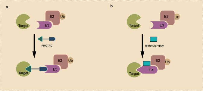
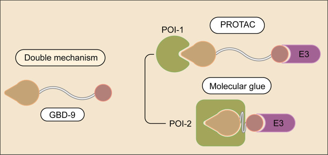
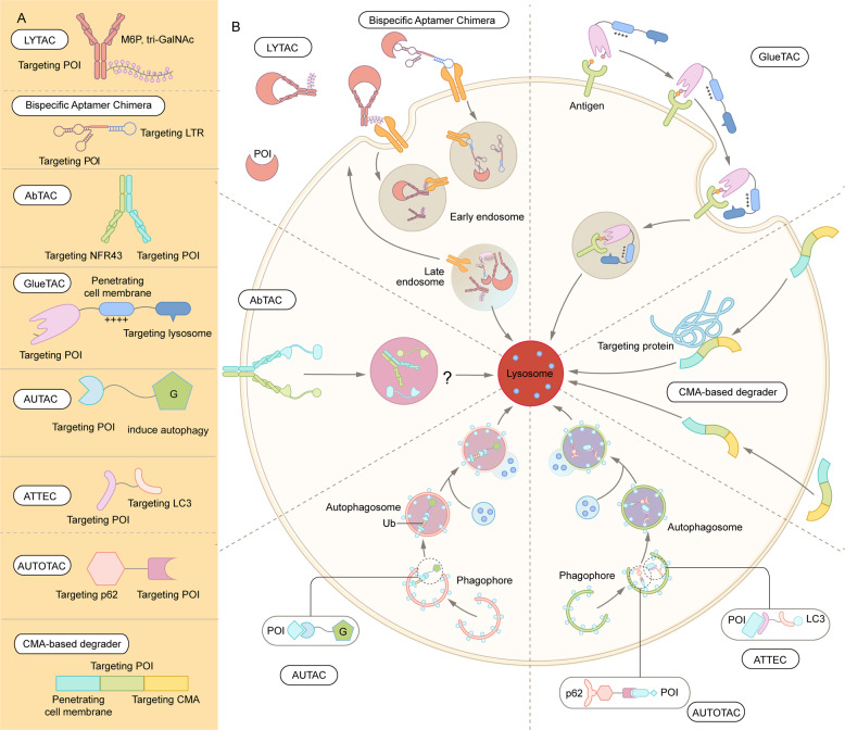
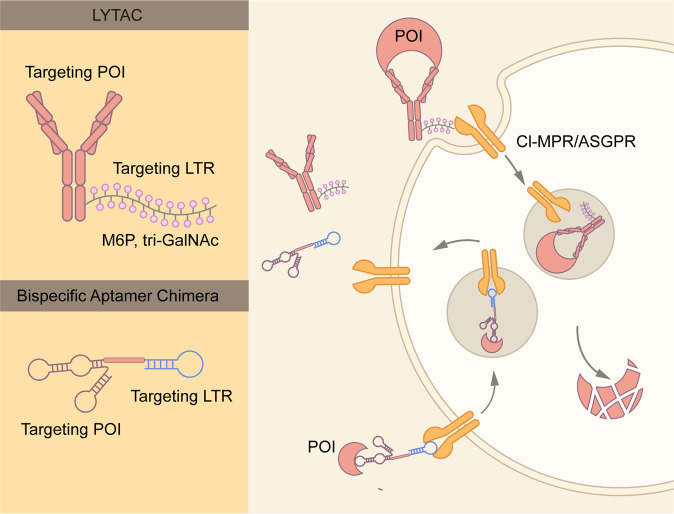
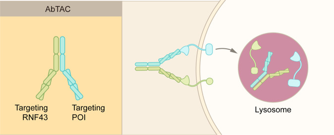
Traditional drug discovery mainly focuses on direct regulation of protein activity. The development and application of protein activity modulators, particularly inhibitors, has been the mainstream in drug development. In recent years, PROteolysis TArgeting Chimeras (PROTAC) technology has emerged as one of the most promising approaches to remove specific disease-associated proteins by exploiting cells' own destruction machinery. In addition to PROTAC, many different targeted protein degradation (TPD) strategies including, but not limited to, molecular glue, Lysosome-Targeting Chimaera (LYTAC), and Antibody-based PROTAC (AbTAC), are emerging. These technologies have not only greatly expanded the scope of TPD, but also provided fresh insights into drug discovery. Here, we summarize recent advances of major TPD technologies, discuss their potential applications, and hope to provide a prime for both biologists and chemists who are interested in this vibrant field.


Conflict of interest statement

The authors declare no competing interests.

Figures

Fig. 1
Fig. 1
Protein degradation via the ubiquitin-proteasome system (UPS). Proteins undergo ubiquitin-dependent degradation by a suite of three enzymes. E1 interacts with E2, and transfers the ubiquitin molecule to E2. E2 interacts with E3-binding substrate and transfers the ubiquitin molecule to the substrate. Repetition of these processes results in polyubiquitination of the substrate, which is subsequently degraded by the 26S proteasome
Fig. 2
Fig. 2
Protein degradation via three distinct lysosome pathways. (1) Cell surface proteins arrive at endosome after endocytosis. They could be degraded by lysosome, or transported to the plasma membrane or other cellular organelles for recycling. (2) In the phagocytic pathway, cells engulf large extracellular particles, such as invading pathogens and dead cells, and then degrade them by lysosome. (3) Misfolded or aggregated proteins, damaged organelles, and intracellular pathogens, are removed by the autophagy–lysosome pathway. There are three different forms of autophagy: macroautophagy, microautophagy, and chaperone-mediated autophagy
Fig. 3
Fig. 3
Representative events in the TPD development. Purple: technologies related with UPS-based technologies; light blue: technologies related with the endosome-lysosome pathway; dark blue: technologies related with the autophagy-lysosome pathway
Fig. 4
Fig. 4
Schematic representation of PROTAC and Molecular Glue. a A PROTAC molecule consists of an E3 ligase-targeting ligand, a linker, and a POI-binding ligand. It simultaneously binds to the POI and the E3 ubiquitin ligase, and induces the polyubiquitination and degradation of the POI. b Molecular glue induces the interaction between a POI and an E3 ubiquitin ligase via binding to the E3 ubiquitin ligase, as illustrated, or the POI. Relative to PROTAC molecules, molecular glues do not have a linker and have a lower MW
Fig. 5
Fig. 5
Schematic representation of a double-mechanism degrader. The molecule binds to one E3 ubiquitin ligase, and can degrade two different POIs via distinct mechanisms
Fig. 6
Fig. 6
Summary of lysosome-dependent protein degradation strategies. A Schematic representation of LYTAC and other degradation technologies via the lysosomal pathway. B LYTAC molecules and Bispecific Aptamer Chimeras degrade membrane proteins and extracellular molecules by engaging a POI and a lysosome-targeting receptor (LTR). AbTAC utilizes a membrane E3 ligases, RNF43, for the degradation of membrane proteins, via a lysosome-dependent manner. GlueTAC utilizes a lysosome-sorting sequence (LSS) to promote the lysosomal degradation. AUTAC, ATTEC and AUTOTAC promote the formation of POI-specific autophagosomes, and subsequently the degradation of the POI via lysosomes. CMA-based degrader harness chaperone-mediated autophagy, rather than macroautophagy, for protein degradation
Fig. 7
Fig. 7
Schematic representation of LYTAC and Bispecific Aptamer Chimera. LYTAC is composed of a small molecule or an antibody conjugated to a ligand that binds to lysosome-targeting receptors (LTRs), such as CI-MPR and ASGPR. Whereas CI-MPR is ubiquitously expressed in all human tissues, ASGPR is only expressed in liver. Thus, ASGPR-base LYTAC could achieve liver specific protein degradation. CI-MPR or ASGPR is endocytosed along with LYTAC molecules and the POI. Whereas the POI is degraded by lysosomes, CI-MPR, or ASGPR is recycled to the plasma membrane for re-use. Bispecific Aptamer Chimera utilizes DNA aptamer to target the LTR and POI
Fig. 8
Fig. 8
Schematic representation of AbTAC. AbTAC utilizes recombinant bispecific antibody to recruit a membrane protein and a membrane-bound E3 ligase, RNF43. The POI is likely degraded via the lysosomes, but not by the proteasomes. However, the exact mechanisms remain to be established
Fig. 9
Fig. 9
Schematic representation of GlueTAC. GlueTAC consists of a covalently-modified nanobody, a Cell-penetrating peptide (CPP), and a lysosome-sorting sequence. The nanobody is responsible for targeting POI, and the CPP induces endocytosis of the GlueTAC-POI complex and subsequent lysosomal degradation
Fig. 10
Fig. 10
Schematic representation of AUTAC. AUTAC molecules consist of a POI-targeting warhead, a linker, and a cGMP-based degradation tag. The degradation tag recruits autophagosomes to degrade cytoplasmic proteins and cellular organelles
Fig. 11
Fig. 11
Schematic representation of ATTEC and AUTOTAC. An ATTEC molecule simultaneously binds LC3 and a POI, while an AUTOTAC molecule binds p62 and a POI. The binding induces the formation of autophagosomes, and subsequent fusion between autophagosomes and lysosomes lead to the POI degradation
Fig. 12
Fig. 12
Schematic representation of CMA-based degrader. CMA-based degrader consists of three modules: a CMA-targeting module, a cell-penetrating peptide, and a POI-targeting module. After the CMA-based degrader entering the cell, it binds the POI and induces chaperone-mediated autophagy

References

    1. Anfinsen CB. Principles that govern the folding of protein chains. Science. 1973;181:223–230. - PubMed
    1. Hanley, S. E. & Cooper, K. F. Sorting nexins in protein homeostasis. Cells. 10, 17 (2020). - PMC - PubMed
    1. de Duve C. The lysosome turns fifty. Nat. Cell Biol. 2005;7:847–849. - PubMed
    1. Dikic I. Proteasomal and autophagic degradation systems. Annu. Rev. Biochem. 2017;86:193–224. - PubMed
    1. Wang X, Robbins J. Proteasomal and lysosomal protein degradation and heart disease. J. Mol. Cell Cardiol. 2014;71:16–24. - PMC - PubMed

Publication types