Clinical Trial
. 2022 May;28(5):1014-1021.
doi: 10.1038/s41591-022-01755-w. Epub 2022 Apr 4.

Intravitreal antisense oligonucleotide sepofarsen in Leber congenital amaurosis type 10: a phase 1b/2 trial

Affiliations
Clinical Trial

Intravitreal antisense oligonucleotide sepofarsen in Leber congenital amaurosis type 10: a phase 1b/2 trial

Stephen R Russell et al. Nat Med. 2022 May.

Abstract

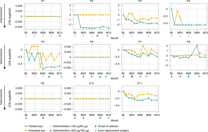
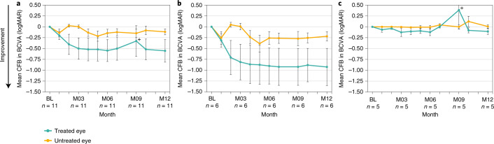
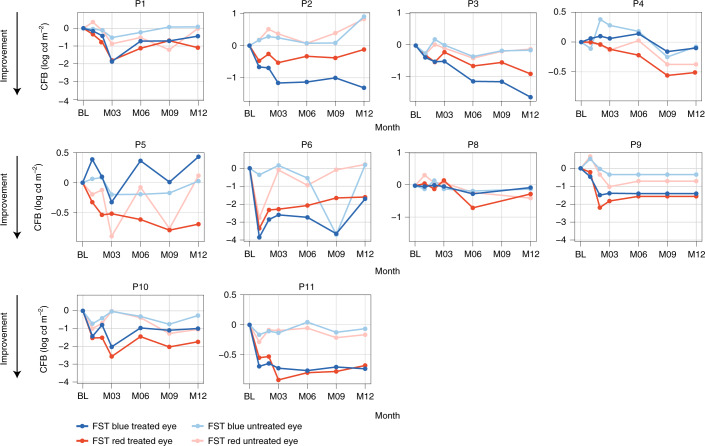
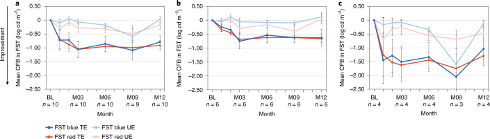
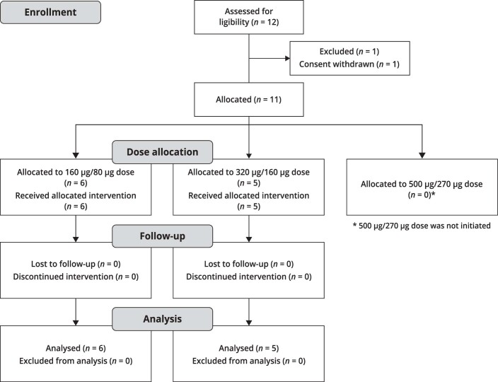
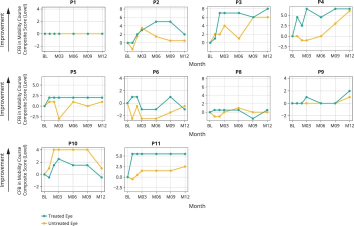
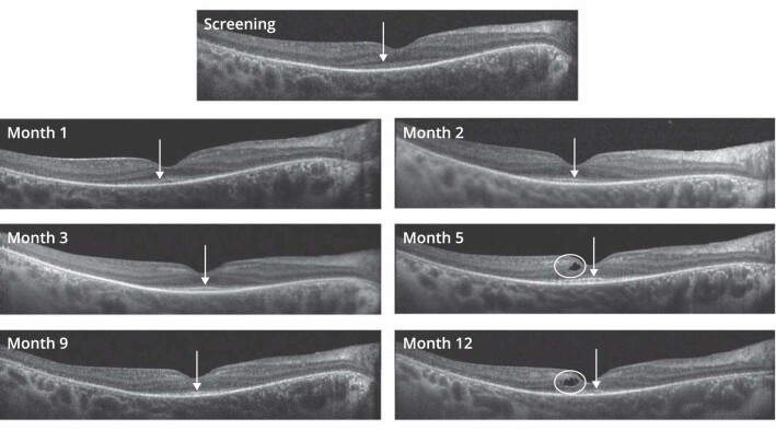
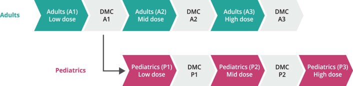
CEP290-associated Leber congenital amaurosis type 10 (LCA10) is a retinal disease resulting in childhood blindness. Sepofarsen is an RNA antisense oligonucleotide targeting the c.2991+1655A>G variant in the CEP290 gene to treat LCA10. In this open-label, phase 1b/2 ( NCT03140969 ), 12-month, multicenter, multiple-dose, dose-escalation trial, six adult patients and five pediatric patients received ≤4 doses of intravitreal sepofarsen into the worse-seeing eye. The primary objective was to evaluate sepofarsen safety and tolerability via the frequency and severity of ocular adverse events (AEs); secondary objectives were to evaluate pharmacokinetics and efficacy via changes in functional outcomes. Six patients received sepofarsen 160 µg/80 µg, and five patients received sepofarsen 320 µg/160 µg. Ten of 11 (90.9%) patients developed ocular AEs in the treated eye (5/6 with 160 µg/80 µg; 5/5 with 320 µg/160 µg) versus one of 11 (9.1%) in the untreated eye; most were mild in severity and dose dependent. Eight patients developed cataracts, of which six (75.0%) were categorized as serious (2/3 with 160 µg/80 µg; 4/5 with 320 µg/160 µg), as lens replacement was required. As the 160-µg/80-µg group showed a better benefit-risk profile, higher doses were discontinued or not initiated. Statistically significant improvements in visual acuity and retinal sensitivity were reported (post hoc analysis). The manageable safety profile and improvements reported in this trial support the continuation of sepofarsen development.


Conflict of interest statement

S.R.R. and A.V.C. report grant funding from ProQR Therapeutics during the conduct of the study. A.V.D. reports grants and other from ProQR Therapeutics during the conduct of the study; personal fees from Medscape and Novartis; grants from Spark Therapeutics and the National Institutes of Health; a patent held by Novartis with royalties paid to Spark Therapeutics and a patent held by the University of Iowa issued to Spark Therapeutics, outside the submitted work. B.P.L. reports grants and non-financial support from ProQR Therapeutics; grants from the Fund for Research Flanders during the conduct of the study; grants from GenSight Therapeutics, IVERIC Bio and Vedere Bio; and grants and non-financial support from Novartis, Spark Therapeutics and RegenXBio, outside the submitted work. C.V.C. reports grants and non-financial support from ProQR Therapeutics, during the conduct of the study, and grants from GenSight Therapeutics, outside the submitted work. A.C.H. reports grants from ProQR Therapeutics both during the conduct of the study and outside the submitted work. E.J. reports grants and non-financial support from ProQR Therapeutics during the conduct of the study and grants from GenSight Therapeutics outside the submitted work. J.D.Z. reports grants from ProQR Therapeutics, during the conduct of the study, and grants from GenSight Therapeutics, outside the submitted work. R.W.J.C. reports other from ProQR Therapeutics, outside the submitted work, and a patent (antisense oligonucleotides) for the treatment of Leber congenital amaurosis licensed to ProQR Therapeutics. P.A. reports share holdings in ProQR Therapeutics. M.E.C. reports grants and personal fees from ProQR Therapeutics, during the conduct of the study, and grants and/or personal fees from Editas Medicine, BridgeBio, PYC and Alia Therapeutics, outside the submitted work. M.R.S., G.P. and A.G. are employees of ProQR Therapeutics. W.D.H., F.A. and D.R. are former employees of ProQR Therapeutics. S.G.J., A.V.D., I.H., M.M., W.P., E.H.S., J.W., A.V.G., A.K.K., C.A.P., A.S., A.J.R., E.V., F.N. and C.H. have nothing to disclose.

Figures

Fig. 1
Fig. 1. Change from baseline in BCVA at month 12 in individual patients (n = 11).
Five of 11 patients (45%) showed a clinically meaningful improvement of at least −0.3 logMAR, and seven of 11 patients (64%) showed an improvement of at least −0.2 logMAR. P1, P2, P6, P9 and P10 were LP at baseline. Different scales of y axis are intended to facilitation data visualization. Eight patients developed cataracts in the treated eye (3/6 in the 160-µg/80-µg dose group and 5/5 in the 320-µg/160-µg dose group) at different time points during the trial (P7 at M3, P8 and P9 at M6, P2 and P5 at M8, P6 and P10 at M9 and P1 at M12). Six cataracts of eight needed lens replacement. These patients regained their pre-cataract visual acuity after surgery. For P8, from M7 onwards, the cataract quickly worsened, with post-capsular opacity grade 1 as well as a cortical opacity grade 2.5 reported at M9, which had substantial effect on BCVA, leading to a decision to perform lens replacement surgery a month later. P5 and P10 lens replacements were performed after M12. The pre-specified baseline (BL) was defined as the average pre-treatment value (that is, average of screening and day 1 (pre-dose) values). CFB, change from baseline; M, month.
Fig. 2
Fig. 2. Mean change from baseline (CFB) to month 12 in BCVA comparing treated and untreated eyes.
Treated and untreated eye groups are shown for the pooled dose groups (n = 11) (a); the low-dose (160-µg/80-µg) group (n = 6) (b); and the mid-dose (320-µg/160-µg) group (n = 5) (c). Bars show s.e.m.; data are summarized in Supplementary Table 1. +Visual acuity changes associated with cataract events occurrence: eight patients developed cataracts in the treated eye (3/6 in the 160-µg/80-µg dose group and 5/5 in the 320-µg/160-µg dose group), and six cataracts required lens replacement. These patients regained their pre-cataract visual acuity after surgery. BL, baseline; M, month.
Fig. 3
Fig. 3. Change from baseline in red and blue FST at month 12 in treated and untreated eyes in individual patients (n = 10).
Order of graphs and different scale of y axis are intended to facilitate data visualization. FST baseline data with the pulse stimulus test were missing for P7, so FST data for this patient were excluded from the efficacy analysis. For P9, M9 and M12 data were imputed using the last observation carried forward because FST was mistakenly tested with a flash stimulus instead of the pulse stimulus at these time points. BL, baseline; CFB, change from baseline; M, month.
Fig. 4
Fig. 4. Mean change from baseline (CFB) to month 12 in blue and red FST comparing treated and untreated eye groups.
Treated and untreated eye groups are shown for the pooled dose groups, n = 10 (a); the low-dose (160-µg/80-µg) group (n = 6) (b); and the mid-dose (320 µg/160 µg) group (n = 4) (c). Bars show s.e.m.; data are summarized in Supplementary Table 1. Enhanced FST response seemed most apparent in patients with visual acuity of light perception; three of five patients (60%) were LP at baseline in the mid-dose (320-µg/160-µg) group, and two of six patients (33%) were LP at baseline in the low-dose (160-µg/80-µg) group. BL, baseline; M, month; TE, treated eyes; UE, untreated eyes.
Extended Data Fig. 1
Extended Data Fig. 1. CONSORT trial flow diagram.
During the enrollment period, 12 patients (6 adults and 6 children) were screened; 1 child withdrew consent before treatment. Of the 11 patients that enrolled, 6 received loading/maintenance doses of sepofarsen of 160µg/80µg and 5 received 320µg/160µg. All patients completed the full 12-month trial, and all were included in the analysis.
Extended Data Fig. 2
Extended Data Fig. 2. Change from baseline in mobility course composite score to Month 12 in treated and untreated eyes in individual patients (n = 10).
Order of graphs and different scale of y-axis are intended to facilitation data visualization. P7 was not included in the mobility course composite score analysis because no baseline data were available. For this patient, at Month 12, the mobility course composite score revealed navigation to 19 of 20 levels for both treated and untreated eyes, suggesting no worsening from the treatment. BL = baseline; CFB = change from baseline; M = month; P = patient; TE = treated eye; UE = untreated eye.
Extended Data Fig. 3
Extended Data Fig. 3. SD-OCT scans along the horizontal meridian crossing the fovea at screening, M1, M2, M3, M5, M9, and M12 for the patient P7.
Arrows: the layer of the EZ line in all panels. Note that whereas the EZ line potentially shows improved definition post-treatment, it also shows some discontinuity at Month 12. Circles: cystoid macular edema in panels M5 and M12; retinal thinning in comparison of screening to M12. SD-OCT images of subject P7 provides examples of cystoid macular edema and retinal thinning. In this subject, these adverse events developed concurrent with reestablishment of the EZ, a feature of normal retinal architecture. SD-OCT = spectral-domain optical coherence tomography; M = month; EZ = ellipsoid zone.
Extended Data Fig. 4
Extended Data Fig. 4. PQ-110-001 trial: Schema for introduction of dose levels.
The safety monitoring committee and/or Sponsor Medical Monitor could decide to de-escalate the dose, hold the dose (delay or skip), or discontinue study drug for an individual subject, in consultation with the Investigator. Subjects who discontinued study drug continued to be followed for safety and efficacy. A = adult subjects; DMC = Data Monitoring Committee; P = pediatric subjects.

References

    1. Kumaran N, Moore AT, Weleber RG, Michaelides M. Leber congenital amaurosis/early-onset severe retinal dystrophy: clinical features, molecular genetics and therapeutic interventions. Br. J. Ophthalmol. 2017;101:1147–1154. doi: 10.1136/bjophthalmol-2016-309975. - DOI - PMC - PubMed
    1. Chacon-Camacho OF, Zenteno JC. Review and update on the molecular basis of Leber congenital amaurosis. World J. Clin. Cases. 2015;3:112–124. doi: 10.12998/wjcc.v3.i2.112. - DOI - PMC - PubMed
    1. Coppieters F, et al. Genetic screening of LCA in Belgium: predominance of CEP290 and identification of potential modifier alleles in AHI1 of CEP290-related phenotypes. Hum. Mutat. 2010;31:E1709–E1766. doi: 10.1002/humu.21336. - DOI - PMC - PubMed
    1. Den Hollander AI, et al. Mutations in the CEP290 (NPHP6) gene are a frequent cause of Leber congenital amaurosis. Am. J. Hum. Genet. 2006;79:556–561. doi: 10.1086/507318. - DOI - PMC - PubMed
    1. Perrault I, et al. Spectrum of NPHP6/CEP290 mutations in Leber congenital amaurosis and delineation of the associated phenotype. Hum. Mutat. 2007;28:416. doi: 10.1002/humu.9485. - DOI - PubMed

Publication types

Supplementary concepts

Associated data