. 2022 Apr 5;13(1):65.
doi: 10.1186/s13244-022-01213-8.

MRI appearances of hepatic epithelioid hemangioendothelioma: a retrospective study of 57 patients

Affiliations

MRI appearances of hepatic epithelioid hemangioendothelioma: a retrospective study of 57 patients

Xiaolei Liu et al. Insights Imaging. .

Abstract

Background: Hepatic epithelioid hemangioendothelioma (HEH) is extremely rare and the MRI features have never been investigated in a large group of patients.

Methods: A retrospective study was designed to review the MRI images of HEH patients. Two radiologists separately evaluated signal intensity (SI) on unenhanced imaging, morphological features, contrast-enhancement pattern at dynamic study. The MRI features were compared between patients with HEH and hepatic metastatic tumor (HMT).

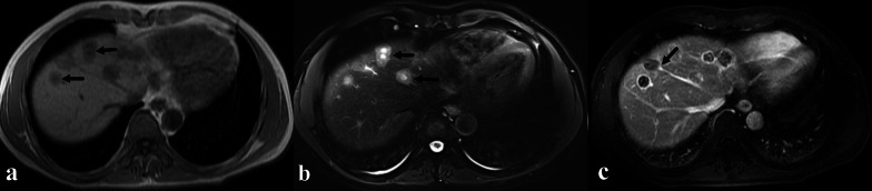
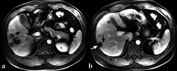
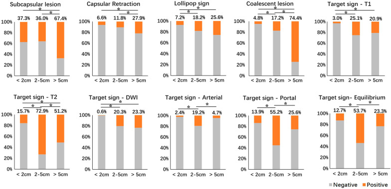
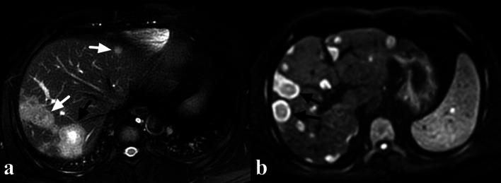
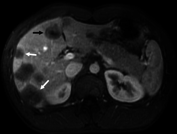
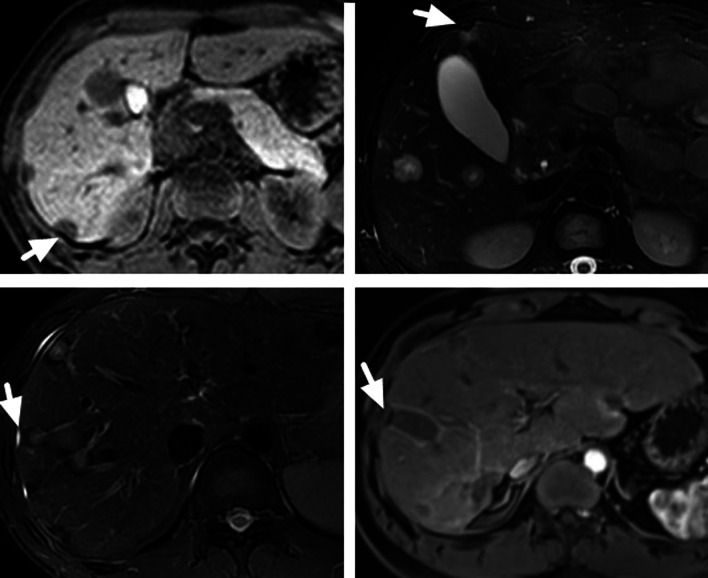
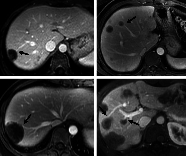
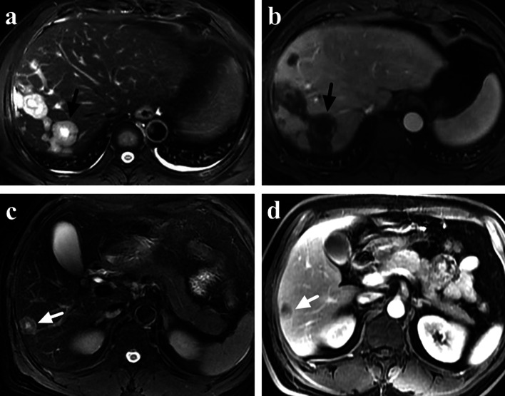
Results: Fifty-seven HEH patients were included in this study and a total of 412 lesions were evaluated. On per-lesion analysis, the rate of coalescent lesion and subcapsular lesion were 18.2% and 39.8%, respectively. Capsular retraction and lollipop sign were observed in 47 lesions (11.4%) and 60 lesions (14.6%), respectively. Large lesions (> 5 cm) had the highest rate of coalescent lesion, subcapsular lesion, capsular retraction and lollipop sign. Target sign appeared in 196 lesions (47.6%) on T2 weighted (T2W) and 146 lesions (35.4%) on portal phase. Medium lesions (2-5 cm) had the highest rate of target sign on both T2W (72.9%) and portal phase (55.2%). On per-patient analysis, compare with HEH patients, HMT patients seldom had the appearance of lollipop sign (66.7% versus 6.4%, p < 0.01), capsular retraction (59.6% versus 3.2%, p < 0.01) and target appearance on both T2Wand portal phase (64.9% versus 12.7%, p < 0.01).

Conclusion: MRI features of HEH correlated with the lesion size. Capsular retraction, lollipop sign and coexistence of target sign on both T2W and portal phase were relatively specific MRI features of HEH, which could be helpful in suggesting the diagnosis.

Keywords: HEH; Hepatic epithelioid hemangioendothelioma; Hepatic tumors; MRI; Rare liver tumors.


Conflict of interest statement

The authors declare that they have no competing interests.

Figures

Fig. 1
Fig. 1
A 42-year-old male HEH patient. a Coalescent lesion with target sign on T1-weighted 3D GRE image, consisted of concentric layers including markedly hypointense in central tumor regions and peripheral zones which were moderately hypointense relative to liver parenchyma (arrows). b Coalescent lesion with target sign on fat-saturated T2-weighted TSE image, consisted of concentric layers including markedly hyperintense in central tumor regions and moderately hyperintense peripheral zone (arrows). c Lollipop sign on portal phase image, consisted of a well-defined tumor (as the “candy”) and an obstructed hepatic vein (as the “stick”) (arrow)
Fig. 2
Fig. 2
A 35-year-old male HEH patient. a Target sign on portal phase image, consisted of concentric layers including markedly hypointense in central tumor regions and peripheral zones which were moderately hypointense relative to liver parenchyma (arrow). b Capsular retraction caused by a subcapsular lesion (white arrow) and lollipop sign, consisted of a well-defined tumor (as the “candy”) and an obstructed portal vein (as the “stick”) (black arrow) on portal phase image
Fig. 3
Fig. 3
The comparison results of different MRI features among lesions of different sizes
Fig. 4
Fig. 4
A 38-year-old male HEH patient. a On fat-saturated T2 weighted image, target sign could be observed on a medium lesion (black arrow), while both small and large lesions failed to show target appearance (white arrow). b Target sign on DWI image (800 b-value), consisted of concentric layers including relatively hyperintense in central tumor regions and peripheral zones which were markedly hyperintense relative to liver parenchyma (arrows)
Fig. 5
Fig. 5
A 22-year-old male HEH patient. On portal phase image, target sign could be observed on a medium lesion (black arrow), while both small and large lesions failed to show target appearance (white arrow)
Fig. 6
Fig. 6
The appearance of capsular retraction on MRI images of 4 HEH patients (arrows)
Fig. 7
Fig. 7
The appearance of lollipop sign on MRI images of 4 HEH patients (arrows)
Fig. 8
Fig. 8
a, b A 40-year-old male HEH patient with coexistence of target appearance on both T2 weighted and portal phase images. a Target sign on fat-saturated T2-weighted image, consisted of concentric layers including markedly hyperintense in central tumor regions and moderately hyperintense peripheral zone (arrow). b Target sign on portal phase image, consisted of concentric layers including markedly hypointense in central tumor regions and peripheral zones which were moderately hypointense relative to liver parenchyma (arrow). c, d A 51-year-old male rectal carcinoma patients with multiple liver metastasis. c A classic target sign on fat-saturated T2-weighted image (arrow). d Heterogeneous enhancement on portal phase image (arrow), instead of target sign

References

    1. Sardaro A, Bardoscia L, Petruzzelli MF, Portaluri M. Epithelioid hemangioendothelioma: an overview and update on a rare vascular tumor. Oncol Rev. 2014;8(2):259. - PMC - PubMed
    1. Weiss SW, Enzinger FM. Epithelioid hemangioendothelioma: a vascular tumor often mistaken for a carcinoma. Cancer. 1982;50(5):970–981. doi: 10.1002/1097-0142(19820901)50:5<970::AID-CNCR2820500527>3.0.CO;2-Z. - DOI - PubMed
    1. Otrock ZK, Al-Kutoubi A, Kattar MM, Zaatari G, Soweid A. Spontaneous complete regression of hepatic epithelioid haemangioendothelioma. Lancet Oncol. 2006;7(5):439–441. doi: 10.1016/S1470-2045(06)70697-0. - DOI - PubMed
    1. Liu XL, Yang ZY. Outcomes of hepatic epithelioid hemangioendothelioma with different managements: a retrospective investigation. Eur Rev Med Pharmacol Sci. 2021;25(12):4274–4282. - PubMed
    1. Makhlouf HR, Ishak KG, Goodman ZD. Epithelioid hemangioendothelioma of the liver: a clinicopathologic study of 137 cases. Cancer. 1999;85(3):562–582. doi: 10.1002/(SICI)1097-0142(19990201)85:3<562::AID-CNCR7>3.0.CO;2-T. - DOI - PubMed

LinkOut - more resources