. 2022 Oct 3;24(10):1689-1699.
doi: 10.1093/neuonc/noac088.

Comprehensive profiling of myxopapillary ependymomas identifies a distinct molecular subtype with relapsing disease

Affiliations

Comprehensive profiling of myxopapillary ependymomas identifies a distinct molecular subtype with relapsing disease

Michael Bockmayr et al. Neuro Oncol. .

Abstract

Background: Myxopapillary ependymoma (MPE) is a heterogeneous disease regarding histopathology and outcome. The underlying molecular biology is poorly understood, and markers that reliably predict the patients' clinical course are unknown.

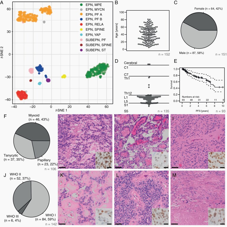
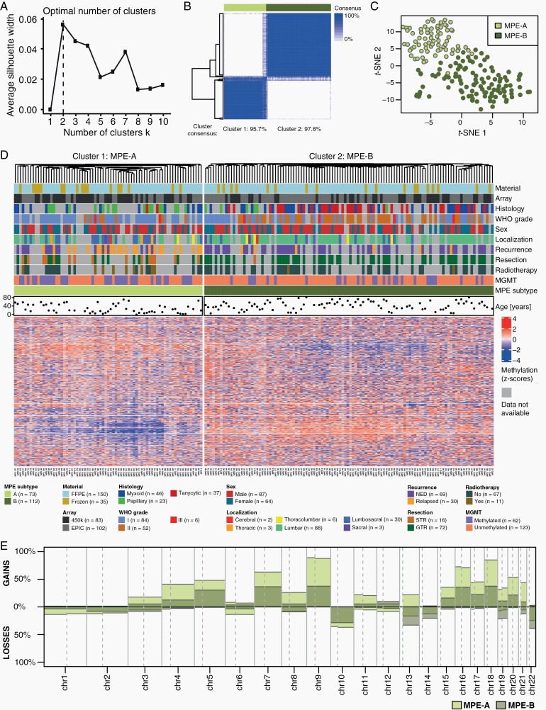
Methods: We assembled a cohort of 185 tumors classified as MPE based on DNA methylation. Methylation patterns, copy number profiles, and MGMT promoter methylation were analyzed for all tumors, 106 tumors were evaluated histomorphologically, and RNA sequencing was performed for 37 cases. Based on methylation profiling, we defined two subtypes MPE-A and MPE-B, and explored associations with epidemiological, clinical, pathological, and molecular characteristics of these tumors.

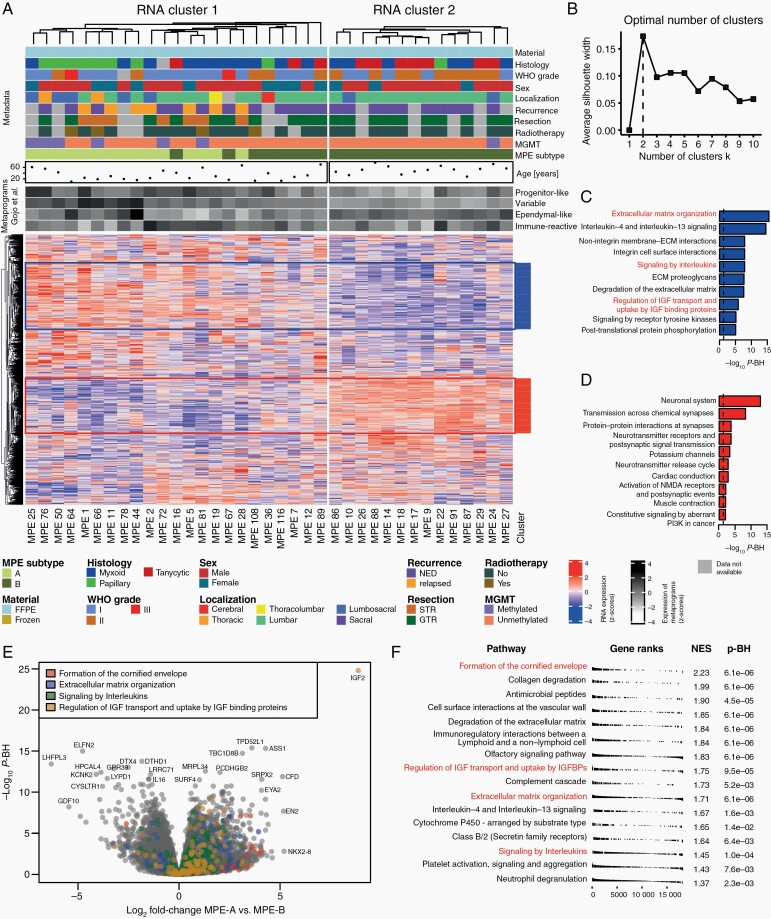
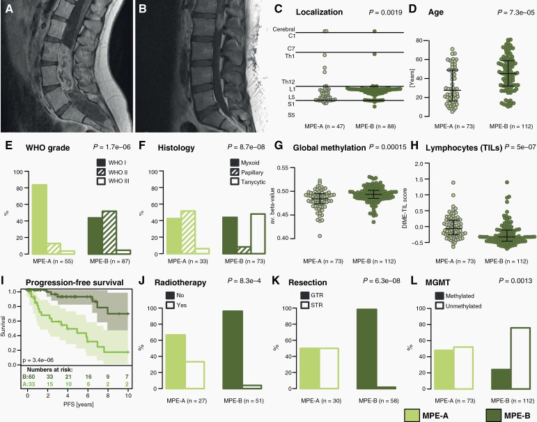
Results: MPE-A occurred at a median age of 27 years and were enriched with tumors demonstrating papillary morphology and MGMT promoter hypermethylation. Half of these tumors could not be totally resected, and 85% relapsed within 10 years. Copy number alterations were more common in MPE-A. RNA sequencing revealed an enrichment for extracellular matrix and immune system-related signatures in MPE-A. MPE-B occurred at a median age of 45 years and included many tumors with a histological diagnosis of WHO grade II and tanycytic morphology. Patients within this subtype had a significantly better outcome with a relapse rate of 33% in 10 years (P = 3.4e-06).

Conclusions: We unraveled the morphological and clinical heterogeneity of MPE by identifying two molecularly distinct subtypes. These subtypes significantly differed in progression-free survival and will likely need different protocols for surveillance and treatment.

Keywords: DNA methylation; RNA sequencing; histology; myxopapillary ependymoma; outcome.


Figures

Fig. 1
Fig. 1
Clinical and histopathological features of myxopapillary ependymoma. (A) T-SNE analysis of the 10 molecular ependymoma types based on methylation profiling. (B) Age distribution of the MPE samples (beeswarm plot). Lines indicate the median, upper and lower quartile. (C) Sex distribution in the MPE cohort. (D) Schematic representation of the MPE tumor localization along the neural axis. (E) Progression-free survival in the MPE cohort. (F-I) Distribution of histological MPE subtypes with typical patterns: myxoid (G), papillary (H), and tanycytic/solid (I) (scale bar = 50 µm). Inset: immunohistochemical staining for HOXB13 (scale bar = 10 µm). (J–M) Distribution of WHO grades in the study cohort with examples of WHO grade I (K), II (L), and III (M) tumors (scale bar = 50 µm). Inset: immunohistochemical staining for HOXB13 (scale bar = 10 µm).
Fig. 2
Fig. 2
Methylation analysis of myxopapillary ependymoma. (A) Determination of the optimal number of methylation-based k-means clusters using the silhouette method. (B) Consensus clustering for the k-means algorithm. The column sidebar shows the k-means cluster for k = 2 with 10 000 CpG sites optimal in (A). (C) T-SNE analysis of methylation data. The colors represent the k-means cluster for k = 2 with 10 000, CpG sites. (D) Cluster analysis with heatmap of methylation data and clinicopathological variables of MPE (n = 185). (E) Overview of chromosome arm-wise copy number alterations in methylation-based subtypes of MPE.
Fig. 3
Fig. 3
RNA sequencing analysis of myxopapillary ependymoma. (A) Heatmap analysis of RNA sequencing data, clinicopathological variables and expression of metaprograms defined by Gojo et al. in 37 MPE. The MPE subtype is based on k-means clustering of methylation data. The optimal number of clusters was determined by the silhouette method (B). Highly correlating gene clusters (R > 0.3, defined by cutting the row dendrogram) with at least 200 genes are annotated and subjected to gene set enrichment analysis (C–D). (E) Volcano plot of differential expression analysis with annotation of four gene sets. (F) Results of gene set enrichment analysis of differentially expressed genes between MPE subtypes.
Fig. 4
Fig. 4
Clinicopathological and radiological features of MPE subtypes. (A–B) Exemplary MRI images of MPE subtype A and B, respectively (T1+CA). (C) Tumor localization of MPE subtypes along the neural axis. (D) Age distribution of MPE subtypes. (E–F) Distribution of WHO grades (2016) and main histological patterns in MPE subtypes. (G–H) Estimation of global DNA methylation level and tumor-infiltrating lymphocytes (TILs) in MPE subtypes. (I) Kaplan-Meier plot comparing PFS between MPE subtypes. (J–L) Distribution of patients having received adjuvant radiotherapy, postoperative resection status, and MGMT promoter methylation in MPE subtypes. Lines in beeswarm diagrams indicate the median, upper, and lower quartile. P-values assess differences between MPE subtypes.

References

    1. Louis DN, Perry A, Wesseling P, et al. The 2021 WHO Classification of Tumors of the Central Nervous System: a summary. Neuro Oncol. 2021;23(8):1231–1251. - PMC - PubMed
    1. Bates JE, Choi G, Milano MT. Myxopapillary ependymoma: a SEER analysis of epidemiology and outcomes. J Neurooncol. 2016;129(2):251–258. - PubMed
    1. Truitt G, Gittleman H, Leece R, et al. Partnership for defining the impact of 12 selected rare CNS tumors: a report from the CBTRUS and the NCI-CONNECT. J Neurooncol. 2019;144(1):53–63. - PMC - PubMed
    1. Weber DC, Wang Y, Miller R, et al. Long-term outcome of patients with spinal myxopapillary ependymoma: treatment results from the MD Anderson Cancer Center and institutions from the Rare Cancer Network. Neuro Oncol. 2015;17(4):588–595. - PMC - PubMed
    1. Pajtler KW, Witt H, Sill M, et al. Molecular classification of ependymal tumors across all CNS compartments, histopathological grades, and age groups. Cancer Cell. 2015;27(5):728–743. - PMC - PubMed

Publication types