. 2022 Jun 1;132(11):e151895.
doi: 10.1172/JCI151895.

Inhibiting SCAP/SREBP exacerbates liver injury and carcinogenesis in murine nonalcoholic steatohepatitis

Affiliations

Inhibiting SCAP/SREBP exacerbates liver injury and carcinogenesis in murine nonalcoholic steatohepatitis

Satoshi Kawamura et al. J Clin Invest. .

Abstract

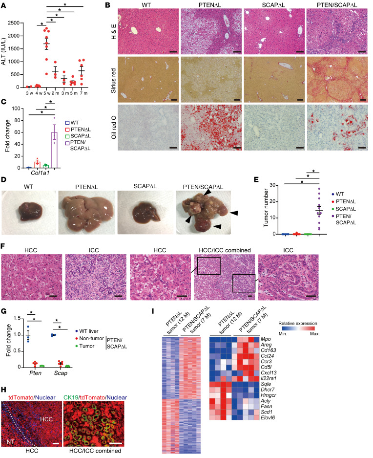
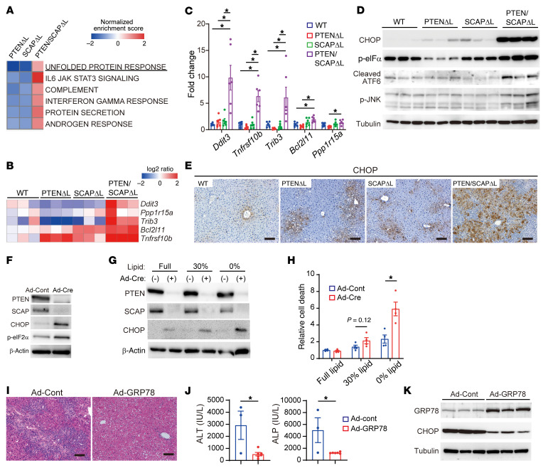
Enhanced de novo lipogenesis mediated by sterol regulatory element-binding proteins (SREBPs) is thought to be involved in nonalcoholic steatohepatitis (NASH) pathogenesis. In this study, we assessed the impact of SREBP inhibition on NASH and liver cancer development in murine models. Unexpectedly, SREBP inhibition via deletion of the SREBP cleavage-activating protein (SCAP) in the liver exacerbated liver injury, fibrosis, and carcinogenesis despite markedly reduced hepatic steatosis. These phenotypes were ameliorated by restoring SREBP function. Transcriptome and lipidome analyses revealed that SCAP/SREBP pathway inhibition altered the fatty acid (FA) composition of phosphatidylcholines due to both impaired FA synthesis and disorganized FA incorporation into phosphatidylcholine via lysophosphatidylcholine acyltransferase 3 (LPCAT3) downregulation, which led to endoplasmic reticulum (ER) stress and hepatocyte injury. Supplementation with phosphatidylcholines significantly improved liver injury and ER stress induced by SCAP deletion. The activity of the SCAP/SREBP/LPCAT3 axis was found to be inversely associated with liver fibrosis severity in human NASH. SREBP inhibition also cooperated with impaired autophagy to trigger liver injury. Thus, excessively strong and broad lipogenesis inhibition was counterproductive for NASH therapy; this will have important clinical implications in NASH treatment.

Keywords: Gastroenterology; Hepatitis.


Figures

Figure 1
Figure 1. Liver-specific PTEN/SCAP double-knockout mice exhibit severe liver injury.
(A) Serum levels of ALT, ALP, total bilirubin, and liver weight in 5-week-old WT, PTENΔL, SCAPΔL, and PTEN/SCAPΔL mice (n = 6 per group). (B and C) H&E (B) and oil red O (C) staining images of livers from mice indicated in A. Right H&E image shows the focal necrosis of liver parenchyma in PTEN/SCAPΔL mice. Scale bars: 50 μm (H&E, furthest right); 100 μm (all others). (D) IHC images of indicated proteins in livers from mice indicated in A. Scale bars: 100 μm. Red arrowheads, Ki67-expressing hepatocytes; yellow arrowheads, hepatocytes expressing SREBP-1 in the cytoplasm but not the nucleus. Enlarged high magnification images of Ki67 and SREBP-1 staining are shown in Supplemental Figure 1B. (E) Cytoplasmic and nuclear protein fractions were extracted from 5-week-old PTENΔL and PTEN/SCAPΔL mouse livers, and the cytoplasmic protein levels of precursor SREBP-1 (p-SREBP1) and nuclear mature form of SREBP-1 (m-SREBP1) were analyzed by WB. (F) WB analyses of indicated proteins in livers from mice indicated in A. (G) Relative expression levels of lipogenic genes determined by real-time PCR in livers from mice indicated in A (n = 6 per group). *P < 0.05. (H) Hepatic TG and total cholesterol content of livers from mice indicated in A (n = 3 per group). (I) Relative expression levels of inflammatory cytokines determined by real-time PCR in livers from mice indicated in A (n = 6 per group). *P < 0.05. All statistical data were assessed using 1-way ANOVA with Tukey’s multiple comparison test (A, G, H, and I) and are presented as mean ± SEM.
Figure 2
Figure 2. Deletion of SCAP in PTENΔL mice induces severe liver fibrosis and accelerates liver cancer development.
(A) Time course of serum ALT in PTEN/SCAPΔL mice (sample sizes by age: 3 weeks, n = 2; 4 weeks, n = 5; 5 weeks, n = 8; 2 months, n = 3; 3 months, n = 3; 5 months, n = 6; 7 months, n = 6). (B) H&E, Sirius red, and oil red O staining images of livers from 5-month-old WT, PTENΔL, SCAPΔL, and PTEN/SCAPΔL mice. Scale bars: 250 μm (Sirius red); 100 μm (all others). (C) Relative expression levels of Col1a1 mRNA determined by real-time PCR in livers from mice indicated in B (n = 3 per group). (D and E) Representative liver images (D) and tumor numbers (E) for 7-month-old mice of each genotype (WT and SCAPΔL, n = 6; PTENΔL and PTEN/SCAPΔL, n = 11). Arrowheads, liver tumors. (F) H&E images of liver tumors from PTEN/SCAPΔL mice. Scale bars: 50 μm. (G) Relative expression levels of Pten and Scap mRNAs by real-time PCR in liver tissues of 7-month-old WT mice and nontumor liver tissues and tumor tissues of 7-month-old PTEN/SCAPΔL mice (WT, n = 3; nontumor and tumor tissues of PTEN/SCAPΔL mice, n = 5). (H) Immunofluorescence staining of tdTomato in HCC (left panel) and double immunofluorescence staining of tdTomato and ductal cell marker CK19 in HCC/ICC combined tumor (right panel) derived from 7-month-old PTEN/SCAPΔL;tdTomato mice. Scale bars: 100 μm. (I) Heatmaps show significant differentially expressed genes between PTENΔL tumors and PTEN/SCAPΔL tumors (PTENΔL, n = 4; PTEN/SCAPΔL, n = 5). Left panel, total differentially expressed genes. Right panel, selected differentially expressed genes. All statistical data were assessed using 1-way ANOVA with Tukey’s multiple comparison test (A, C, E, and G). Data are presented as mean ± SEM. *P < 0.05.
Figure 3
Figure 3. Restoration of SREBP function in PTEN/SCAPΔL mice ameliorates liver injury, fibrosis, and carcinogenesis.
(A and B) H&E images of livers (A) and serum levels of ALT and ALP (B) for 5-week-old PTEN/SCAPΔL and PTEN/SCAPΔL;S1aTg mice. Scale bars: 100 μm. (PTEN/SCAPΔL, n = 5; PTEN/SCAPΔL;S1aTg, n = 6). (C) Relative expression levels of lipogenic genes determined by real-time PCR in livers of 5-week-old WT, PTEN/SCAPΔL, and PTEN/SCAPΔL;S1aTg mice (n = 3 per group). (D) H&E and Sirius red images of livers from 5-month-old PTEN/SCAPΔL and PTEN/SCAPΔL;S1aTg mice. Scale bars: 100 μm. Bar graph shows Sirius red–positive area (n = 3 per group). (E) Relative expression levels of Col1a1 mRNA determined by real-time PCR in livers from 5-month-old PTEN/SCAPΔL and PTEN/SCAPΔL;S1aTg mice (n = 3 per group). (F) Representative liver images and tumor numbers for 7-month-old PTEN/SCAPΔL and PTEN/SCAPΔL;S1aTg mice (PTEN/SCAPΔL, n = 11; PTEN/SCAPΔL;S1aTg, n = 8). Arrowheads indicate liver tumors. (GJ) We intravenously injected 4-week-old PTEN/SCAPΔL mice with 1.5 × 10¹¹ genome copies of AAV-control, AAV-nSREBP-1c, or AAV-nSREBP-2 and analyzed 1 week after injection (AAV-control, n = 3 for each experiment; AAV-nSREBP-1c and AAV-nSREBP-2, n = 5). (G and I) H&E images of livers and serum ALT. Scale bars: 100 μm. (H and J) Relative expression levels of inflammatory cytokines and lipogenic genes determined by real-time PCR. Statistical data in B, D, and EJ were assessed using Student’s t test and in C using 1-way ANOVA with Tukey’s multiple comparison test. Data are presented as mean ± SEM. *P < 0.05.
Figure 4
Figure 4. ER stress is involved in liver injury in PTEN/SCAPΔL mice.
(A) Pathway analysis of RNA-Seq data. Pathways upregulated in 5-week-old PTEN/SCAPΔL mouse livers are shown. (B) Relative expression levels of ER stress–responsive genes involved in cell death determined by RNA-Seq. Data are expressed as log2 ratio compared with WT mice. (C) Relative expression levels of genes indicated in B were analyzed using real-time PCR (n = 6 per group). (D and E) WB analyses of ER stress markers (D) and IHC images of CHOP (E) for livers of 5-week-old WT, PTENΔL, SCAPΔL, and PTEN/SCAPΔL mice. Scale bars: 100 μm. (F) Primary hepatocytes isolated from Ptenfl/fl/Scapfl/fl mice were infected with Ad-Cont or Ad-Cre. The indicated proteins were assessed by WB 96 hours after infection. (G and H) At 24 hours after infection of Ptenfl/fl/Scapfl/fl hepatocytes with Ad-Cont or Ad-Cre, culture media were changed to normal FBS media, moderately delipidated FBS (30% lipid compared with normal FBS), or completely delipidated FBS (0% lipid). At 72 hours, expression levels of indicated proteins were determined by WB analyses (G). At 96 hours, cell death was assessed using the Cell Death Detection ELISA Kit (n = 4 per group) (H). (IK) Effects of GRP78 overexpression in PTEN/SCAPΔL mouse livers. We intravenously injected 4-week-old PTEN/SCAPΔL mice with 1 × 10⁹ PFU of Ad-Cont or Ad-GRP78. One week later, liver injury was assessed by H&E staining. (I) and serum ALT (Ad-Cont, n = 3; Ad-GRP78, n = 6) (J). Scale bars: 100 μm. Expression levels of GRP78 and CHOP in the liver were determined by WB analyses (K). Statistical data were assessed using 1-way ANOVA with Tukey’s multiple comparisons test (C) or Student’s t test (H and J). Data are presented as mean ± SEM. *P < 0.05.
Figure 5
Figure 5. Comprehensive lipidomic analyses identified altered phospholipid composition in PTEN/SCAPΔL mice.
(A) Liver FA contents of 5-week-old WT, PTENΔL, SCAPΔL, and PTEN/SCAPΔL mice analyzed by GC-MS. Data are expressed as fold changes relative to the average in WT mice (n = 3 per group). *P < 0.05, WT versus PTENΔL; P < 0.05, PTENΔL versus PTEN/SCAPΔL; P < 0.05, SCAPΔL versus PTEN/SCAPΔL. Right panel shows enlargement of the lower range of the data. (B) Relative expression levels of lipogenesis genes determined by RNA-Seq. (C) Hierarchical clustering analyses of LC-MS results for liver tissues from 5-week-old WT, PTENΔL, SCAPΔL, and PTEN/SCAPΔL mice. Bar graph shows relative amounts of PC species (n = 3 per group). *P < 0.05 compared with WT mice. (D) ER fractions extracted from 3 livers of 5-week-old WT or PTEN/SCAPΔL mice were pooled and analyzed for FA composition of PCs using LC-MS. (E) Model of membrane fluidity. A double bond in the unsaturated FA results in a bend in the string of carbon that gives the ER membrane a fluid character. (F) Primary hepatocytes isolated from Ptenfl/fl/Scapfl/fl mice were infected with Ad-Cont or Ad-Cre, and PTEN/SCAPΔ/Δ hepatocytes were treated with ER-targeting liposomes enriched for PC(16:0_20:4) and PC(18:0_20:4) or saline (control). At 96 hours, the indicated proteins were assessed by WB. (GI) We orally administered a PC cocktail or vehicle to 4-week-old PTEN/SCAPΔL mice once daily; liver injury was assessed 1 week later. (G) H&E images of livers. Scale bars: 100 μm. (H) ALT and ALP serum levels (means ± SEM, n = 7 per group). *P < 0.05. (I) WB analysis of CHOP protein in the liver. Statistical data were assessed using 1-way ANOVA with Tukey’s multiple comparison test (A), Dunnett’s test (C), and Student’s t test (H).
Figure 6
Figure 6. SREBP dysfunction–mediated downregulation of LPCAT3 is associated with ER stress.
(A) FA composition of PCs in 5-week-old PTEN/SCAPΔL and PTEN/SCAPΔL;S1aTg mouse livers (n = 3 per group). (B and C) Relative expression levels of LPCAT family members determined by RNA-Seq (B) and real-time PCR (C) in 5-week-old mice of each genotype (n = 6 per group). (D) Relative expression levels of LPCAT family members in 5-week-old mice of each genotype by real-time PCR (n = 3 per group). (E) Expression levels of indicated proteins determined by WB. (F) Ptenfl/fl/Scapfl/fl hepatocytes were infected with Ad-Cont or Ad-Cre. At 96 hours, expression levels of Lpcat3 were determined by real-time PCR (n = 3 per group). (G) Primary hepatocytes from PTEN/SCAPΔL mice were infected with Ad-Cont (MOI = 30) or Ad-LPCAT3 at indicated MOI. At 96 hours, p-eIF2α expression levels were determined by WB. Experiments were performed in medium containing normal FBS (left) or moderately delipidated FBS (30% lipid) (right). (H) Relative expression levels of indicated genes in livers from 5-week-old mice of each genotype by real-time PCR (n = 6 per group). (I) Expression levels of indicated genes by real-time PCR (n = 3 per group). (J) Relative expression levels of LXR target genes in hepatocytes indicated in F by real-time PCR (n = 3 per group). (K) PTEN/SCAPΔL hepatocytes were treated with 5 μM GW3965 or vehicle. At 48 hours, expression levels of indicated genes were analyzed by real-time PCR (n = 6 per group). Statistical data were assessed using Student’s t test (A, F, J and K) and 1-way ANOVA with Tukey’s multiple comparisons test (C, D, H, and I). Data are presented as mean ± SEM. *P < 0.05.
Figure 7
Figure 7. Analyses of human samples.
(A) Hepatic expression levels of SREBF1, SCAP, and LPCAT3 according to the fibrosis stage in European NAFLD cohort (F0, n = 38; F1, n = 47; F2, n = 53; F3, n = 54; F4, n = 14). Box plot shows the mean (horizontal line), interquartile range (box), 10th and 90th percentiles (whiskers), and outliers outside the 10th and 90th percentiles (dots). The decreasing tendencies of gene expression levels across the fibrosis stage were assessed using the Jonckheere-Terpstra trend test. (B) Left panels show scatterplots of the hepatic expression levels of SREBF1 and LPCAT3, and right panels show Spearman’s rank-correlation matrix (upper panels, European cohort; lower panels, Japanese cohort).*P < 0.05.
Figure 8
Figure 8. Deletion of SCAP cooperated with mTOR activation to trigger liver injury.
(A) WB analyses of the indicated proteins for livers from 5-week-old WT, PTENΔL, SCAPΔL, and PTEN/SCAPΔL mice. (B) Ptenfl/fl and Ptenfl/fl/Scapfl/fl primary hepatocytes were infected with Ad-Cont or Ad-Cre. At 96 hours, the indicated proteins were assessed by WB. (C) IHC images of p62 for livers from mice indicated in A. Scale bars: 100 μm. (D) Ptenfl/fl/Scapfl/fl primary hepatocytes were infected with Ad-Cont or Ad-Cre. At 96 hours, the indicated proteins were assessed by WB. (EG) Effects of mTOR inhibitor (PP242) administration on PTEN/SCAPΔL mouse livers. We orally administered PP242 (60 mg/kg) or vehicle control to 4-week-old PTEN/SCAPΔL mice once daily and assessed liver injury 1 week later. (E) H&E staining images, ALT and ALP serum levels, and liver weight are shown (vehicle, n = 7; PP242, n = 5). Scale bars: 100 μm (F) Relative expression levels of inflammatory cytokines analyzed by real-time PCR (vehicle, n = 5; PP242, n = 5). (G) WB analyses of the indicated liver proteins. (HJ) Effects of double knockout of ATG5 and SCAP on the liver. (H) H&E images of livers and serum ALT in 2-month-old ATG5ΔL and SCAP/ATG5ΔL mice are shown (ATG5ΔL, n = 6; SCAP/ATG5ΔL, n = 8). Scale bars: 100 μm. (I) WB analyses of indicated proteins in livers. (J) Representative liver images and tumor numbers in 10-month-old ATG5ΔL and SCAP/ATG5ΔL mice (ATG5ΔL, n = 10; SCAP/ATG5ΔL, n = 13). Arrowheads indicate liver tumors. (K) Schematic representation of our proposed model. All statistical data were assessed using Student’s t test (E, F, H, and J). Data are presented as mean ± SEM. *P < 0.05.

Comment in

References

    1. Friedman SL, et al. Mechanisms of NAFLD development and therapeutic strategies. Nat Med. 2018;24(7):908–922. doi: 10.1038/s41591-018-0104-9. - DOI - PMC - PubMed
    1. Donnelly KL, et al. Sources of fatty acids stored in liver and secreted via lipoproteins in patients with nonalcoholic fatty liver disease. J Clin Invest. 2005;115(5):1343–1351. doi: 10.1172/JCI23621. - DOI - PMC - PubMed
    1. Moon YA. The SCAP/SREBP pathway: a mediator of hepatic steatosis. Endocrinol Metab (Seoul) 2017;32(1):6–10. doi: 10.3803/EnM.2017.32.1.6. - DOI - PMC - PubMed
    1. Marra F, Svegliati-Baroni G. Lipotoxicity and the gut-liver axis in NASH pathogenesis. J Hepatol. 2018;68(2):280–295. doi: 10.1016/j.jhep.2017.11.014. - DOI - PubMed
    1. Enooku K, et al. Altered serum acylcarnitine profile is associated with the status of nonalcoholic fatty liver disease (NAFLD) and NAFLD-related hepatocellular carcinoma. Sci Rep. 2019;9(1):10663. doi: 10.1038/s41598-019-47216-2. - DOI - PMC - PubMed

Publication types

MeSH terms

Substances