

Patterns of white and gray structural abnormality associated with paediatric demyelinating disorders
- PMID: 35381508
- PMCID: PMC8980471
- DOI: 10.1016/j.nicl.2022.103001
Patterns of white and gray structural abnormality associated with paediatric demyelinating disorders
Abstract
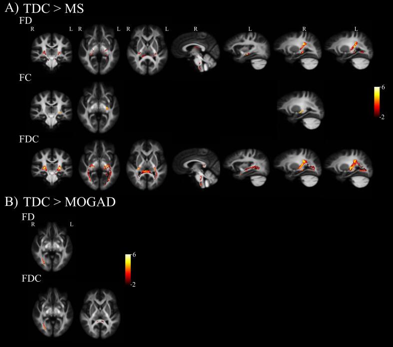
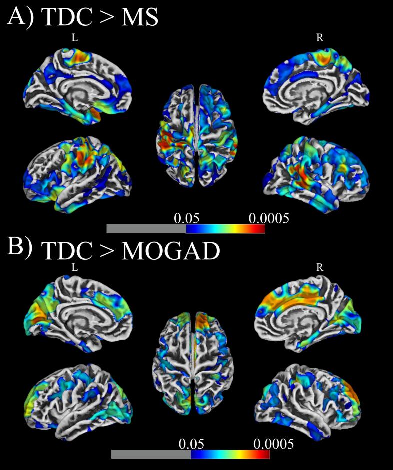
The impact of multiple sclerosis (MS) and myelin oligodendrocyte glycoprotein (MOG) - associated disorders (MOGAD) on brain structure in youth remains poorly understood. Reductions in cortical mantle thickness on structural MRI and abnormal diffusion-based white matter metrics (e.g., diffusion tensor parameters) have been well documented in MS but not in MOGAD. Characterizing structural abnormalities found in children with these disorders can help clarify the differences and similarities in their impact on neuroanatomy. Importantly, while MS and MOGAD affect the entire CNS, the visual pathway is of particular interest in both groups, as most patients have evidence for clinical or subclinical involvement of the anterior visual pathway. Thus, the visual pathway is of key interest in analyses of structural abnormalities in these disorders and may distinguish MOGAD from MS patients. In this study we collected MRI data on 18 MS patients, 14 MOGAD patients and 26 age- and sex-matched typically developing children (TDC). Full-brain group differences in fixel diffusion measures (fibre-bundle populations) and cortical thickness measures were tested using age and sex as covariates. Visual pathway analysis was performed by extracting mean diffusion measures within lesion free optic radiations, cortical thickness within the visual cortex, and retinal nerve fibre layer (RNFL) and ganglion cell layer thickness measures from optical coherence tomography (OCT). Fixel based analysis (FBA) revealed MS patients have widespread abnormal white matter within the corticospinal tract, inferior longitudinal fasciculus, and optic radiations, while within MOGAD patients, non-lesional impact on white matter was found primarily in the right optic radiation. Cortical thickness measures were reduced predominately in the temporal and parietal lobes in MS patients and in frontal, cingulate and visual cortices in MOGAD patients. Additionally, our findings of associations between reduced RNFLT and axonal density in MOGAD and TORT in MS patients in the optic radiations imply widespread axonal and myelin damage in the visual pathway, respectively. Overall, our approach of combining FBA, cortical thickness and OCT measures has helped evaluate similarities and differences in brain structure in MS and MOGAD patients in comparison to TDC.
Keywords: Cortical thickness; Diffusion; Fixel-based analysis; Multiple sclerosis; Myelin oligodendrocyte glycoprotein associated disorders; Optic neuritis.
Copyright © 2022. Published by Elsevier Inc.
Conflict of interest statement
The authors declare that they have no known competing financial interests or personal relationships that could have appeared to influence the work reported in this paper.
Figures
References
-
- Ades-Aron B., Veraart J., Kochunov P., McGuire S., Sherman P., Kellner E., Novikov D.S., Fieremans E. Evaluation of the accuracy and precision of the diffusion parameter EStImation with Gibbs and NoisE removal pipeline. Neuroimage. 2018;183:532–543. doi: 10.1016/j.neuroimage.2018.07.066. - DOI - PMC - PubMed
-
- Aliotta R., Cox J.L., Donohue K., Weinstock-Guttman B., Yeh E.A., Polak P., Dwyer M.G., Zivadinov R. Tract-based spatial statistics analysis of diffusion-tensor imaging data in pediatric- and adult-onset multiple sclerosis. Hum. Brain Mapp. 2014;35:53–60. doi: 10.1002/hbm.22148. - DOI - PMC - PubMed
Publication types
MeSH terms
LinkOut - more resources
Full Text Sources
Medical
