

Advances in nailfold capillaroscopic analysis in systemic sclerosis
- PMID: 35382238
- PMCID: PMC8892861
- DOI: 10.1177/2397198318757699
Advances in nailfold capillaroscopic analysis in systemic sclerosis
Abstract
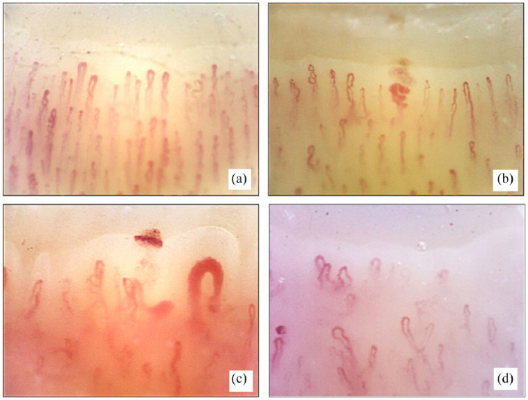
Systemic sclerosis is an autoimmune connective tissue disease characterized by early and persistent microvascular impairment which leads to functional and organic manifestations, with progressive fibrosis of the skin and internal organs. Morphological and functional assessment of the peripheral microvasculature is a must, not only for diagnosis but also for the prognosis and therapeutical follow-up of systemic sclerosis patients, as reported in recent studies. Nailfold videocapillaroscopy is the validated technique for the study of scleroderma microangiopathy as it is able to detect peripheral microvascular morphology and both classify and score the capillary abnormalities into different microangiopathy patterns ('Early', 'Active' and 'Late'). Indeed, the possibility to early diagnose and follow the microvascular changes and the safety of the technique have made nailfold videocapillaroscopy a mandatory tool for patient evaluation and included its assessment in the new systemic sclerosis classification criteria. Important links between nailfold videocapillaroscopy patterns and systemic sclerosis clinical manifestations have been described.
Keywords: Systemic sclerosis; connective tissue diseases; diagnostic tools; microangiopathy; nailfold videocapillaroscopy; scleroderma patterns.
© The Author(s) 2018.
Conflict of interest statement
Declaration of conflicting interests: The author(s) declared no potential conflicts of interest with respect to the research, authorship, and/or publication of this article.
Figures
References
-
- Varga J, Trojanowska M, Kuwana M. Pathogenesis of systemic sclerosis: recent insights of molecular and cellular mechanisms and therapeutic opportunities. J Scleroderma Relat Disord 2017; 2: 135–234, e7–e11.
-
- Cutolo M, Smith V. State of the art on nailfold capillaroscopy: a reliable diagnostic tool and putative biomarker in rheumatology? Rheumatology 2013; 52(11): 1933–1940. - PubMed
-
- Cutolo M, Pizzorni C, Tuccio M, et al. Nailfold videocapillaroscopic patterns and serum autoantibodies in systemic sclerosis. Rheumatology 2004; 43(6): 719–726. - PubMed
-
- Cutolo M, Sulli A, Pizzorni C, et al. Nailfold videocapillaroscopy assessment of microvascular damage in systemic sclerosis. J Rheumatol 2000; 27(1): 155–160. - PubMed
-
- Maricq HR, LeRoy EC. Patterns of finger capillary abnormalities in connective tissue disease by ‘wide-field’ microscopy. Arthritis Rheum 1973; 16(5): 619–628. - PubMed
Publication types
LinkOut - more resources
Full Text Sources