. 2022 Apr 5;19(1):78.
doi: 10.1186/s12974-022-02433-x.

Macrophage elastase (MMP12) critically contributes to the development of subretinal fibrosis

Affiliations

Macrophage elastase (MMP12) critically contributes to the development of subretinal fibrosis

Caijiao Yi et al. J Neuroinflammation. .

Abstract

Background: Macular subretinal fibrosis is the end-stage complication of neovascular age-related macular degeneration (nAMD). We previously developed a mouse model of two-stage laser-induced subretinal fibrosis that mimics closely the dynamic course of macular fibrosis in nAMD patients. This study was aimed to understand the molecular mechanism of subretinal fibrosis.

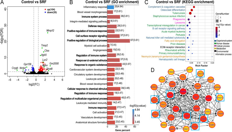
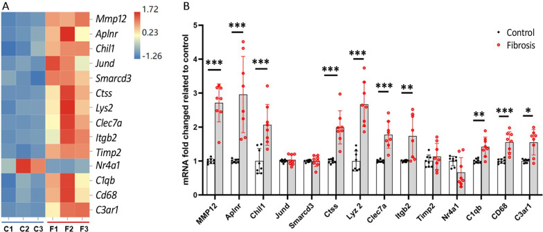
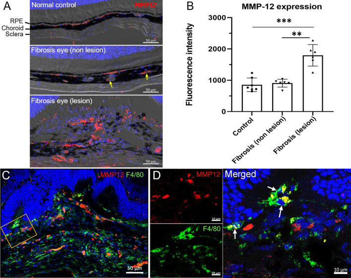
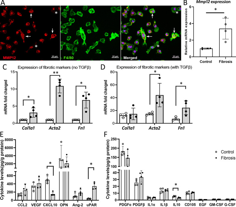
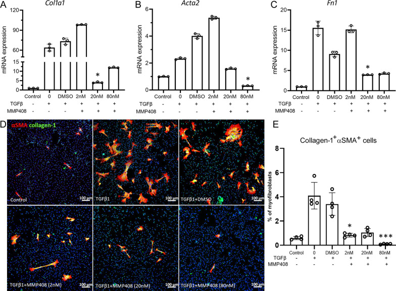
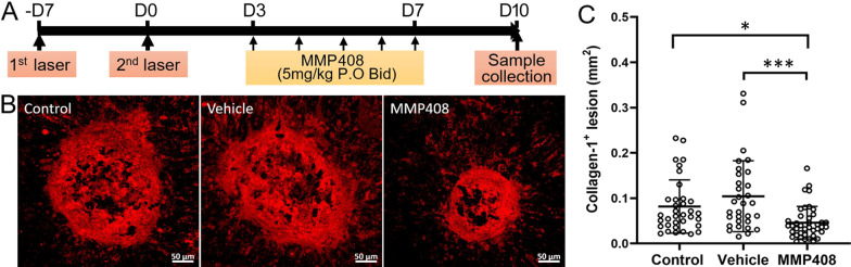
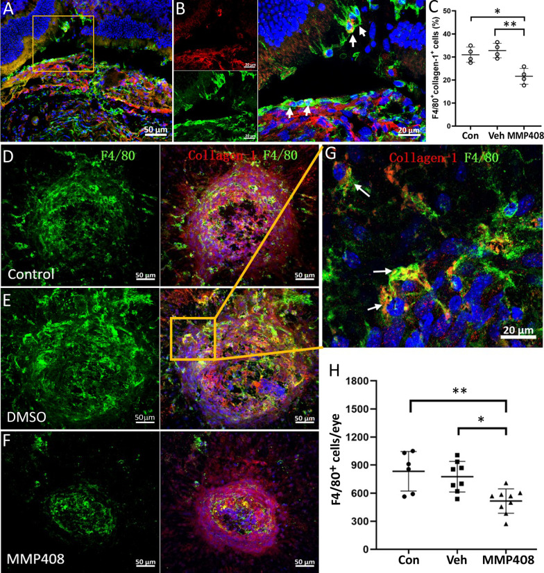
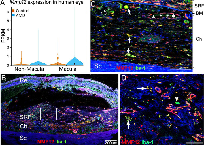
Methods: Subretinal fibrosis was induced in C57BL/6J mice using the two-stage laser-induced protocol. Twenty days later, eyes were collected and processed for RNA sequencing (RNA-seq) analysis. DESeq2 was used to determine the differentially expressed genes (DEGs). Gene Ontology (GO) and KEGG were used to analyze the enriched pathways. The expression of the selected DEGs including Mmp12 was verified by qPCR. The expression of MMP12 in subretinal fibrosis of mouse and nAMD donor eyes was examined by immunofluorescence and confocal microscopy. The expression of collagen 1, αSMA and fibronectin and cytokines in bone marrow-derived macrophages from control and subretinal fibrosis mice were examined by qPCR, immunocytochemistry and Luminex multiplex cytokine assay. The MMP12 specific inhibitor MMP408 was used to evaluate the effect of MMP12 on TGFβ-induced macrophage-to-myofibroblast transition (MMT) in vitro and its role in subretinal fibrosis in vivo.

Results: RNA-seq analysis of RPE-choroid from subretinal fibrosis eyes uncovered 139 DEGs (fold change log2(fc) ≥ 0.5, FDR < 0.05), including 104 up-regulated and 35 were down-regulated genes. The top 25 enrichment GO terms were related to inflammation, blood vessels/cardiovascular development and angiogenesis. One of the most significantly upregulated genes, Mmp12, contributed to 12 of the top 25 GO terms. Higher levels of MMP12 were detected in subretinal fibrotic lesions in nAMD patients and the mouse model, including in F4/80+ or Iba1+ macrophages. BMDMs from subretinal fibrosis mice expressed higher levels of MMP12, collagen-1, αSMA and fibronectin. MMP408 dose-dependently suppressed TGFβ-induced MMT in BMDMs. In vivo treatment with MMP408 (5 mg/kg) significantly reduced subretinal fibrosis accompanied by reduced F4/80+ macrophage infiltration.

Conclusions: MMP12 critically contributes to the development of subretinal fibrosis, partially through promoting MMT.

Keywords: Age-related macular degeneration; Inflammation; Macular fibrosis; Matrix metalloproteinase-12; RNA sequencing.


Conflict of interest statement

The authors declare that they have no competing interests.

Figures

Fig. 1
Fig. 1
RNA-seq transcriptome of RPE/choroid from control and subretinal fibrosis mice. RNA-seq was conducted in RPE/choroidal tissue from control and subretinal fibrosis mice. The differentially expressed genes (DEGs) were identified as FDR < 0.05, log2(fc) ≥ 0.5, and count > 50 in at least 2 samples. A Volcano plot showing the comparison of DEGs from control and subretinal fibrosis mice. The green dots represent the most significantly regulated genes. n = 3 mice in each group. B The top 25 enriched functions from GO analysis of DEGs. Bold font indicating the significantly enriched GO terms that Mmp12 is involved in. Numbers beside the bar indicate the number of DEG and q value (in bracket). C DEG enriched pathways from KEGG analysis. D The network image shows the top 30 highest degree genes calculated by the Cytohubba plugin of Cytoscape. The eight genes (blue boxes) were selected for further qRT-PCR verification
Fig. 2
Fig. 2
Verification of selected DEGs in RPE-choroid from control and subretinal fibrosis mice via qRT-PCR. A Heat map showing the expression levels of selected DEGs in RPE-choroid from control and subretinal fibrosis eyes in RNA-seq analysis. C = control, F = fibrosis. Data showing as FPKM, n = 3 mice. B qRT-PCR analysis of selected 14 notable DEGs expressions in RPE-choroid from control and subretinal fibrosis eyes. Mean ± SD, n = 8 mice, *p < 0.05, **p < 0.01, ***p < 0.001. Mann–Whitney test
Fig. 3
Fig. 3
MMP12 expression in mouse eyes with/without subretinal fibrosis. A Representative confocal images showing MMP12 (red) expression in the choroid/sclera of eyes from normal and subretinal fibrosis (5 days after the second laser) mice. Yellow arrows indicate MMP12+ cells in choroidal blood vessels. B Dot/bar figure showing fluorescence intensity of MMP12 in the choroid of normal eyes, non-lesion site and fibrotic lesions of fibrosis eyes. Mean ± SD, n = 6–7 eyes. **p < 0.01, ***p < 0.001, Kruskal–Wallis with Dunn’s multiple comparisons test. C Confocal image from a subretinal fibrosis eye showing MMP12 (red) and F4/80 (green). A high magnification view of the yellow rectangle area, including each channel, is shown in (D). Arrows indicate F4/80+MMP12+ cells
Fig. 4
Fig. 4
The expression of MMP12 and pro-fibrotic markers in bone marrow-derived macrophages (BMDMs) from control and subretinal fibrosis mice. BMDMs were cultured with/without 10 ng/ml recombinant TGF-β1 for 96 h, the cells were collected for immunocytochemistry or qRT-PCR, and supernatants were used for Luminex multiplex cytokine assay. A BMDMs from normal mice were stained for MMP12 (red) and F4/80 (green), imaged by confocal microscopy. Arrows indicating MMP12high in F4/80+ cells. Asterisks indicating MMP12low in F4/80+ cells. B qRT-PCR analysis of Mmp12 expression in BMDMs from control and subretinal fibrosis mice (20 days after the second laser). C, D qRT-PCR analysis of fibrotic marker genes (Col1a1, Acta2, Fn1) in untreated BMDMs (C) and TGFβ1 treated BMDMs (D) from control and subretinal fibrosis mice. E, F Luminex bead-based assay of the production of pro-fibrotic cytokines by BMDMs from normal and subretinal fibrosis mice. Mean ± SD, n = 4 mice, *p < 0.05, **p < 0.01, Mann–Whitney test
Fig. 5
Fig. 5
The effect of MMP408 on macrophage-to-myofibroblast transition (MMT). BMDMs from normal mice were treated with TGFβ1 (10 ng/ml) with or without different concentrations of MMP408 (2 nM/ml, 20 nM/ml and 80 nM/ml) or DMSO for 96 h. Cells were collected for qRT-PCR or immunocytochemistry. AC The mRNA expression levels of Col1a1 (A), Acta2 (B) and Fn1 (C). Mean ± SD, n = 3, *p < 0.05 compared with DMSO group. Kruskal–Wallis with Dunn’s multiple comparisons test. D Representative confocal images of BMDMs stained for α-SMA (red) and collagen-1(green) from different treatment groups. Scale bar = 100 µm. E Dot/bar figure showing the percentages of collagen-1+ α-SMA + cells in different groups. Mean ± SD, n = 4, **p < 0.01, ***p < 0.001. Kruskal–Wallis with Dunn’s multiple comparisons test
Fig. 6
Fig. 6
The effect of MMP408 on subretinal fibrosis. A Schematic view of the study design. Mice were treated with MMP408 (5 mg/kg twice daily, gavage) or vehicle three days after the second laser for 5 days. Eyes were collected on day 10 for immunohistochemistry. Subretinal fibrosis mice that received no treatment were considered as a treatment-naïve control group. B Representative confocal images of RPE/choroid flatmounts stained for collagen-1 from control non-treatment group, vehicle group and MMP408 group. Scale bar = 50 µm. C Quantitative analysis of collagen-1+ lesion area in different groups. Mean ± SD, n = 31–40 lesions per group from 8 to 10 eyes, *p < 0.05, **p < 0.01. One-way ANOVA followed by Tukey’s multiple comparison’s test
Fig. 7
Fig. 7
The effect of MMP408 on macrophage accumulation in subretinal fibrosis. A Cryosections from subretinal fibrosis eyes were stained for collagen-1 (red) and F4/80 (green) and imaged by confocal microscopy. B Magnified area of box in (A) showing F4/80+MMP12+ cells (arrows) inside and around the subretinal fibrotic lesion. C The percentage of F4/80+Collagen-1+ cells among all F4/80+ macrophages. DH Representative confocal images of RPE/choroid flatmounts stained for collagen-1 (red) and F4/80 (green) from control non-treatment group (D), vehicle (DMSO) group (E) and MMP408 group (F). Scale bar = 50 µm. G Magnified area of box in (E) showing F4/80+collagen-1+ cells (arrows) in the fibrotic lesion. H Quantitative analysis of the total F4/80+ cells in RPE-choroid flatmount in different groups. Mean ± SD, n = 6–9 eyes from 4–5 mice, *p < 0.05, **p < 0.01. Kruskal–Wallis with Dunn’s multiple comparisons test
Fig. 8
Fig. 8
The expression of MMP-12 in human AMD and macular fibrosis. A The mRNA expression levels of Mmp12 in non-macular and macular RPE-choroid from non-AMD controls and AMD (Bulk RNA-Seq data from NCBI GEO database, ID: GSE135092). Mean ± SD, (AMD macula, n = 13; AMD non-macula, n = 10; non-AMD macula, n = 33; non-AMD non-macula, n = 36). B A confocal image from human nAMD with macular fibrosis showing MMP12 (red) and Iba-1 (green) in the choroid and subretinal lesion. C High magnification image showing Iba-1+MMP12+ cells (arrows), Iba-1+MMP12 cells (open arrow) in the choroid and MMP12+ cells in choroidal blood vessels (asterisks). D Magnified area of box in (B) showing Iba-1+MMP12+cells (arrows), Iba-1+MMP12cells (arrowhead) in the macular fibrotic lesion. Re = retina, SRF = subretinal fibrotic lesion, Ch = choroid, BM = Bruch’s membrane. Scale bars in C and D = 50 µm

References

    1. Mitchell P, Liew G, Gopinath B, Wong TY. Age-related macular degeneration. Lancet. 2018;392(10153):1147–1159. doi: 10.1016/S0140-6736(18)31550-2. - DOI - PubMed
    1. Lim LS, Mitchell P, Seddon JM, Holz FG, Wong TY. Age-related macular degeneration. Lancet. 2012;379(9827):1728–1738. doi: 10.1016/S0140-6736(12)60282-7. - DOI - PubMed
    1. de Juan Jr. E, Machemer R. Vitreous surgery for hemorrhagic and fibrous complications of age-related macular degeneration. Am J Ophthalmol. 1988;105(1):25–29. doi: 10.1016/0002-9394(88)90116-X. - DOI - PubMed
    1. Bloch SB, Lund-Andersen H, Sander B, Larsen M. Subfoveal fibrosis in eyes with neovascular age-related macular degeneration treated with intravitreal ranibizumab. Am J Ophthalmol. 2013;156(1):116–24.e1. doi: 10.1016/j.ajo.2013.02.012. - DOI - PubMed
    1. Rofagha S, Bhisitkul RB, Boyer DS, Sadda SR, Zhang K, Group S-US. Seven-year outcomes in ranibizumab-treated patients in ANCHOR, MARINA, and HORIZON: a multicenter cohort study (SEVEN-UP) Ophthalmology. 2013;120(11):2292–2299. doi: 10.1016/j.ophtha.2013.03.046. - DOI - PubMed

Substances