. 2022 Apr 5;5(1):318.
doi: 10.1038/s42003-022-03265-4.

The medial occipital longitudinal tract supports early stage encoding of visuospatial information

Affiliations

The medial occipital longitudinal tract supports early stage encoding of visuospatial information

Ahmad Beyh et al. Commun Biol. .

Abstract

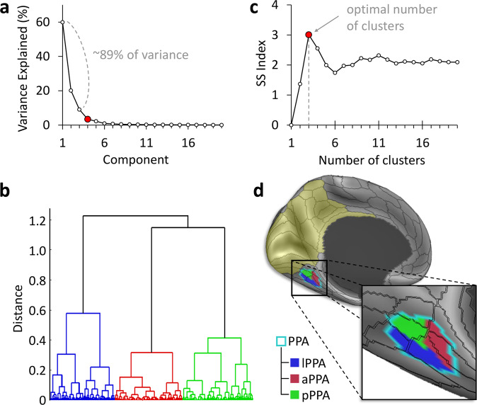
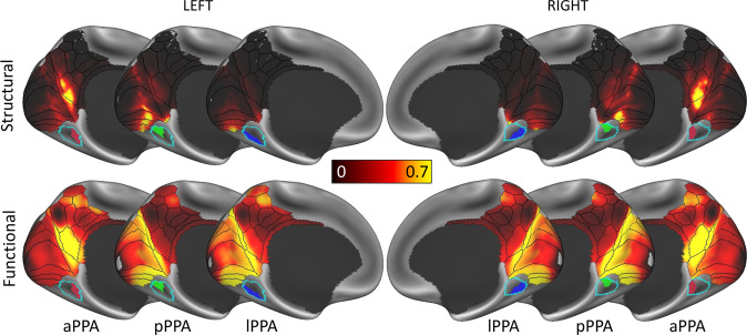
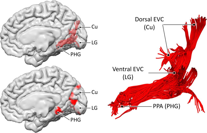
Visuospatial learning depends on the parahippocampal place area (PPA), a functionally heterogenous area which current visuospatial processing models place downstream from parietal cortex and only from area V4 of early visual cortex (EVC). However, evidence for anatomical connections between the PPA and other EVC areas is inconsistent, and these connections are not discussed in current models. Through a data-driven analysis based on diffusion MRI tractography, we present evidence that the PPA sits at the confluence of two white matter systems. The first conveys information from the retrosplenial complex to the anterior PPA and runs within the cingulum bundle. The second system connects all peripheral EVC areas to the posterior PPA and corresponds to the medial occipital longitudinal tract (MOLT), a white matter pathway that is distinct from the cingulum and that we describe here in detail. Based on further functional connectivity analysis and meta-analytic data, we propose that the MOLT supports early stage encoding of visuospatial information by allowing direct reciprocal exchange between the PPA and EVC. Our findings may improve symptom interpretation in stroke and tumour patients with damage to the medial occipito-temporal region and call for revisiting current visuospatial processing models.


Conflict of interest statement

The authors declare no competing interests.

Figures

Fig. 1
Fig. 1. Density of tractography projections from medial occipital and parietal regions to the medial temporal lobe.
The upper panel shows the three large ROIs used to select streamlines between the medial temporal lobe (MTL) on one end, and early visual cortex (EVC), medial parietal cortex (MPC), and retrosplenial complex (RSC) on the other end. The parahippocampal place area (PPA), which is located in the posterior MTL, is also delineated with a green contour. The plots in the lower panel represent the mean density of streamline terminations in the left and right MTL according to its longitudinal y-axis. Projections from both EVC (red lines) or the RSC/MPC (blue dashed lines) are displayed as the mean and standard deviation (shaded areas) across 200 subjects. While the EVC connections show a strong preference for the posterior MTL, especially the parahippocampal place area (PPA), the RSC/MPC connections show posterior and anterior peaks. Within the PPA, the anterior portion receives high density projections from both ROIs, while the posterior portion receives projection preferentially from EVC. The PPA’s most posterior and anterior coordinates are indicated with the vertical green lines within the plots. Coordinates correspond to the MNI template. The main ROIs are based on the MMP atlas shown as black contours, and the PPA is based on.
Fig. 2
Fig. 2. Clustering analysis of the PPA reveals multiple anatomical subunits.
Data-driven clustering of the parahippocampal place area (PPA) based on average structural connectivity in 200 subjects. a Principal component analysis (PCA) based on the PPA’s connectivity to a region encompassing early visual cortex (EVC), the retrosplenial complex (RSC), and medial parietal cortex (MPC) (yellow tint) resulted in three principal components. b Hierarchical agglomerative clustering grouped PPA surface vertices with similar PCA coefficients. c The highest separation vs. spread (SS) index objectively determined the optimal number of clusters. d The resulting anterior, posterior, and lateral PPA clusters are shown on the inflated brain surface.
Fig. 3
Fig. 3. PPA clusters share similar structural and functional connectivity profiles.
Structural and functional connectivity of the three parahippocampal place area (PPA) clusters. The anterior cluster (aPPA) is preferentially connected to retrosplenial complex and corresponds to the parieto-medial-temporal branch of the dorsal visual stream. The posterior cluster (pPPA) is preferentially connected to the anterior medial occipital lobe, i.e., peripheral representations within early visual cortex (EVC). The lateral cluster (lPPA) is more ambiguous but is preferentially connected to EVC. This is discussed in detail in Supplementary Note 1 (also see Supplementary Figs. 1–3).
Fig. 4
Fig. 4. Encoding and retrieval in the PPA.
These plots are based on meta-analytic maps, obtained from NeuroSynth, of locations associated with the terms ‘encoding’ and ‘retrieval’. The maximum z-statistic is plotted along the posterior-anterior axis of the parahippocampal place area (PPA).
Fig. 5
Fig. 5. The medial occipital longitudinal tract (MOLT).
Tractography reconstruction of the medial occipital longitudinal tract (MOLT) in an example HCP participant. The MOLT is an occipito-temporal white matter pathway that stems from the anterior cuneus (Cu) and lingual gyrus (LG) and terminates in the posterior parahippocampal gyrus (PHG). In the medial occipital lobe, it projects onto peripheral visual field representations within early visual cortex (EVC), while its temporal lobe terminations overlap the posterior parahippocampal place area (PPA). Further visual representations of the MOLT are available in Supplementary Fig. 5, and descriptive statistics are presented in Supplementary Table 1.
Fig. 6
Fig. 6. Macrostructural and microstructural assessment of the MOLT.
These box charts summarise the comparisons between the MOLT’s components within each hemisphere and across hemispheres in 200 subjects. a In both hemispheres, the lingual gyrus (LG) component has a larger volume, distributes to a wider occipital surface, and has a lower fibre density (as indicated by its lower HMOA) compared to the cuneus (Cu) component (values were derived using Eq. 1). b Both the Cu and LG components of the MOLT tend to exhibit a slight lateralisation towards the right hemisphere (values derived using Eq. 2). Within the box charts, the notch and shaded bar represent the median value, the edges of the box correspond to the upper and lower quartiles, and the whiskers extend to the maximum and minimum non-outlier values. Asterisks indicate that the mean of the corresponding distribution is significantly different from zero after Bonferroni correction. Detailed descriptions of these metrics are available in the Methods section. Full descriptive statistics of these metrics and comparisons are reported in Supplementary Tables 2 and 3.

References

    1. O’Keefe J, Dostrovsky J. The hippocampus as a spatial map: Preliminary evidence from unit activity in the freely-moving rat. Brain Res. 1971;34:171–175. - PubMed
    1. Burgess N, Maguire EA, O’Keefe J. The human hippocampus and spatial and episodic memory. Neuron. 2002;35:625–641. - PubMed
    1. Maguire EA, et al. Navigation-related structural change in the hippocampi of taxi drivers. Proc. Natl Acad. Sci. USA. 2000;97:4398–4403. - PMC - PubMed
    1. Maguire EA, Frith CD, Burgess N, Donnett JG, O’Keefe J. Knowing where things are: Parahippocampal involvement in encoding object locations in virtual large-scale space. J. Cogn. Neurosci. 1998;10:61–76. - PubMed
    1. Owen AM, Milner B, Petrides M, Evans AC. A specific role for the right parahippocampal gyrus in the retrieval of object-location: A positron emission tomography study. J. Cogn. Neurosci. 1996;8:588–602. - PubMed

Publication types