

The lymphatic system throughout history: From hieroglyphic translations to state of the art radiological techniques
- PMID: 35383381
- PMCID: PMC9542037
- DOI: 10.1002/ca.23867
The lymphatic system throughout history: From hieroglyphic translations to state of the art radiological techniques
Abstract
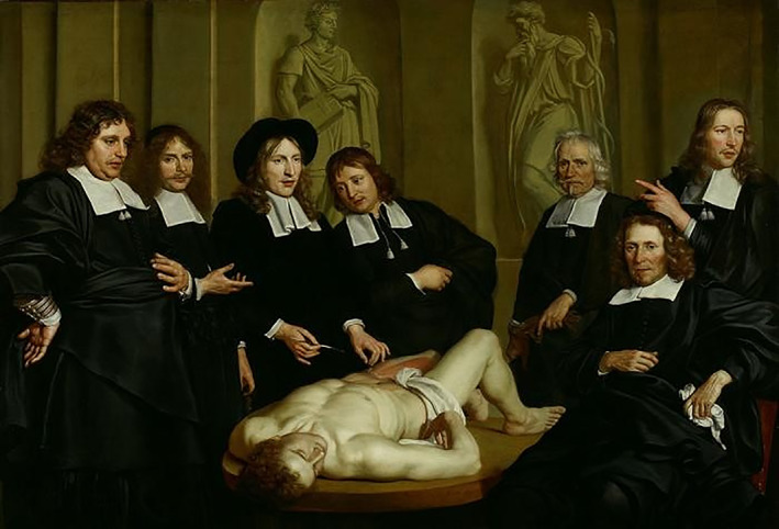
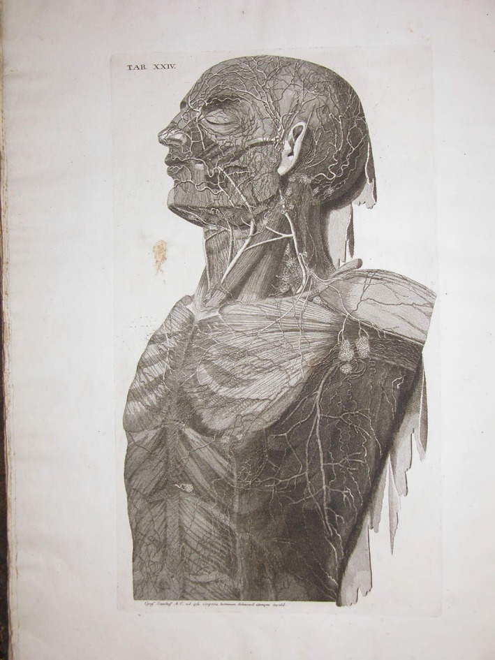
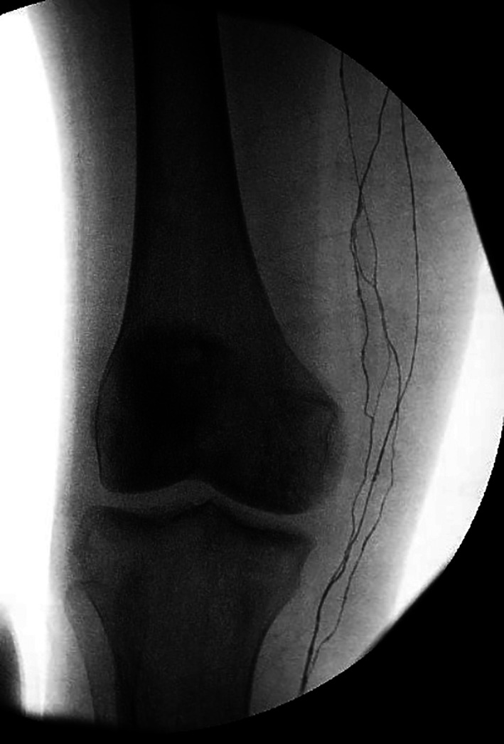
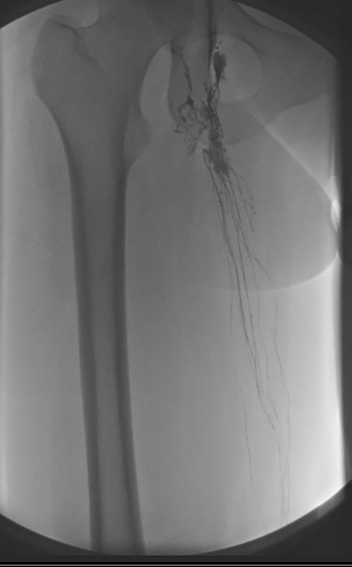
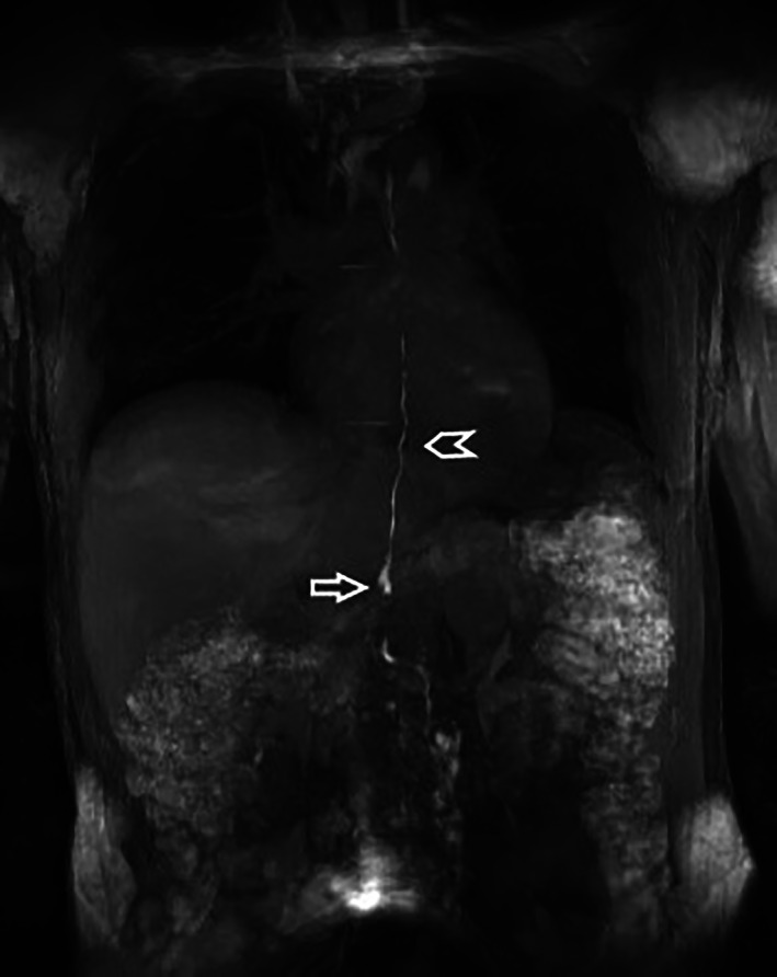
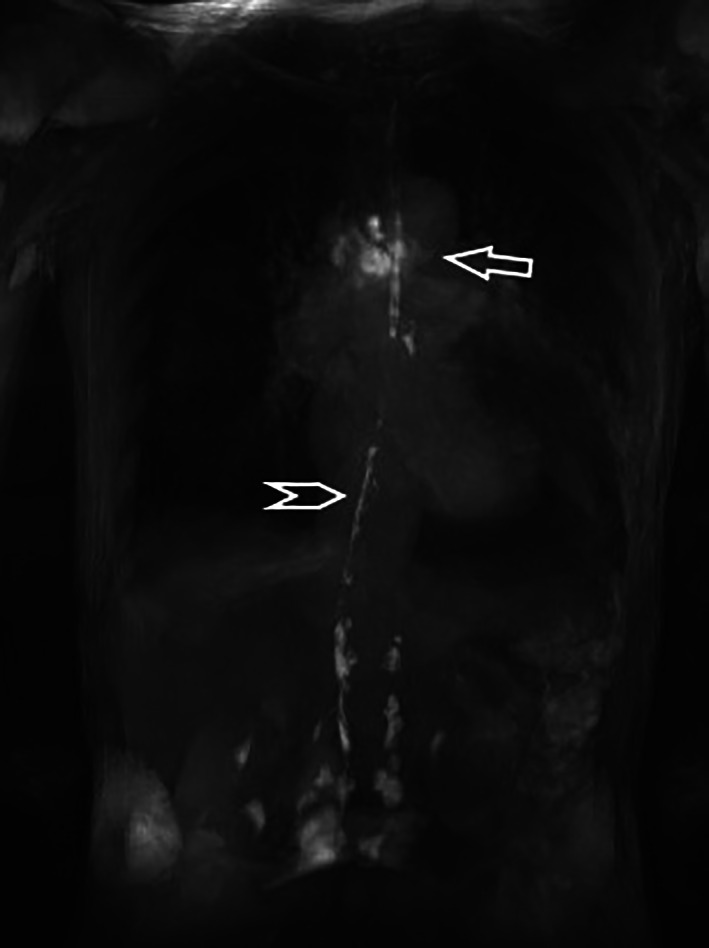
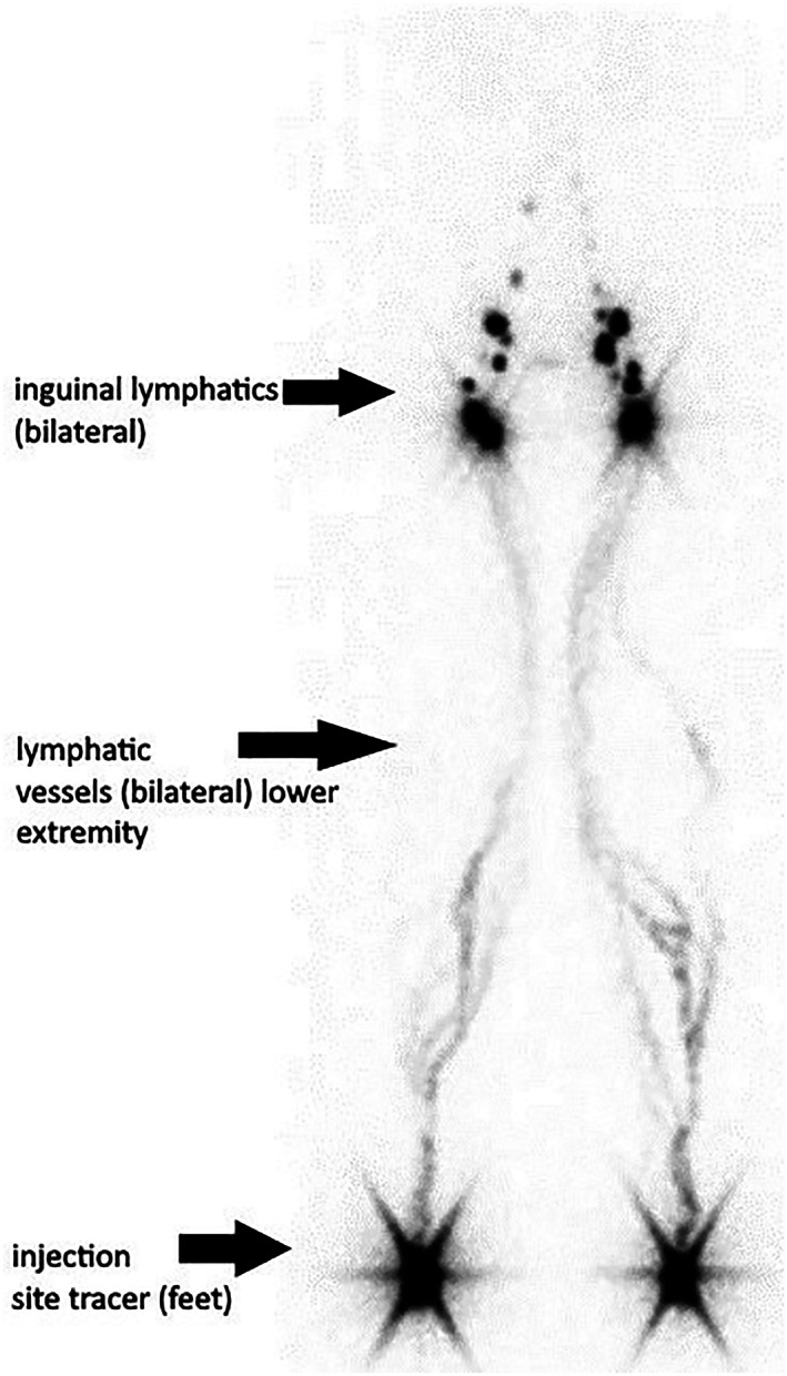
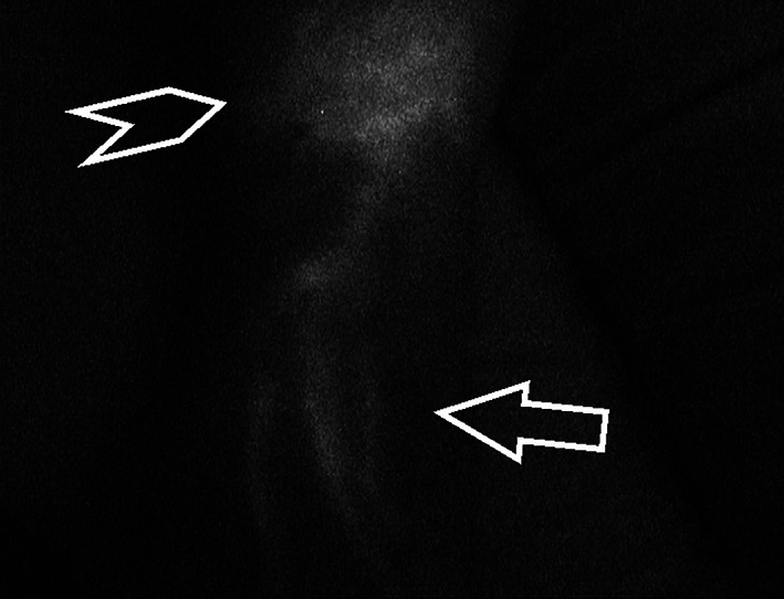
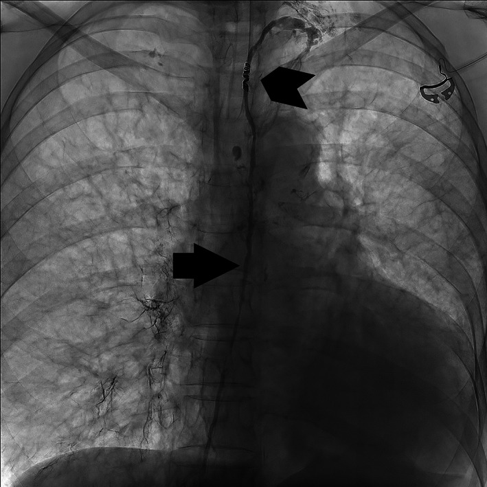
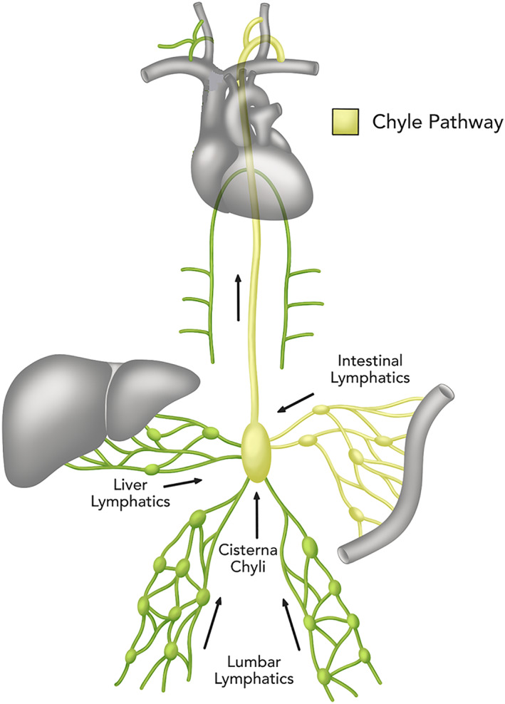
A comprehensive lymphatic system is indispensable for a well-functioning body; it is integral to the immune system and is also interrelated with the digestive system and fluid homeostasis. The main difficulty in examining the lymphatic system is its fine-meshed structure. This remains a challenge, leaving patients with uninterpreted symptoms and a dearth of potential therapies. We review the history of the lymphatic system up to the present with the aim of improving current knowledge. Several findings described throughout history have made fundamental contributions to elucidating the lymphatic system. The first contributions were made by the ancient Egyptians and the ancient Greeks. Vesalius obtained new insights by dissecting corpses. Thereafter, Ruysch (1638-1731) gained an understanding of lymphatic flow. In 1784, Mascagni published his illustration of the whole lymphatic network. The introduction of radiological lymphography revolutionized knowledge of the lymphatic system. Pedal lymphangiography was first described by Monteiro (1931) and Kinmonth (1952). Lymphoscintigraphy (nuclear medicine), magnetic resonance imaging, and near-infrared fluorescence lymphography further improved visualization of the lymphatic system. The innovative dynamic contrast-enhanced magnetic resonance lymphangiography (DCMRL) transformed understanding of the central lymphatic system, enabling central lymphatic flow disorders in patients to be diagnosed and even allowing for therapeutic planning. From the perspective of the history of lymph visualization, DCMRL has ample potential for identifying specific causes of debilitating symptoms in patients with central lymphatic system abnormalities and even allows for therapeutic planning.
Keywords: dynamic MR lymphangiography; lymphatic system; lymphography; therapeutic embolization.
© 2022 The Authors. Clinical Anatomy published by Wiley Periodicals LLC on behalf of American Association for Clinical Anatomists and the British Association for Clinical Anatomists.
Figures
References
-
- Absinta, M. , Ha, S. K. , Nair, G. , Sati, P. , Luciano, N. J. , Palisoc, M. , Louveau, A. , Zaghloul, K. A. , Pittaluga, S. , Kipnis, J. , & Reich, D. S. (2017). Human and nonhuman primate meninges harbor lymphatic vessels that can be visualized noninvasively by MRI. eLife, 6, e29738. - PMC - PubMed
-
- Ambrose, C. T. (2006). Immunology's first priority dispute—An account of the 17th‐century Rudbeck‐Bartholin feud. Cellular Immunology, 242(1), 1–8. - PubMed
-
- Ambrose, C. T. (2007). Rudbeck's complaint: A 17th‐century Latin letter relating to basic immunology. Scandinavian Journal of Immunology, 66, 486–493. - PubMed
-
- Andrews, R. K. , & Gardiner, E. E. (2014). Lymphomania. Blood, 123(20), 3057–3058. - PubMed
-
- Androutsos, G. , Marketos, S. , & Marketos, S. (1994). In memoriam Miltiades Papamiltiades (1910–1987): Eminent Greek anatomist and ambassador of Franco‐Hellenic friendship. Surgical and Radiologic Anatomy, 16(4), 445–448. - PubMed
Publication types
MeSH terms
Substances
LinkOut - more resources
Full Text Sources
Medical
