. 2022 Jul;42(7):1605-1617.
doi: 10.1111/liv.15270. Epub 2022 Apr 12.

An MMP-degraded and cross-linked fragment of type III collagen as a non-invasive biomarker of hepatic fibrosis resolution

Affiliations

An MMP-degraded and cross-linked fragment of type III collagen as a non-invasive biomarker of hepatic fibrosis resolution

Martin Pehrsson et al. Liver Int. 2022 Jul.

Abstract

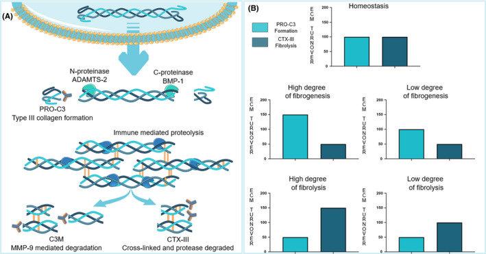
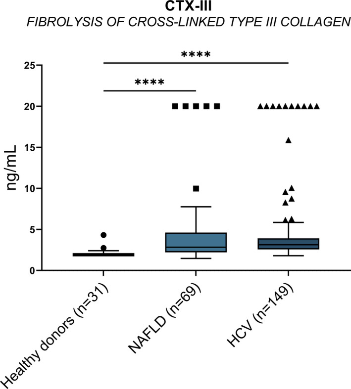
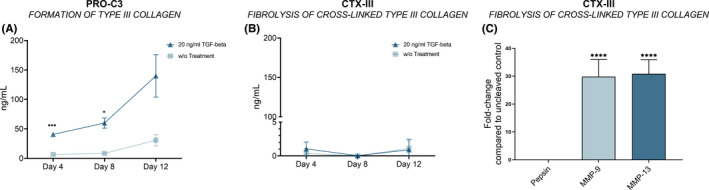
Background and aims: Liver fibrosis results from a prolonged wound healing response to continued injury with excessive production of extracellular proteins. In patients with chronic liver disease, the monitoring of liver fibrosis dynamics is of high interest. Whilst markers of fibrogenesis exist, markers of hepatic fibrosis resolution remain an unmet clinical need. Thus, we sought to develop an assay quantifying a circulating proteolytic fragment of cross-linked type III collagen as a biomarker of fibrolysis, testing its utility in two clinical cohorts of liver fibrosis of distinct aetiology and regressing endotype METHODS: We used a monoclonal antibody targeting the C-telopeptide of type III collagen following C-proteinase cleavage to develop and validate a neo-epitope-specific enzyme-linked immunosorbent assay (CTX-III). A potential fibrosis resolution marker, CTX-III, was measured in two clinical cohorts of patients with obesity-associated non-alcoholic fatty liver disease undergoing bariatric surgery or hepatitis C virus infection from a clinical trial study evaluating the anti-fibrotic effect of farglitazar.

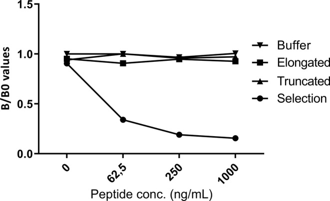
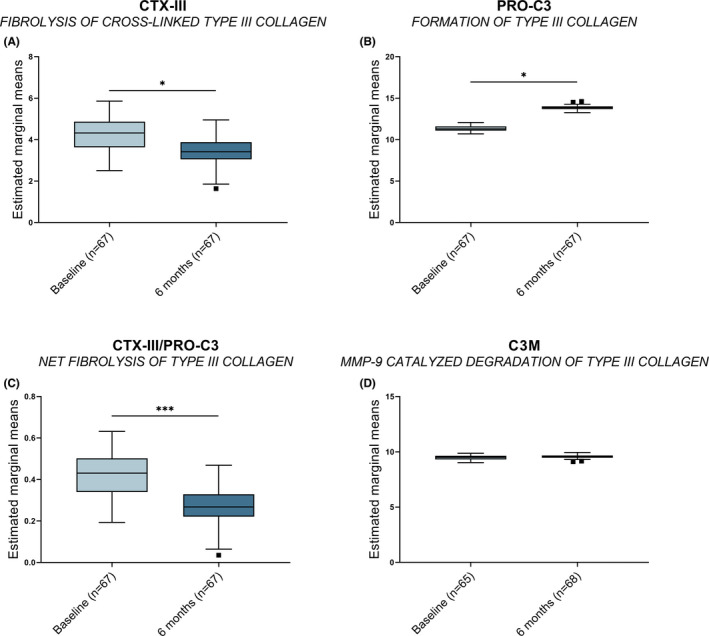
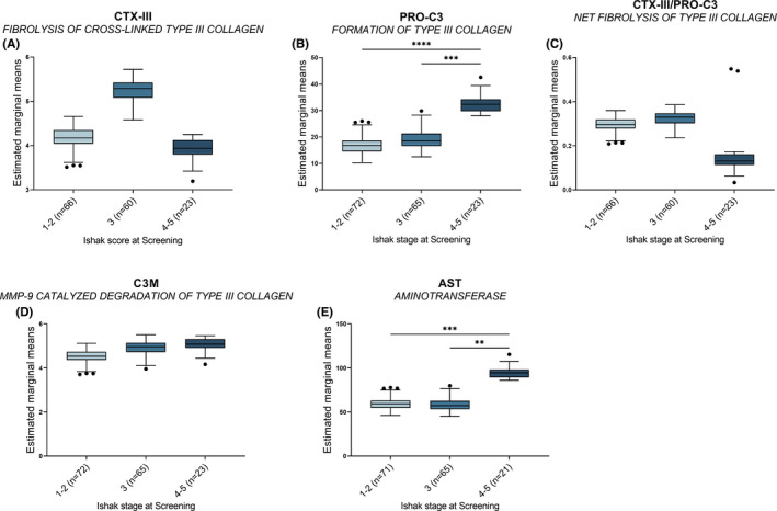
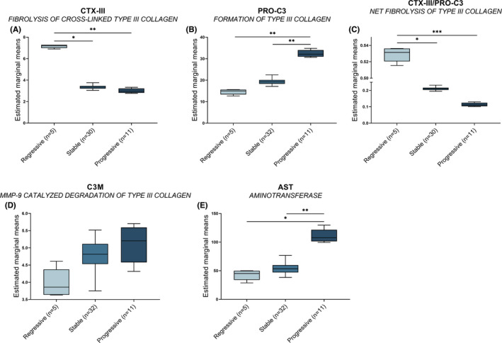
Results: CTX-III was robust and specific for the targeted neo-epitope with good reproducibility in EDTA plasma. We assessed type III collagen remodelling using a panel of biomarkers, including a type III collagen formation marker (PRO-C3), degradation (C3M), and CTX-III (fibrolysis). Net fibrolysis was increased in patients with non-alcoholic fatty liver disease following bariatric surgery (p < .001). Moreover, net fibrolysis identified spontaneous fibrotic regressors from stable and progressors (p < .05 and p < .001) among hepatitis C virus infection patients.

Conclusion: Circulating CTX-III as a marker of fibrolysis indicates the biomarker's beneficial use in assessing hepatic fibrosis resolution.

Keywords: collagen cross-linking; fibrosis resolution; hepatic fibrosis; non-invasive biomarkers.


Conflict of interest statement

Martin Pehrsson, Shu Sun, Ida Falk Villesen, Mette Juul Nielsen and Joachim Høg Mortensen are employed at Nordic Bioscience A/S, a company involved in the discovery and development of biochemical biomarkers. Tina Manon‐Jensen, Anne‐Christine Bay‐Jensen, Diana Julie Leeming and Morten Asser Karsdal are employed at and own stocks in Nordic Bioscience A/S. Helena Castañé, Jorge Joven, Keyur Patel and Zachary Goodman have no competing interests.

Figures

FIGURE 1
FIGURE 1
The balance between fibrogenesis and fibrolysis. (A) The procollagen is released to the extracellular environment following intracellular collagen α‐chain trimerisation, with subsequent proteolytic removal of the pro‐peptides by N‐ and C‐proteinases. The collagen monomers are incorporated into the collagen fibrils within the extracellular matrix by forming intra‐ and inter‐molecular enzymatic cross‐links. Immune‐mediated proteolysis of either newly synthesised collagen monomers or the enzymatically cross‐linked collagen multimers results in specific collagen fragments release. Dependent on the protease, formation biomarkers (PRO‐C3) or immune‐mediated proteolysis are generated (C3M or CTX‐III). (B) Measuring type III collagen formation and degradation markers, various endotypes of either fibrogenesis or fibrolysis may be identified
FIGURE 2
FIGURE 2
Monoclonal antibody specificity. Evaluation of NBH‐242 monoclonal antibody specificity in an indirect competitive ELISA. Reactivity towards the selection‐, elongated, truncated, immunogenic‐ peptide, and buffer was tested
FIGURE 3
FIGURE 3
The biological relevance of the CTX‐III biomarker. Plasma CTX‐III was plotted for healthy donors and patients with NAFLD (Cohort 1) or HCV (Cohort 2). The data is presented as a Tukey plot calculating the statistical difference between the healthy donors and NAFLD or HepC by Kruskal‐Wallis using Dunn's to correct for multiple comparisons. The statistical significance is depicted as ****p < .0001
FIGURE 4
FIGURE 4
Bariatric surgery modifies circulating biomarkers. The estimated marginal means of CTX‐III (A), PRO‐C3 (B), net fibrolysis (C), and C3M (D) at baseline and the 6‐month follow‐up in patients with NAFLD. The estimated marginal means were obtained by ANCOVA using patient BMI at baseline as a covariate. Data is presented as a Tukey plot calculating the statistical difference between baseline and the 6‐month follow‐up by Mann–Whitney. The statistical significance is depicted as *p < .05, and ***p < .001
FIGURE 5
FIGURE 5
Fibrosis severity is associated with fibrogenesis. The estimated marginal means of CTX‐III (A), PRO‐C3 (B), net fibrolysis (C), and C3M (D) was plotted for the Ishak stage at the initial histological screening of patients with HCV‐associated liver fibrosis. The estimated marginal means were obtained by ANCOVA using patient BMI at screening as a covariate. Data is presented as a Tukey plot calculating the statistical difference between the stages by Kruskal‐Wallis using Dunn's test to correct for multiple comparisons. The statistical significance is depicted as: **p < .01, ***p < .001, and ****p < .0001
FIGURE 6
FIGURE 6
Fibrogenetic and fibrolytic biomarkers differentiate endotypes. The estimated marginal means of CTX‐III (A), PRO‐C3 (B), net fibrolysis (C), and C3M (D) was plotted for the three spontaneous endotypes: regressive, stable, and progressive. The estimated marginal means were obtained by ANCOVA using patient BMI at screening as a covariate. Data is presented as a Tukey plot calculating the statistical difference between the stages by Kruskal‐Wallis using Dunn's test to correct for multiple comparisons. The statistical significance is depicted as *p < .05, **p < .01, and ***p < .001
FIGURE 7
FIGURE 7
In vitro formation and proteolytic degradation of type III collagen. (A) Depicts the measured PRO‐C3 levels at day 4, 8, and 12 in the supernatant of the SIAJ cell model without or with 20 ng/ml of TGF‐β1 stimulation. (B) The CTX‐III levels measured in the SIAJ supernatants are plotted for days 4, 8, and 12 for HSCs without or with 20 ng/ml of TGF‐β1 stimulation. (C) The fold‐change of CTX‐III levels was calculated from the uncleaved media control measured in the supernatants following pepsin, MMP‐9, or MMP‐13 cleavage of the HSC matrix at day 12. The statistical significance was calculated by two‐way‐ANOVA applying Dunnet's correcting for multiple comparisons (A and B), or one‐way ANOVA applying Sidak correcting for multiple comparisons (C) and depicted as *p < .05, **p < .01, ***p < .001, and ****p < .0001

Similar articles

Cited by

References

    1. Asrani SK, Devarbhavi H, Eaton J, Kamath PS. Burden of liver diseases in the world. J Hepatol. 2019;70:151‐171. doi:10.1016/j.jhep.2018.09.014 - DOI - PubMed
    1. Lackner C, Spindelboeck W, Haybaeck J, et al. Histological parameters and alcohol abstinence determine long‐term prognosis in patients with alcoholic liver disease. J Hepatol. 2017;66:610‐618. doi:10.1016/j.jhep.2016.11.011 - DOI - PubMed
    1. Nallagangula KS, Nagaraj SK, Venkataswamy L, Chandrappa M. Liver fibrosis: a compilation on the biomarkers status and their significance during disease progression. Futur Sci OA. 2018;4:FSO250. doi:10.4155/fsoa-2017-0083 - DOI - PMC - PubMed
    1. Karsdal MA, Daniels SJ, Holm Nielsen S, et al. Collagen biology and non‐invasive biomarkers of liver fibrosis. Liver Int. 2020;40:1‐15. doi:10.1111/liv.14390 - DOI - PubMed
    1. Standish RA, Cholongitas E, Dhillon A, Burroughs AK, Dhillon AP. An appraisal of the histopathological assessment of liver fibrosis. Gut. 2006;55:569‐578. doi:10.1136/gut.2005.084475 - DOI - PMC - PubMed

Publication types