. 2022 Dec;24(4):437-451.
doi: 10.1007/s12017-022-08709-y. Epub 2022 Apr 6.

Interleukin-1 Mediates Ischemic Brain Injury via Induction of IL-17A in γδ T Cells and CXCL1 in Astrocytes

Affiliations

Interleukin-1 Mediates Ischemic Brain Injury via Induction of IL-17A in γδ T Cells and CXCL1 in Astrocytes

Ines Sophie Schädlich et al. Neuromolecular Med. 2022 Dec.

Abstract

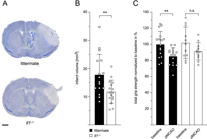
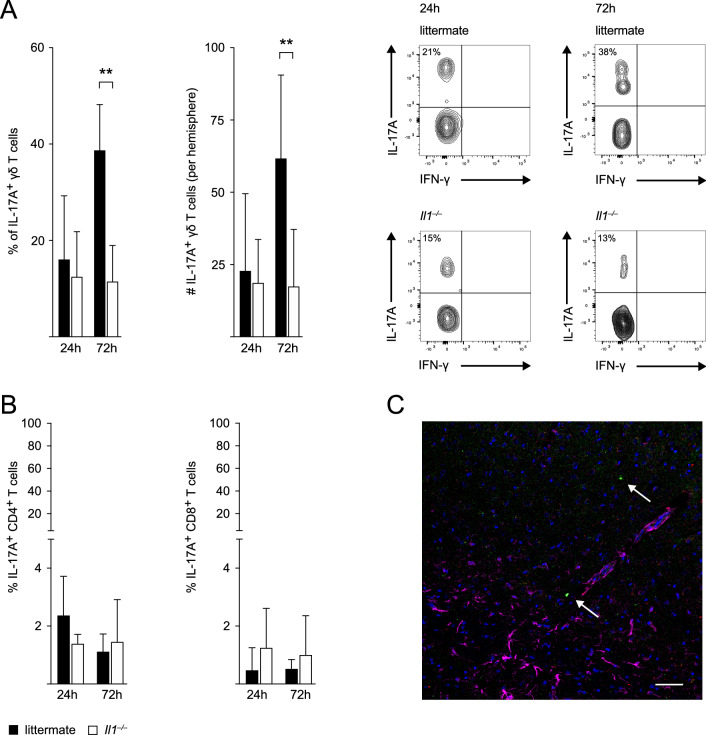
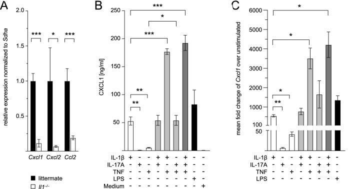
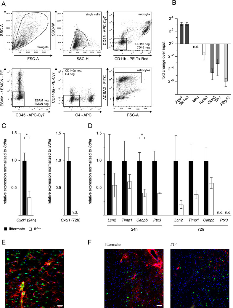
As a prototypical proinflammatory cytokine, interleukin-1 (IL-1) exacerbates the early post-stroke inflammation, whereas its neutralization is protective. To further investigate the underlying cell-type-specific IL-1 effects, we subjected IL-1 (α/β) knockout (Il1-/-) and wildtype (WT) littermate mice to permanent middle cerebral artery occlusion (pMCAO) and assessed immune cell infiltration and cytokine production in the ischemic hemisphere by flow cytometry 24 h and 72 h after stroke. Il1-/- mice showed smaller infarcts and reduced neutrophil infiltration into the ischemic brain. We identified γδ T cells and astrocytes as target cells of IL-1 signaling-mediated neutrophil recruitment. First, IL-1-induced IL-17A production in γδ T cells in vivo, and IL-17A enhanced the expression of the main neutrophil attracting chemokine CXCL1 by astrocytes in the presence of tumor necrosis factor (TNF) in vitro. Second, IL-1 itself was a potent activator of astrocytic CXCL1 production in vitro. By employing a novel FACS sorting strategy for the acute isolation of astrocytes from ischemic brains, we confirmed that IL-1 is pivotal for Cxcl1 upregulation in astrocytes in vivo. Our results underscore the pleiotropic effects of IL-1 on immune and non-immune cells within the CNS to mount and amplify the post-stroke inflammatory response.

Keywords: Astrocytes; CXCL1; Inflammation; Interleukin-1; Interleukin-17A; Ischemic stroke; γδ T cells.


Conflict of interest statement

All authors declare that the research was conducted in the absence of any commercial or financial relationships that could be construed as a potential conflict of interest.

Figures

Fig. 1
Fig. 1
Infarct volumes and motorsensory deficits are reduced in Il1−/− mice post-stroke. A Representative toluidine blue-stained sections for infarct volume analysis of WT littermates and Il1−/− mice 24 h after pMCAO. Scale bar indicates 1 mm. B Infarct volumes in WT littermates and Il1−/− mice 24 h after pMCAO, n = 16/group, unpaired Student’s t-test. (C) Total grip strength in WT littermates (n = 16) and Il1−/− mice (n = 11) 24 h after pMCAO relative to their total grip strength at baseline, which was normalized to 100%, paired Student’s t-test. Data are presented as mean ± SD, **p ≤ 0.01
Fig. 2
Fig. 2
Neutrophil infiltration into the ischemic hemisphere is reduced in Il1−/− mice. A Gating strategy: Microglia were gated as CD45intermediateCD11b+ cells. Neutrophils were gated as CD45highSSChighCD11b+Ly6G+, dendritic cells as CD45highSSChighLy6GlowMHCII+CD11c+ and macrophages as CD45highSSChighLy6GlowCD11b+F4/80+. Lymphocyte populations were gated as CD45highSSClow. B cells were excluded via B220, and NK cells were identified as CD45highSSClowCD3NK1.1+. CD3+ T cells were divided into CD45highSSClowCD3+γδTCR+ γδ T cells, CD45highSSClowCD3+γδTCRCD4+ CD4+ T cells and CD45highSSClowCD3+γδTCRCD8+ CD8+ T cells. Respective parent populations are indicated above the plots. B Neutrophil cell counts (#) in the ischemic hemisphere of WT littermates and Il1−/− mice 24 h and 72 h after pMCAO and representative flow cytometry plots of CD45highSSChighCD11b+Ly6G+ neutrophils. C Frequency of CD45highSSChighCD11b+Ly6G+ neutrophils of all CD45+ cells in cervical lymph nodes 24 h and 72 h after pMCAO. D Absolute numbers of microglia and macrophages in the ischemic hemisphere of WT littermates and Il1−/− mice 24 h and 72 h after pMCAO. E Absolute numbers of γδ, CD4+ and CD8+ T cells in the ischemic hemisphere of WT littermates and Il1−/− mice 24 h and 72 h after pMCAO. All data are presented as mean ± SD, n = 4–7/group, Mann Whitney test, *p ≤ 0.05, **p ≤ 0.01
Fig. 3
Fig. 3
IL-17A production by γδ T cells is reduced in Il1−/− mice following stroke. A Proportion of IL-17A producing γδ T cells of all γδ T cells and absolute numbers (#) of IL-17A positive γδ T cells in the ischemic hemisphere of WT littermates and Il1−/− mice 24 h and 72 h after pMCAO (n = 5–7/group) and representative flow cytometry plots of IL-17A+ γδ T cells (CD45highSSClowCD3+γδTCR+). B Proportion of IL-17A producing CD4+ and CD8+ T cells of all CD4+ and CD8+ T cells, respectively, in the ischemic hemisphere of WT littermates and Il1−/− mice 24 h and 72 h after pMCAO (n = 5–7/group). C Immunohistochemistry of brain sections from Tcrd-H2BeGFP mice 24 h after tMCAO for visualization of GFP (green) and CD3 (AlexaFluor-555, red) double-positive γδ T cells (arrows) and GFAP (AlexaFluor-647, pink) positive astrocytes in the ischemic penumbra. 10 × magnification, scale bar indicates 50 µm. Data are presented as mean ± SD, Mann Whitney test, **p ≤ 0.01
Fig. 4
Fig. 4
Cxcl1 levels are reduced in the ischemic hemisphere of Il1−/− mice, and astrocytes produce CXCL1 upon stimulation with IL-1ß in vitro. A Expression of Cxcl1, Cxcl2, and Ccl2 in whole-brain mRNA from the ischemic hemisphere of n = 5 Il1−/− mice normalized to levels in n = 3 WT littermate mice (normalized to 1) 24 h after pMCAO, data are presented as mean ± SEM, unpaired Student’s t-test. B CXCL1 levels in the supernatant of cultivated astrocytes 24 h after stimulation with IL-1ß, IL-17A, TNF, LPS and medium and combinations of two or all three cytokines, respectively, n = 4 independent experiments. Data are presented as mean ± SD, repeated measures one-way ANOVA. C Fold change of Cxcl1 expression in astrocytes stimulated with IL-1ß, IL-17A, TNF, LPS and combinations of two or all three cytokines, respectively, over unstimulated astrocytes, n = 5 independent experiments. Data are presented as mean ± SEM, repeated measures one-way ANOVA. *p ≤ 0.05, **p ≤ 0.01, ***p ≤ 0.001
Fig. 5
Fig. 5
Cxcl1 expression is reduced in astrocytes from Il1−/− mice in vivo. A Gating strategy for FACS sorting of astrocytes. B Fold change over input material of astrocyte markers Aqp4 and Slc1a3, oligodendrocyte marker Mog, neuronal marker Tubb3, endothelial cell markers Cldn5 and Tie1, and microglia marker P2ry12 in n = 8 astrocyte samples sorted from the contralateral hemisphere of  n= 8 Il1−/− mice and n = 10 WT littermates at both time points after pMCAO. C Cxcl1 expression 24 h and 72 h after pMCAO. Cxcl1 expression at 24 h in n = 3 astrocyte samples sorted from the ischemic hemisphere of n = 6 Il1−/− mice relative to the expression in n  = 3 samples from n = 6 WT littermates, which was normalized to 1. Cxcl1 expression at 72 h in n  = 4 astrocyte samples sorted from the ischemic hemisphere of n = 7 Il1−/− mice relative to the expression in n = 4 samples from n = 9 WT littermates, which was normalized to 1. D Expression of reactivity markers lipocalin 2 (Lcn2), tissue inhibitor of metalloproteinase 1 (Timp1), CCAAT/enhancer-binding protein beta (Cebpb) and pentraxin 3 (Ptx3) 24 h and 72 h after pMCAO in n = 3 astrocyte samples sorted from the ischemic hemisphere of n = 6 Il1−/− mice relative to the expression in n = 3 samples from n = 6 WT littermates and n = 4 astrocyte samples sorted from the ischemic hemisphere of n = 7 Il1−/− mice relative to the expression in n = 4 samples from n = 9 WT littermates, respectively. E Immunofluorescent co-staining for GFAP (Cy3, red), CXCL1 (AlexaFluor-488, green), and DAPI (blue) in WT littermate mice 12 h after pMCAO, 40 × magnification, scale bar indicates 20 µm. F Immunofluorescent staining for GFAP (Cy3, red), Iba1 (AlexaFluor-488, green), and DAPI (blue) in the penumbra of WT littermates and Il1−/− mice 24 h after pMCAO, 40 × magnification, scale bar indicates 50 µm. All astrocyte gene expression data are presented as mean ± SEM, unpaired Student’s t-test, *p ≤ 0.05

References

    1. Allen C, Thornton P, Denes A, McColl BW, Pierozynski A, Monestier M, Pinteaux E, Rothwell NJ, Allan SM. Neutrophil cerebrovascular transmigration triggers rapid neurotoxicity through release of proteases associated with decondensed DNA. The Journal of Immunology. 2012;189(1):381–392. doi: 10.4049/jimmunol.1200409. - DOI - PMC - PubMed
    1. Arumugam TV, Chan SL, Jo DG, Yilmaz G, Tang SC, Cheng A, Gleichmann M, Okun E, Dixit VD, Chigurupati S, Mughal MR, Ouyang X, Miele L, Magnus T, Poosala S, Granger DN, Mattson MP. Gamma secretase-mediated Notch signaling worsens brain damage and functional outcome in ischemic stroke. Nature Medicine. 2006;12(6):621–623. doi: 10.1038/nm1403. - DOI - PubMed
    1. Bach A, Clausen BH, Møller M, Vestergaard B, Chi CN, Round A, Sørensen PL, Nissen KB, Kastrup JS, Gajhede M, Jemth P, Kristensen AS, Lundström P, Lambertsen KL, Strømgaard K. A high-affinity, dimeric inhibitor of PSD-95 bivalently interacts with PDZ1-2 and protects against ischemic brain damage. Proceedings of the National Academy of Sciences. 2012;109(9):3317–3322. doi: 10.1073/pnas.1113761109. - DOI - PMC - PubMed
    1. Batiuk MY, De Vin F, Duqué SI, Li C, Saito T, Saido T, Fiers M, Belgard TG, Holt MG. An immunoaffinity-based method for isolating ultrapure adult astrocytes based on ATP1B2 targeting by the ACSA-2 antibody. Journal of Biological Chemistry. 2017;292(21):8874–8891. doi: 10.1074/jbc.M116.765313. - DOI - PMC - PubMed
    1. Boutin H, LeFeuvre RA, Horai R, Asano M, Iwakura Y, Rothwell NJ. Role of IL-1α and IL-1β in ischemic brain damage. Journal of Neuroscience. 2001;21(15):5528–5534. doi: 10.1523/jneurosci.21-15-05528.2001. - DOI - PMC - PubMed

Publication types