

Histopathology of Cerebral Microinfarcts and Microbleeds in Spontaneous Intracerebral Hemorrhage
- PMID: 35384634
- PMCID: PMC9995541
- DOI: 10.1007/s12975-022-01016-5
Histopathology of Cerebral Microinfarcts and Microbleeds in Spontaneous Intracerebral Hemorrhage
Abstract
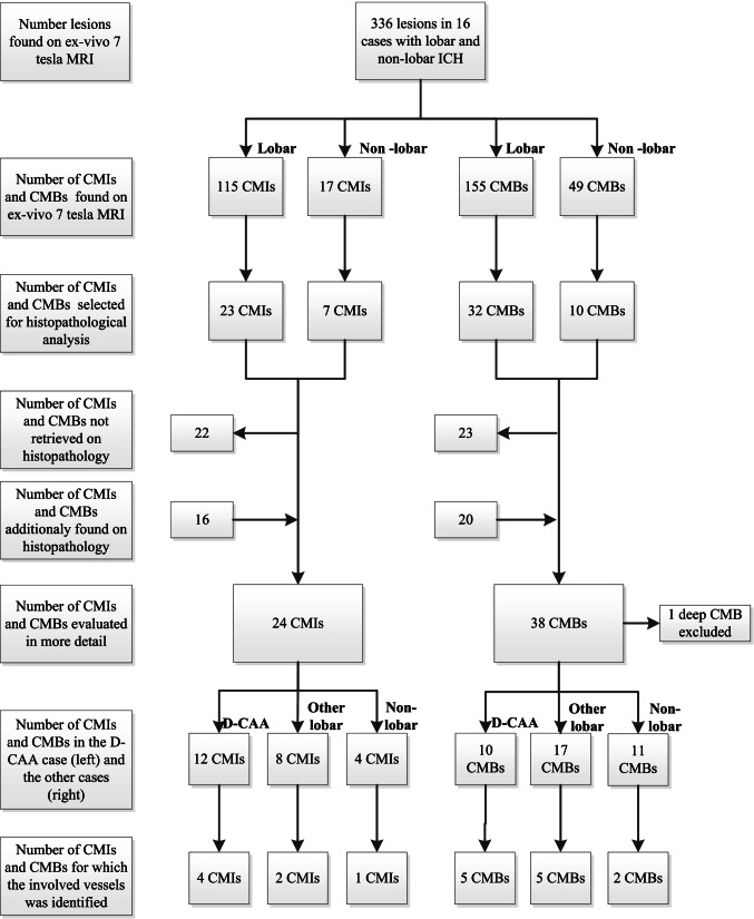
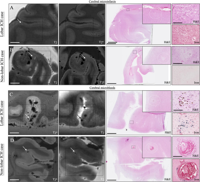
In patients with spontaneous intracerebral hemorrhage caused by different vasculopathies, cerebral microinfarcts have the same aspect on MRI and the same applies to cerebral microbleeds. It is unclear what pathological changes underlie these cerebral microinfarcts and cerebral microbleeds. In the current study, we explored the histopathological substrate of these lesions by investigating the brain tissue of 20 patients (median age at death 77 years) who died from ICH (9 lobar, 11 non-lobar) with a combination of post-mortem 7-T MRI and histopathological analysis. We identified 132 CMIs and 204 CMBs in 15 patients on MRI, with higher numbers of CMIs in lobar ICH patients and similar numbers of CMBs. On histopathology, CMIs and CMBs were in lobar ICH more often located in the superficial than in the deep layers of the cortex, and in non-lobar ICH more often in the deeper layers. We found a tendency towards more severe CAA scores in lobar ICH patients. Other histopathological characteristics were comparable between lobar and non-lobar ICH patients. Although CMIs and CMBs were found in different segments of the cortex in lobar ICH compared to non-lobar ICH patients, otherwise similar histopathological features of cortical CMIs and CMBs distant from the ICH suggest shared pathophysiological mechanisms in lobar and non-lobar ICH caused by different vasculopathies.
Keywords: Cerebral amyloid angiopathy; Histopathology; Spontaneous intracerebral hemorrhage; Ultra-high-field MRI.
© 2022. The Author(s).
Conflict of interest statement
The authors declare no competing interests.
Figures
References
-
- Rodrigues MA, Samarasekera N, Lerpiniere C, Humphreys C, McCarron MO, White PM, et al. The Edinburgh CT and genetic diagnostic criteria for lobar intracerebral haemorrhage associated with cerebral amyloid angiopathy: model development and diagnostic test accuracy study. The Lancet Neurology. The Author(s). Published by Elsevier Ltd. This is an Open Access article under the CC BY 4.0 license; 2018;17:232–40. - PMC - PubMed
Publication types
MeSH terms
LinkOut - more resources
Full Text Sources