. 2022 Jun 29;10(3):e0221221.
doi: 10.1128/spectrum.02212-21. Epub 2022 Apr 6.

TIM-1 Augments Cellular Entry of Ebola Virus Species and Mutants, Which Is Blocked by Recombinant TIM-1 Protein

Affiliations

TIM-1 Augments Cellular Entry of Ebola Virus Species and Mutants, Which Is Blocked by Recombinant TIM-1 Protein

Min Zhang et al. Microbiol Spectr. .

Abstract

Ebola virus, a member of the Filoviridae family, utilizes the attachment factors on host cells to support its entry and cause severe tissue damage. TIM-1 has been identified as a predominant attachment factor via interaction with phosphatidylserine (PS) localized on the viral envelope and glycoprotein (GP). In this study, we give the first demonstration that TIM-1 enhances the cellular entry of three species of Ebola virus, as well as those harboring GP mutations (A82V, T544I, and A82V T544I). Furthermore, two TIM-1 variants (i.e., TIM-1-359aa and TIM-1-364aa) had comparable effects on promoting Zaire Ebola virus (EBOV) attachment, internalization, and infection. Importantly, recombinant TIM-1 ectodomain (ECD) protein could decrease the infectivity of Ebola virus and display synergistic inhibitory effects with ADI-15946, a monoclonal antibody with broad neutralizing activity to Ebola virus. Of note, EBOV strains harboring GP mutations (K510E and D552N), which were refractory to antibody treatment, were still sensitive to TIM-1 protein-mediated impairment of infectivity, indicating that TIM-1 protein may represent an alternative therapeutic regimen when antibody evasion occurs. IMPORTANCE The viral genome has acquired numerous mutations with the potential to increase transmission during the 2013-to-2016 outbreak of Ebola virus. EBOV strains harboring GP mutations (A82V, T544I, and A82V T544I), which have been identified to increase viral infectivity in humans, have attracted our attention. Herein, we give the first report that polymorphic TIM-1 enhances the infectivity of three species of Ebola virus, as well as those harboring GP mutations (A82V, T544I, and A82V T544I). We show that recombinant TIM-1 ECD protein could decrease the infectivity of Ebola virus with or without a point mutation and displays synergistic inhibitory effects with ADI-15946. Furthermore, TIM-1 protein potently blocked cell entry of antibody-evading Ebola virus species. These findings highlight the role of TIM-1 in Ebola virus infection and indicate that TIM-1 protein represents a potential therapeutic avenue for Ebola virus and its mutated species.

Keywords: Ebola virus; recombinant TIM-1 protein; variant surface glycoprotein.


Conflict of interest statement

The authors declare no conflict of interest.

Figures
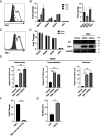
FIG 1
FIG 1
Ectopic expression of TIM-1 enhances Ebola virus cell entry. (A) Surface TIM-1 expression at 36 h following transfection. 293T cells were transfected with 2 μg of empty plasmid (solid gray histogram) or increasing amounts of TIM-1 (black dotted line, 0.5 μg; black dashed line, 1 μg; black solid line, 2 μg). (B) Cell entry of the transfected cells by pSG3.Δenv.cmvFluc (left panel) and VSV-ΔG* (right panel) bearing EBOV, BDBV, SUDV, and VSV GPs. Relative luciferase intensity was detected at 48 h following infection. Data are the fold increase of viral entry into TIM-1-expressing cells compared with cells transfected with empty vector. VSV-pseudotyped virus was used to infect 293T cells transfected with 2 μg TIM-1 and empty vector. (C) Polymorphic TIM-1 expression at 36 h following transfection with 2 μg TIM-1-359aa and TIM-1-364aa. (D) Cell entry of transfected cells by pSG3.Δenv.cmvFluc bearing EBOV, BDBV, SUDV, and VSV GPs (left panel) and cell entry of transfected cells by pSG3.Δenv.cmvFluc bearing EBOV GP by the Western blot method, with GP detected by monoclonal antibody mab114 (right panel). (E) TIM-1 enhances EBOV attachment, internalization, and infection. 293T cells were transfected with an empty vector or plasmids encoding polymorphic TIM-1 for 36 h. EBOV pseudovirus was incubated with cells at 4°C for 30 min and washed with PBS three times (attachment, left panel). After being washed with PBS to remove unbound EBOV particles, cells were transferred to 37°C for 2 h to allow virus internalization and treated with proteinase K to remove uninternalized virions (internalization, middle panel), and then cells were directly challenged with EBOV for 48 h (infection, right panel). Total RNA was extracted and used for quantification of EBOV GP by qRT-PCR. (F) Function comparison of TIM-1 and TIM-1 bearing mutations in the PS binding pocket (TIM-1-N114A D115A) in EBOV cell entry. (G) Function of annexin V to compete for cellular entry of pSG3.Δenv.cmvFluc bearing EBOV and VSV GPs. **, P < 0.01; ***, P < 0.001.
FIG 2
FIG 2
EBOV strains bearing variants of GPs that elicit enhanced infectivity are enhanced by TIM-1 expression. (A) Western blots of lentiviral particles pseudotyped with variant EBOV GPs probed with mab114. The ratio of GP to p24 (GP: p24) was calculated by the Chemiscope 6000 analysis system. In panels B, C, D, and E, the GPs present on virions used here were normalized according to the results of Western blots. (B) Cell entry of the 293T cells by EBOV and EBOV bearing variant GPs. Data points represent independent experiments with three independent viral stocks; each data point represents a normalized transduction. Data are the fold increase of viral entry into TIM-1-expressing (C), Axl-expressing (D), DC-SIGN-expressing (E), and LSECtin-expressing 293T cells (F) compared with vector-expressing 293T cells. *, P < 0.05; **, P < 0.01; ***, P < 0.001.
FIG 3
FIG 3
Identification functional properties of TIM-1 protein. TIM-1 proteins inhibit Ebola virus cell entry. (A) Neutralization of EBOV, BDBV, and SUDV by dilutions of TIM-1 ECD. (B) TIM-1 ECD competes for EBOV entry to 293T cells that were transfected with polymorphic TIM-1. (C) TIM-1 ECD competes for EBOV and mutated EBOV cell entry. (D) Function of TIM-1 ECD, TIM-1 IgV, and TIM-1 ΔIgV protein to compete for EBOV entry. (E) Binding affinity of recombinant TIM-1 ECD, TIM-1 IgV, and TIM-1 ΔIgV to PS and PC. *, P < 0.05; **, P < 0.01.
FIG 4
FIG 4
Inhibition of EBOV infection by TIM-1 ECD in different cell lines. (Left panel) Flow cytometric detection of TIM-1, Axl, and DC-SIGN expression on the surface of Huh7.5.1 cells (A), A549 cells (B), Axl-expressing 293T cells (C), TIM-1-expressing 293T cells (D), DC-SIGN-expressing cells (E), and 293T cells (G) using PE-conjugated anti-TIM, PE-conjugated anti-Axl, and APC-conjugated anti-DC-SIGN antibodies. The expression level of LSECtin in LSECtin-expressing 293T cells (F) was detected by RT-qPCR. (Right panel) TIM-1 ECD protein impeded EBOV entry into Huh7.5.1 cells (A), A549 cells (B), Axl-expressing 293T cells (C), TIM-1-expressing 293T cells (D), DC-SIGN-expressing cells (E), LSECtin-expressing 293T cells (F), and 293T cells (G). *, P < 0.05; **, P < 0.01; ***, P < 0.001.
FIG 5
FIG 5
Analysis of synergistic effects of TIM-1 protein and ADI-15946 on impeding cell entry of virus. The inhibition was evaluated by varying one substrate at a fixed concentration of another. (A, B, C) Cooperative function of TIM-1 protein and ADI-15946 on impeding cell entry of EBOV (A), BDBV (B), SUDV (C) when the doses of TIM-1 ECD were titrated. (D, E, and F) Cooperative function when the doses of ADI-15946 were titrated. (G) TIM-1 ECD inhibits EBOV attachment, but ADI-15946 does not. EBOV pseudovirus was incubated with TIM-1 ECD (left panel) or ADI-15946 (right panel) at 37°C for 1 h before being added to TIM-1-expressing cells. Then, the cell-virus blend was transferred to 4°C for 30 min to allow virus attachment, and cells were washed with PBS three times. Total RNA was extracted and used for quantification of EBOV GP by RT-qPCR. *, P < 0.05; **, P < 0.01; ***, P < 0.001.
FIG 6
FIG 6
TIM-1 protein potently blocks cell entry of antibody-evading Ebola virus bearing GP mutations. (A) Inhibition of ADI-15946 (left panel) and TIM-1 ECD (right panel) to EBOV and EBOV bearing escape variant GP-K510E of ADI-15946. (B) Inhibition of MIL77-2 (left panel) and TIM-1 ECD (right panel) to EBOV and EBOV bearing escape variant GP-D552N of MIL77-2. *, P < 0.05; ***, P < 0.001.

References

    1. Nicastri E, Kobinger G, Vairo F, Montaldo C, Mboera LEG, Ansunama R, Zumla A, Ippolito G. 2019. Ebola virus disease: epidemiology, clinical features, management, and prevention. Infect Dis Clin North Am 33:953–976. doi:10.1016/j.idc.2019.08.005. - DOI - PubMed
    1. Fan P, Chi X, Liu G, Zhang G, Chen Z, Liu Y, Fang T, Li J, Banadyga L, He S, Yu C, Qiu X, Chen W. 2020. Potent neutralizing monoclonal antibodies against Ebola virus isolated from vaccinated donors. MAbs 12:1742457. doi:10.1080/19420862.2020.1742457. - DOI - PMC - PubMed
    1. Olejnik J, Forero A, Deflubé LR, Hume AJ, Manhart WA, Nishida A, Marzi A, Katze MG, Ebihara H, Rasmussen AL, Mühlberger E. 2017. Ebolaviruses associated with differential pathogenicity induce distinct host responses in human macrophages. J Virol 91:e00179-17. doi:10.1128/JVI.00179-17. - DOI - PMC - PubMed
    1. Ahmad MD, Usman M, Khan A, Imran M. 2016. Optimal control analysis of Ebola disease with control strategies of quarantine and vaccination. Infect Dis Poverty 5:72. doi:10.1186/s40249-016-0161-6. - DOI - PMC - PubMed
    1. Hoffmann M, Crone L, Dietzel E, Paijo J, González-Hernández M, Nehlmeier I, Kalinke U, Becker S, Pöhlmann S. 2017. A polymorphism within the internal fusion loop of the Ebola virus glycoprotein modulates host cell entry. J Virol 91:e00177-17. doi:10.1128/JVI.00177-17. - DOI - PMC - PubMed

Publication types

Substances