. 2022 May 1;322(5):C1011-C1021.
doi: 10.1152/ajpcell.00454.2021. Epub 2022 Apr 6.

Control of coronary vascular resistance by eicosanoids via a novel GPCR

Affiliations

Control of coronary vascular resistance by eicosanoids via a novel GPCR

Nabil J Alkayed et al. Am J Physiol Cell Physiol. .

Erratum in

Abstract

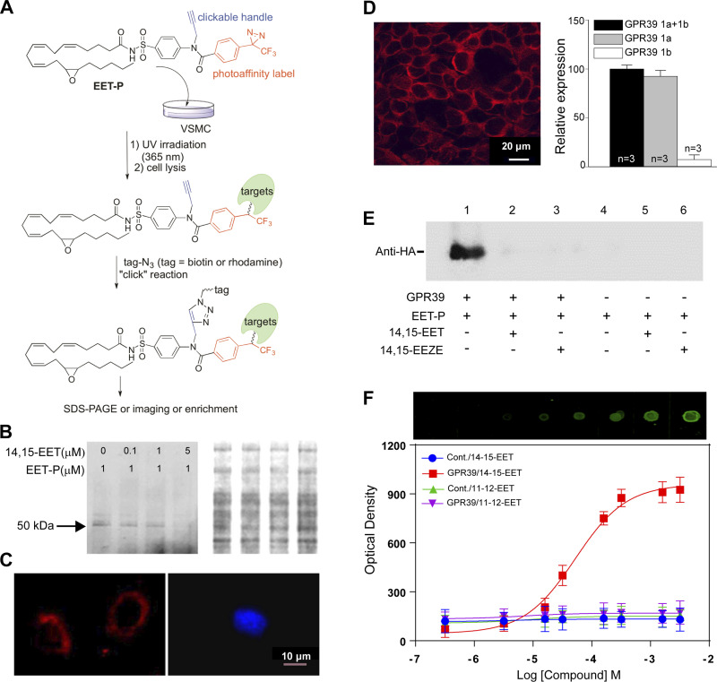
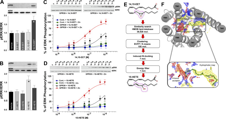
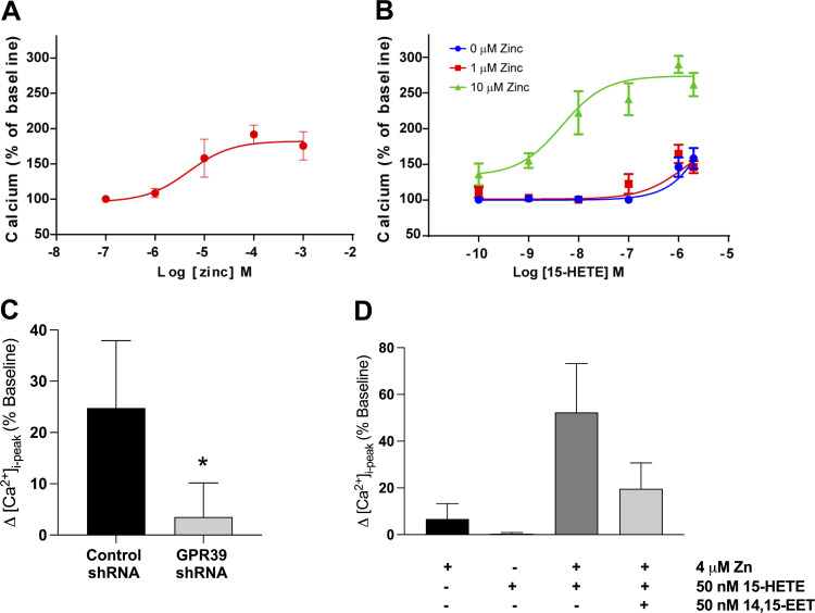
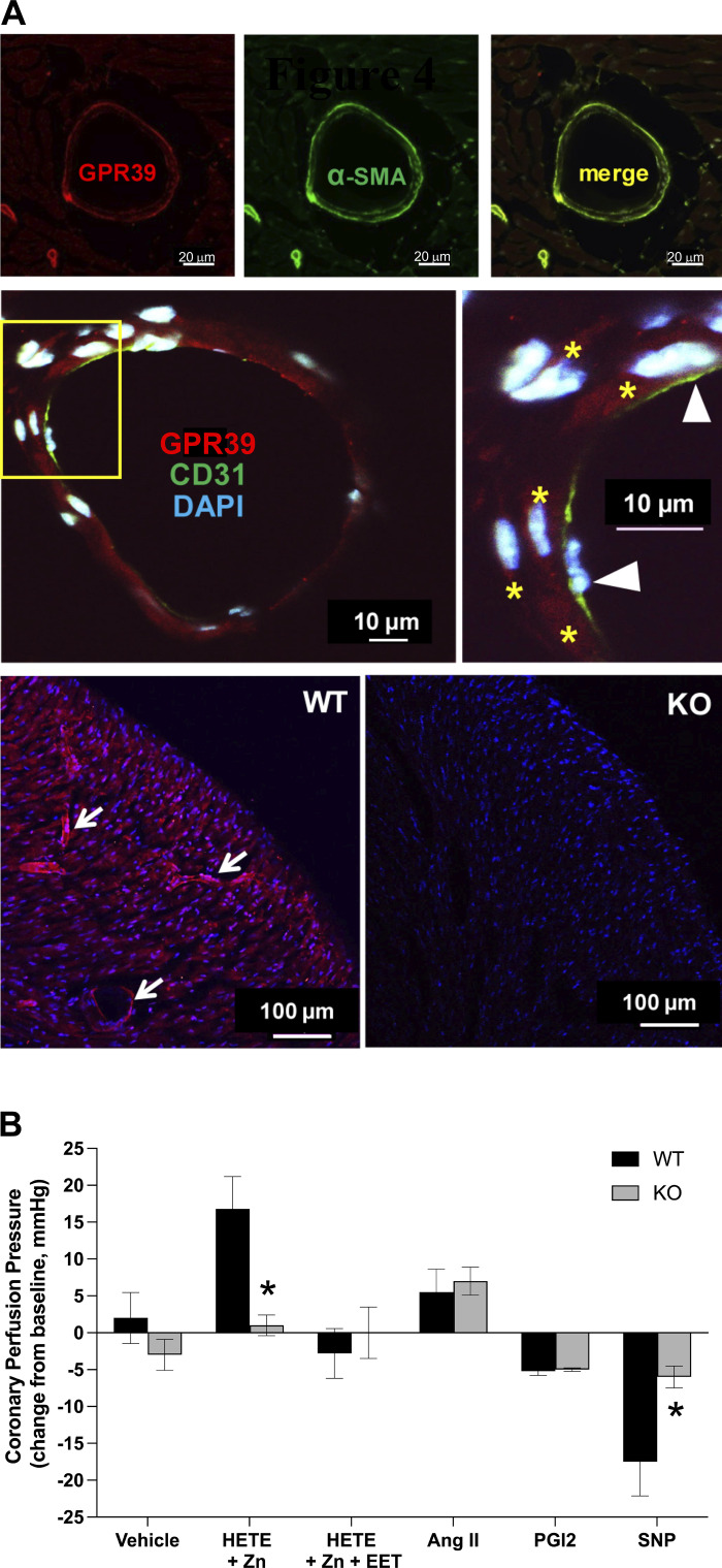
Arachidonic acid metabolites epoxyeicosatrienoates (EETs) and hydroxyeicosatetraenoates (HETEs) are important regulators of myocardial blood flow and coronary vascular resistance (CVR), but their mechanisms of action are not fully understood. We applied a chemoproteomics strategy using a clickable photoaffinity probe to identify G protein-coupled receptor 39 (GPR39) as a microvascular smooth muscle cell (mVSMC) receptor selective for two endogenous eicosanoids, 15-HETE and 14,15-EET, which act on the receptor to oppose each other's activity. The former increases mVSMC intracellular calcium via GPR39 and augments coronary microvascular resistance, and the latter inhibits these actions. Furthermore, we find that the efficacy of both ligands is potentiated by zinc acting as an allosteric modulator. Measurements of coronary perfusion pressure (CPP) in GPR39-null hearts using the Langendorff preparation indicate the receptor senses these eicosanoids to regulate microvascular tone. These results implicate GPR39 as an eicosanoid receptor and key regulator of myocardial tissue perfusion. Our findings will have a major impact on understanding the roles of eicosanoids in cardiovascular physiology and disease and provide an opportunity for the development of novel GPR39-targeting therapies for cardiovascular disease.

Keywords: EETs; GPCR; GPR39; HETEs; eicosanoids.


Conflict of interest statement

This research involves technology of which N. J. Alkayed, S. Kaul, and S. Nagarajan are co-inventors and which has been licensed, in part by OHSU, to Vasocardea. OHSU and S. Kaul have a financial interest in Vasocardea, a company that may have a commercial interest in the results of this research and technology. This potential conflict of interest has been reviewed and managed by Oregon Health and Science University. None of the other authors has any conflicts of interest, financial or otherwise, to disclose.

This article is part of the special collection "Advances in GPCRs: Structure, Mechanisms, Disease, and Pharmacology." Wei Kong, MD, PhD, and Jinpeng Sun, PhD, served as Guest Editors of this collection.

Figures

Figure 1.
Figure 1.
Purification and validation of GPR39 as a putative receptor for 14,15-EET. A: chemoproteomics strategy to identify 14,15-EET receptor in mVSMCs. B: photo-crosslinking GPR39 with EET-P in mVSMCs. Left: representative SDS-PAGE gel revealing the presence of a ∼50 kDa band. 14,15-EET pretreatment reduced protein binding to EET-P in a dose-dependent manner. Right: total protein determined by Coomassie Blue staining of the same gel. C: representative confocal images demonstrating binding of EET-P to outer surface of mVSMCs (left, red). Pretreatment with 1 μM 14,15-EET for 10 min prevented EET-P surface binding (right; blue color is nuclear stain DAPI). D: a representative immunofluorescent confocal image illustrating GPR39 expression (left, red) in primary cultured mouse heart mVSMCs (scale bar = 20 μm), and expression of GPR39 1a, but not 1b, confirmed by qPCR (right), n = 3. E: a representative Western blot of EET-P cross linking to epitope-tagged GPR39 1a in transfected HEK cells. HEK cells were treated with 5 μM 14,15-EET or 14,15-EEZE for 10 min, 1 μM EET-P for 15 min, and then irradiated with UV for 5 min at 4oC. After clicking with biotin and purification with streptavidin Dynabeads, protein extracts were probed by anti-HA antibody. F: a representative dot-blot assay and quantification of dose-dependent binding of 14,15-EET, but not 11,12-EET, to membrane protein extracts from HEK-293 cells stably expressing GPR39 1a, but not untransfected control cells (top, raw image, and optical density quantification, bottom; n = 3). EET, epoxyeicosatrienoate; EET-P, epoxyeicosatrienoate probe; GPR39, G protein-coupled receptor 39; HEK, human embryonic kidney; mVSMCs, microvascular smooth muscle cells; 14,15-EEZE, 14,15-epoxyeicosa-5(Z)-enoic acid.
Figure 2.
Figure 2.
Functional activation and modeling of GPR39 ligand binding. ERK phosphorylation induced by 1 min treatment with 1 μM of each one of four regioisomers of EETs (A) or HETEs (B) , n = 3 for each regioisomer (A, *P < 0.05; B, *P < 0.05). Total was used as a protein loading control. Dose-dependent ERK phosphorylation induced by 1 min treatment with either 14,15-EET (C; n = 3) or 15-HETE (D; n = 3) in HEK cells expressing GPR39 1a, but not untransfected cells, in the presence and absence of 1 μM zinc. E: virtual screening of 14,15-EET-like compounds from HMDB database revealed comparable energies for 15-HETE and 14,15-EET. The 14,15-EET and 15-HETE structures exhibit a high degree of structural similarity. The dotted red line around carbons 13–15 highlights the structural difference between the two eicosanoids. F: GPR39 7TM core binding pocket and an enlarged view of the predicted binding pose of 14,15-EET. Blue, red, and yellow maps correspond to hydrogen bond donor, hydrogen bond acceptor, and hydrophobic areas within the binding pocket, respectively. N-terminal residues contributing to minor pocket are not shown for the sake of clarity. The carboxylate group of 14,15-EET interacts with polar residues in TM6, and the lipid portion interacts with the hydrophobic site formed at the minor-binding pocket, as shown in site mapping. EET, epoxyeicosatrienoate; ERK, extracellular signal-regulated kinase; GPR39, G protein-coupled receptor 39; HETE, hydroxyeicosatetraenoate; HMDB, human metabolome database; TM6, transmembrane domain 6.
Figure 3.
Figure 3.
A: concentration-dependent increase in [Ca2+]i in mVSMCs in response to extracellular zinc (n = 5 for each concentration). B: dose-dependent increase in mVSMC [Ca2+]i in response to 15-HETE with and without zinc (n = 5–7 for each concentration). C: summary of [Ca2+]i response to 1 μM 15-HETE in mVSMCs treated with a lentivirus containing either a scrambled or GPR39-targeting shRNA for 72 h (n = 5, *P = 0.0122). D: summary of changes in mVSMCs [Ca2+]i in response to 50 nM of 14,15-EET and 15-HETE, separately and in combination, and with and without zinc (n = 4 for zinc alone, and n = 6). When applied alone, neither 4 μM zinc nor 50 nM 15-HETE had an effect on [Ca2+]i. Zinc (4 μM) potentiates the effect of 15-HETE (50 nM) on [Ca2+]i in mVSMCs, and the increase in mVSMCs [Ca2+]i by 15-HETE is abolished by pretreatment with 14,15-EET (*P < 0.00011). EET, epoxyeicosatrienoate; GPR39, G protein-coupled receptor 39; HETE, hydroxyeicosatetraenoate; mVSMCs, microvascular smooth muscle cells.
Figure 4.
Figure 4.
Microvascular localization and function of GPR39 in mouse heart. A: immunofluorescent imaging of GPR39 shows microvascular pattern of expression in mouse heart (top left, red; scale bar = 20 μm), which colocalizes with VSMC markers α-smooth muscle actin (α-SMA, green, top middle and right; scale bar = 20 μm), but not endothelial marker CD31 (middle; green and arrow heads in magnified view; yellow stars point to GPR39-positive mVSMCs within vascular wall in red; blue is DAPI nuclear staining; scale bar = 10 μm). The bottom panel shows GPR39 immunoreactivity in wild-type (WT, left; arrows point to immuno-reactive microvessels in red) and GPR39 knockout (KO; blue is nuclear stain DAPI) mouse heart tissue sections (scale bar = 100 μm). B: changes in coronary perfusion pressure, at a constant flow rate, in response to infusion of 15-HETE (1 μM; in presence of 10 µM zinc), 15-HETE plus 14,15-EET (1 μM; in presence of 10 µM zinc), prostaglandin I2 (PGI2, 200 nM), angiotensin II (AngII, 100 nM), sodium nitroprusside (SNP; 10 µM), or vehicle in isolated mouse heart preparation from WT and GPR39 KO mice (n = 5, *P < 0.05). EET, epoxyeicosatrienoate; GPR39, G protein-coupled receptor 39; HETE, hydroxyeicosatetraenoate; mVSMCs, microvascular smooth muscle cells.

References

    1. Muller JM, Davis MJ, Chilian WM. Integrated regulation of pressure and flow in the coronary microcirculation. Cardiovasc Res 32: 668–678, 1996. - PubMed
    1. Capdevila JH, Falck JR, Harris RC. Cytochrome P450 and arachidonic acid bioactivation. Molecular and functional properties of the arachidonate monooxygenase. J Lipid Res 41: 163–181, 2000. - PubMed
    1. Roman RJ. P-450 metabolites of arachidonic acid in the control of cardiovascular function. Physiol Rev 82: 131–185, 2002. doi: 10.1152/physrev.00021.2001. - DOI - PubMed
    1. Campbell WB, Gebremedhin D, Pratt PF, Harder DR. Identification of epoxyeicosatrienoic acids as endothelium-derived hyperpolarizing factors. Circ Res 78: 415–423, 1996. doi: 10.1161/01.res.78.3.415. - DOI - PubMed
    1. Fisslthaler B, Popp R, Kiss L, Potente M, Harder DR, Fleming I, Busse R. Cytochrome P450 2C is an EDHF synthase in coronary arteries. Nature 401: 493–497, 1999. doi: 10.1038/46816. - DOI - PubMed

Publication types

MeSH terms