. 2022 Jun 27;61(26):e202200269.
doi: 10.1002/anie.202200269. Epub 2022 Apr 29.

Structure-Based Evolution of G Protein-Biased μ-Opioid Receptor Agonists

Affiliations

Structure-Based Evolution of G Protein-Biased μ-Opioid Receptor Agonists

Haoqing Wang et al. Angew Chem Int Ed Engl. .

Abstract

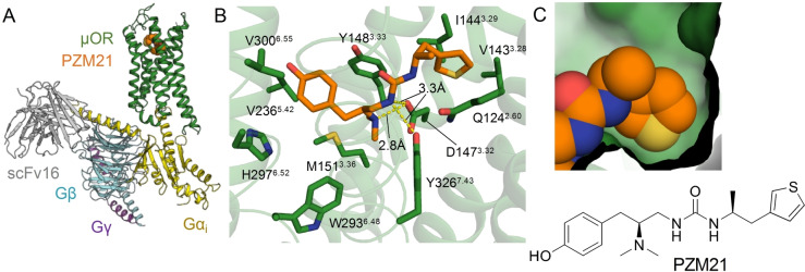
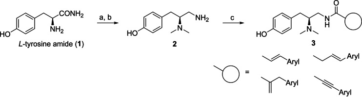
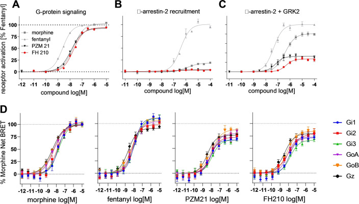
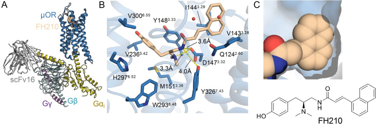
The μ-opioid receptor (μOR) is the major target for opioid analgesics. Activation of μOR initiates signaling through G protein pathways as well as through β-arrestin recruitment. μOR agonists that are biased towards G protein signaling pathways demonstrate diminished side effects. PZM21, discovered by computational docking, is a G protein biased μOR agonist. Here we report the cryoEM structure of PZM21 bound μOR in complex with Gi protein. Structure-based evolution led to multiple PZM21 analogs with more pronounced Gi protein bias and increased lipophilicity to improve CNS penetration. Among them, FH210 shows extremely low potency and efficacy for arrestin recruitment. We further determined the cryoEM structure of FH210 bound to μOR in complex with Gi protein and confirmed its expected binding pose. The structural and pharmacological studies reveal a potential mechanism to reduce β-arrestin recruitment by the μOR, and hold promise for developing next-generation analgesics with fewer adverse effects.

Keywords: Drug Discovery; GPCRs; Medicinal Chemistry; Opioids; cryoEM Structure.


Conflict of interest statement

B.K.K. and P.G. are co‐founders of Epiodyne. B.K.K is a co‐founder of and consultant for ConformetRx.

Figures

Figure 1
Figure 1
A) Structure of the μOR‐Gi complex bound to PZM21 colored by subunit. Green, μOR; orange, PZM21; gold, Gαi; cyan, Gβ; purple Gγ; gray scFv16. B) View of PZM21 in the binding pocket. PZM21 forms polar contacts to D1473.32 and Y3267.43 and the phenol group is near H2976.52 suggesting that PZM21 forms a water‐mediated interaction with H2976.52, as observed in previous structures of the μOR. C) Surface presentation of the thiophene group of PZM21 in the lipophilic vestibule of the μOR.
Scheme 1
Scheme 1
Synthesis of PZM21 analogs. Reagents and conditions: a) formaldehyde, sodium triacetoxyborohydride, water, acetonitrile, −10 °C, 5 min (>99 %); b) borane–THF complex, THF, 0 °C to reflux, 6 h (43 %), c) carboxylic acid, BOP or PyBOP, triethylamine, DMF, r.t., 0.5–24 h (56–95 %).
Figure 2
Figure 2
Functional activity of morphine, fentanyl, PZM21 and FH210 at the μOR. A) G‐protein signaling measured by IP accumulation assay in cells transfected with the chimeric G protein Gqi. B), C) β‐arrestin‐2 recruitment measured by PathHunter assay, with (C) and without (B) GRK2 co‐transfection. D) Gi/o/z subtypes activation measured by BRET‐based assay.
Figure 3
Figure 3
A) Structure of the μOR‐Gi complex bound to FH210 colored by subunit. Blue, μOR; beige, FH210; gold, Gαi; cyan, Gβ; purple Gγ; gray scFv16. B) View of FH210 in the binding pocket. Blue, μOR; beige, FH210. FH210 forms polar contacts with D1473.32 and Y3267.43 and shows proximity of the phenol group to H2976.52 suggesting that the ligand forms a water mediated interaction to H2976.52. C) Surface representation of the naphthyl group of FH210 in the lipophilic vestibule of the μOR.
Figure 4
Figure 4
Comparison of μOR bound to PZM21 and FH210. A), B) Contact surface area (red) between the thiophene of PZM21 (orange) and the PZM21‐μOR cryoEM complex (green), a=124 Å2; and between the naphthyl of FH210 (beige) and the FH210‐μOR cryoEM complex (blue), a=155 Å2. Contact surface area was calculated with a cut‐off value of 2 Å using UCSF Chimera. C) Representative structures extracted from MD simulations of PZM21‐μOR and FH210‐μOR. The simulations show a more compact lipophilic vestibule for FH210 bound μOR. The distances are measured between the Cα atoms of the labeled residues. The histogram plots on the right shows the distance distribution over all simulations. The bar chart at the bottom of each plot shows the mean and SEM, with the dots representing the individual values. These statistics are based on 6 individual simulations and the first 500 ns of each simulation was not included in these analyses.

References

    1. Volkow N. D., Blanco C., Mol. Psychiatry 2021, 26, 218–233. - PMC - PubMed
    1. Che T., Dwivedi-Agnihotri H., Shukla A. K., Roth B. L., Sci. Signaling 2021, 14, eaav0320. - PMC - PubMed
    1. Manglik A., Lin H., Aryal D. K., McCorvy J. D., Dengler D., Corder G., Levit A., Kling R. C., Bernat V., Hübner H., Huang X.-P., Sassano M. F., Giguère P. M., Löber S., Da D., Scherrer G., Kobilka B. K., Gmeiner P., Roth B. L., Shoichet B. K., Nature 2016, 537, 185–190. - PMC - PubMed
    1. Kudla L., Bugno R., Skupio U., Wiktorowska L., Solecki W., Wojtas A., Golembiowska K., Zádor F., Benyhe S., Buda S., Makuch W., Przewlocka B., Bojarski A. J., Przewlocki R., Br. J. Pharmacol. 2019, 176, 4434–4445. - PMC - PubMed
    1. Gillis A., Gondin A. B., Kliewer A., Sanchez J., Lim H. D., Alamein C., Manandhar P., Santiago M., Fritzwanker S., Schmidel F., Katte T. A., Reekie T., Grimsey N. L., Kassiou M., Kellam B., Krasel C., Halls M. L., Connor M., Lane J. R., Schulz S., Christie M. J., Canals M., Sci. Signaling 2020, 13, eaaz3140. - PubMed

Publication types

MeSH terms

LinkOut - more resources