. 2022 Apr 5;39(1):110610.
doi: 10.1016/j.celrep.2022.110610.

Augmin-dependent microtubule self-organization drives kinetochore fiber maturation in mammals

Affiliations

Augmin-dependent microtubule self-organization drives kinetochore fiber maturation in mammals

Ana C Almeida et al. Cell Rep. .

Abstract

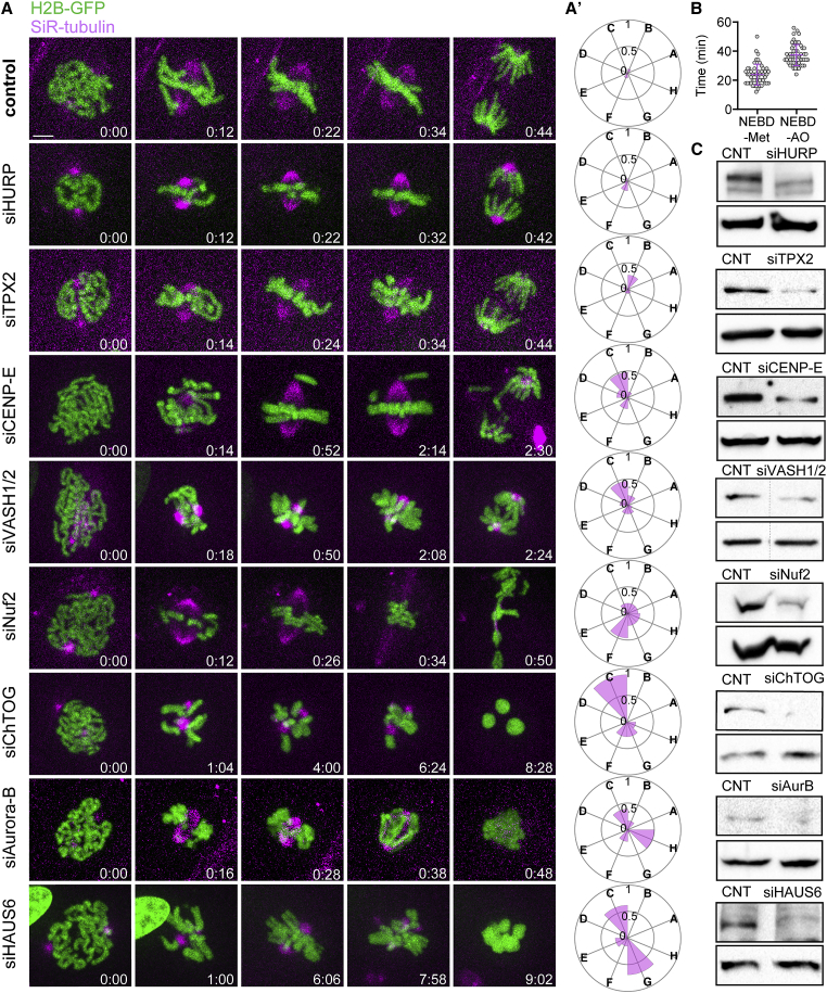
Chromosome segregation in mammals relies on the maturation of a thick bundle of kinetochore-attached microtubules known as k-fiber. How k-fibers mature from initial kinetochore microtubule attachments remains a fundamental question. By combining molecular perturbations and phenotypic analyses in Indian muntjac fibroblasts containing the lowest known diploid chromosome number in mammals (2N = 6) and distinctively large kinetochores, with fixed/live-cell super-resolution coherent-hybrid stimulated emission depletion (CH-STED) nanoscopy and laser microsurgery, we demonstrate a key role for augmin in kinetochore microtubule self-organization and maturation, regardless of pioneer centrosomal microtubules. In doing so, augmin promotes kinetochore and interpolar microtubule turnover and poleward flux. Tracking of microtubule growth events within individual k-fibers reveals a wide angular dispersion, consistent with augmin-mediated branched microtubule nucleation. Augmin depletion reduces the frequency of kinetochore microtubule growth events and hampers efficient repair after acute k-fiber injury by laser microsurgery. Together, these findings underscore the contribution of augmin-mediated microtubule amplification for k-fiber self-organization and maturation in mammals.

Keywords: CP: Cell biology; Indian muntjac; augmin; coherent-hybrid STED; k-fiber; kinetochore; laser microsurgery; microtubules; mitosis; mitotic spindle; super-resolution.


Conflict of interest statement

Declaration of interests The authors declare no competing interests.

Figures

None
Graphical abstract
Figure 1
Figure 1
A live-cell RNAi screen in Indian muntjac fibroblasts identifies augmin as a critical spindle assembly factor required for chromosome segregation (A) Schematic representation of the mitotic screen performed in IM fibroblasts. Mitotic timings (T. NEBD-AO) were determined and genes blindly clustered based on the probability of occurrence of eight binary features: A, incomplete congression and faster mitosis; B, incomplete chromosome and normal mitotic duration; C, incomplete congression and prolonged mitosis; D, congression delay; E, metaphase delay; F, anaphase lagging chromosomes; G, mitotic death; and H, cytokinesis failure. (B) Dendrogram highlighting the hierarchical relationships between 10 distinct clusters (I–X) and few orphan proteins based on phenotypic similarities and respective frequencies. The severity of the defects increases from left to right. Euclidean distance was used as the distance metric to compare the phenotypical fingerprints.
Figure 2
Figure 2
Augmin depletion is one of the most deleterious conditions for mitosis in Indian muntjac fibroblasts (A) Examples of the phenotypic analysis performed by live-cell spinning-disk confocal microscopy in IM fibroblasts: siHURP (n = 17 cells), siTPX2 (n = 26 cells), siCENP-E (n = 25 cells), siVASH1/2 (n = 26 cells), siNuf2 (n = 26 cells), siChTOG (n = 18 cells), siAurora B (n = 18 cells), and siHAUS6 (n = 13 cells). Mock transfection (lipofectamine only) was used as control (n = 52 cells). Scale bar: 5 μm. Time is shown as hours:minutes. (A′) Radar plots illustrating the phenotypic fingerprints reflecting the probability of occurrence of the eight analyzed features, A–G, for the depletions shown in Figure 1A. Zero corresponds to a null event and 1 to all cells displaying a certain event. (B) Mitotic timings from NEBD-metaphase and NEBD-AO in control cells. Data pooled from three independent experiments. Error bars indicate mean ± SD. (C) Validation of RNAi efficiency by immunoblotting with specific antibodies against each target protein (upper band), except for VASH1/2, where only anti-VASH1 was used, and Nuf2, where anti-Hec1 was used. The bottom band corresponds to anti-GAPDH (siTPX2, siMad2, siVASH1/2, siNuf2, and siHAUS6), anti-α-tubulin (siHURP, siCENP-E, and siAuroraB), or anti-vinculin (siChTOG), which were used as loading controls. CNT, control.
Figure 3
Figure 3
Augmin contributes to k-fiber and interpolar microtubule formation Images of control and HAUS6-depleted cells acquired by CH-STED nanoscopy. (A) Immunofluorescence of IM fibroblasts using paraformaldehyde and glutaraldehyde fixation. DAPI, α-tubulin, and ACA are shown in inverted grayscale. Astral MT tracks are represented in magenta (tracing). Scale bar: 5 μm. (B) Quantification of astral MT length (n = 665 control astral MTs/10 cells; n = 750 siHAUS6 astral MTs/11 cells). The boxplot determines the interquartile range; the line inside the box represents the median; data pooled from three independent experiments and analyzed using an unpaired t test; ∗∗∗∗p ≤ 0.0001. (C) Immunofluorescence of IM fibroblasts using cold methanol fixation. β-Tubulin (magenta) and PRC1 (green). Scale bar: 5 μm. (D) 3D representations of mitotic spindles in control and HAUS6-depleted cells, illustrating KT surfaces 1 and 2 (magenta), as well as the plates that define the measurement volumes, corresponding to interpolar MTs (ipMTs, green) and k-fibers (kMTs) plus ipMTs (cyan). (E) Quantification of ipMTs and kMTs in control and HAUS6-depleted cells. Proportion relative to control levels is represented for HAUS6-depleted cells (n = 14 control cells; n = 12 siHAUS6 cells). Data pooled from two independent experiments and analyzed using an unpaired t test; ∗∗∗∗p ≤ 0.0001.
Figure 4
Figure 4
Augmin sustains centrosome-independent microtubule self-organization from kinetochores (A–D) (A) CH-STED images of IM cells stably expressing GFP-Centrin-1 (magenta) and 2xGFP-CENP-A (magenta) treated with centrinone for 8 days with or without HAUS6 RNAi for 72 h. Cells were treated with the MT-depolymerizing drug nocodazole for 2 h, followed by drug washout and fixation after 2, 5, and 10 min. α-Tubulin (green) and DAPI (inverted gray scale). Insets show 2.5× magnification of selected regions with KT and nucleated MTs (grayscale for single channels of 2xGFP-CENP-A and α-tubulin). The experimental setup is described in (A’). The percentage of KTs with MTs and overall MT length are represented in (B) and (C), respectively (control 2’, n = 13 cells/154 MTs; control 5’, n = 20 cells/350 MTs; control 10’, n = 19 cells/402 MTs; siHAUS6 2’, n = 16 cells/91 MTs; siHAUS6 5’, n = 14 cells/176 MTs; siHAUS6 10’, n = 14 cells/254 MTs). The ratio of polymerized tubulin at KTs relative to the overall cytoplasmatic pool 2 min after nocodazole washout is shown in (D) (control 2’, n = 41 KTs; siHAUS6 2’, n = 53 KTs). Data pooled from three independent experiments, analyzed using a Mann-Whitney test; the boxplot determines the interquartile range and the line inside the box represents the median (C); mean ± SD (B and D); ns, not significant; p ≤ 0.05; ∗∗p ≤ 0.01; ∗∗∗∗p ≤ 0.0001. Scale bar: 5 μm.
Figure 5
Figure 5
Augmin promotes kinetochore microtubule turnover and poleward flux (A–K) (A) Pre-recording snapshots of control, HAUS6-, and Ndc80-depleted IM fibroblasts stably expressing 2xGFP-CENP-A (magenta) and EB3-Halo tag conjugated with JF646 (green), imaged by confocal microscopy. Scale bar: 5 μm. (A′) Collapsed kymographs of live CH-STED recordings (time lapse: 8 s; pixel size, 40 nm). Graphical sketches on the right highlight chromosome movement over time (tracing); P, pole (green); KT, kinetochore (magenta); EB3 accumulation at KT is shown in green. Quantitative analysis of chromosome anti-poleward (B) and poleward (C) velocities, chromosome oscillatory amplitude (D), and period (E). Fraction of EB3 accumulation at KT per minute (approximately one period) was measured from track data in (F), and KT-to-pole distance determined in (G). Horizontal bar, 1 μm; vertical bar, 10 s (n = 8 control cells, n = 9 siHAUS6 cells, and n = 8 siNdc80 cells). (H) Examples of control and partial HAUS6-depleted metaphase cells displaying photoactivatable PA-GFP-α-tubulin (inverted grayscale), GFP-Centrin-1 (inverted grayscale), and labeled with 50 nM SiR-DNA to visualize chromosomes (inverted grayscale). Pre-PA, frame immediately before photoactivation; PA, frame immediately after photoactivation. Scale bar: 5 μm. (I) Normalized fluorescence dissipation after photoactivation (FDAPA) curves of control and partial HAUS6-depleted cells. Whole lines show double exponential curve fittings (R2 > 0.98), and error bars show 95% confidence interval for each time point. (J) Table showing the calculated MT percentages and turnover values for control and partial HAUS6-depleted cells (n = 20 control cells; n = 11 siHAUS6 cells). (K) MT flux velocity (n = 21 control cells; n = 16 siHAUS6 cells). Each data point represents one measurement; data pooled from at least three independent experiments and analyzed using a Mann-Whitney test (B, C, F, G) or an unpaired t test (D, J, K); Error bars indicate mean ± SD; ns, not significant; p ≤ 0.05; ∗∗p ≤ 0.01; ∗∗∗p < 0.001; ∗∗∗∗p ≤ 0.0001.
Figure 6
Figure 6
Microtubule growth within individual k-fibers shows a wide angular dispersion and requires augmin (A) CH-STED images of control, HAUS6-, and Ndc80-depleted cells stained with α-tubulin, ACA (cyan), and DAPI (white; opacity 15%). Temporal color code tool on Fiji was used to match each α-tubulin z plane to a different color. Scale bar: 5 μm. Insets show the maximum-intensity projection of relevant z planes highlighting the presence/absence of k-fibers (α-tubulin, inverted grayscale). Scale bar: 1 μm. (B) IM fibroblasts stably expressing 2xGFP-CENP-A (magenta) and EB3-Halo tag conjugated with JF646 (green) were used to track MT polymerization events within one k-fiber by live CH-STED microscopy (time lapse, 750 ms; pixel size, 40 nm). Images on the top show a pre-recording snapshot (confocal) of control, HAUS6-, and Ndc80-depleted cells. Images below show chromo-projections of the time-lapse movie of fluorescently labeled EB3 over time and CENP-A contours. A limited time-window of 12 s (five frames) was selected, allowing a fine time-discrimination of MT growing events within a k-fiber. Scale bar: 500 nm. (C) Frequency count of EB3 comets’ angular dispersion relative to the k-fiber axis in control cells (n = 17 cells). (D) Corresponding collapsed kymographs of control, HAUS6-, and Ndc80-depleted cells from (B). Graphical sketches on the right highlight detected EB3 comets’ trajectories; KT, kinetochore (magenta); kMTs, green; non-kMTs, light brown. Vertical bar, 10 s; horizontal bar, 1 μm. (E) Number of EB3 growing events per KT (control, n = 46 kMT comets/n = 21 non-kMT comets/n = 12 cells; siHAUS6, n = 38 kMT comets/n = 41 non-kMT comets/n = 15 cells; siNdc80, n = 23 kMT comets/n = 41 non-kMT comets/n = 10 cells). Error bars indicate mean ± SD, ns, not significant; ∗∗p ≤ 0.01; ∗∗∗p < 0.001. (F) Distance between KT pairs upon stable expression of 2xGFP-CENP-A (control n = 34 cells; siHAUS6 n = 36 cells; siNdc80 n = 28 cells). Data pooled from at least three independent experiments and analyzed using an unpaired t test (E, F); Error bars indicate mean ± SD; ns, not significant; p ≤ 0.05.
Figure 7
Figure 7
Augmin is required for microtubule amplification from pre-existing kinetochore microtubules (A) Schematic summary of the laser microsurgery-based k-fiber injury/repair assay in control cells (see STAR Methods for details). (B) Spinning-disk confocal images of total k-fiber severing in control IM fibroblasts stably expressing GFP-α-tubulin (green) and mScarlet-CENP-A (magenta). Insets show the analyzed k-fibers (GFP-α-tubulin, inverted grayscale). Magenta arrowhead indicates ablated k-fiber. Scale bar: 5 μm (left); 1 μm (right). Time is shown as minutes:seconds. (C) Partial k-fiber damage was confirmed by correlative live-cell spinning-disk confocal microscopy (left) and CH-STED nanoscopy (right) upon fixation and immunostaining of the damaged cell (α-tubulin and DAPI shown in inverted grayscale). 0 s corresponds to the first frame after k-fiber ablation. Magenta arrowhead indicates partially ablated k-fiber. Scale bar: 5 μm. (D) Immunoblot analysis of cell lysates treated with control or partial HAUS6 RNAi (asterisk indicates the band of interest; ∼26% depletion) and α-tubulin was used as loading control (bottom). (E) Control and partial HAUS6-depleted IM fibroblasts illustrating MT recovery after partial k-fiber laser ablation. Yellow dashed rectangle indicates the injured k-fiber. Scale bar: 5 μm. Insets show the analyzed k-fibers (GFP-α-tubulin, inverted grayscale). −10 s represents the maximum time before partial k-fiber ablation. Magenta arrowhead points to the ablated k-fiber portion at time zero (first frame after laser ablation). Scale bar: 1 μm. Time is shown as minutes:seconds. (F) Kinetics of fluorescence recovery after surgery (FRAS) was determined as a proxy for k-fiber recovery, in controls and after partial HAUS6-depletion by RNAi. Whole lines show a single exponential fitting curve. Each data point represents the mean ±95% confidence interval. ∗∗p ≤ 0.01. (G) Fluorescence recovery from partial k-fiber ablation within the first 30 s after surgery, in control and partial HAUS6-depleted cells (n = 12 control cells; n = 13 siHAUS6 cells). Data pooled from at least three independent experiments; non-linear fit (F) Mann-Whitney test (G); Error bars indicate mean ± SD; p ≤ 0.05.

References

    1. Aillaud C., Bosc C., Peris L., Bosson A., Heemeryck P., Van Dijk J., Le Friec J., Boulan B., Vossier F., Sanman L.E., et al. Vasohibins/SVBP are tubulin carboxypeptidases (TCPs) that regulate neuron differentiation. Science. 2017;358:1448–1453. doi: 10.1126/science.aao4165. - DOI - PubMed
    1. Al-Bassam J., Chang F. Regulation of microtubule dynamics by TOG-domain proteins XMAP215/Dis1 and CLASP. Trends Cell Biol. 2011;21:604–614. doi: 10.1016/j.tcb.2011.06.007. - DOI - PMC - PubMed
    1. Alfaro-Aco R., Thawani A., Petry S. Biochemical reconstitution of branching microtubule nucleation. Elife. 2020;9:e4979. doi: 10.7554/eLife.49797. - DOI - PMC - PubMed
    1. Almeida A.C., Drpic D., Okada N., Bravo J., Madureira M., Maiato H. Functional dissection of mitosis using immortalized fibroblasts from the Indian muntjac, a placental mammal with only three chromosomes. Methods Mol. Biol. 2020;2101:247–266. doi: 10.1007/978-1-0716-0219-5_16. - DOI - PubMed
    1. Bajer A.S., Mole-Bajer J. Reorganization of microtubules in endosperm cells and cell fragments of the higher plant Haemanthus in vivo. J. Cell Biol. 1986;102:263–281. doi: 10.1083/jcb.102.1.263. - DOI - PMC - PubMed

Publication types