Automated Pupillometry for Prediction of Electroencephalographic Reactivity in Critically Ill Patients: A Prospective Cohort Study
- PMID: 35386412
- PMCID: PMC8977520
- DOI: 10.3389/fneur.2022.867603
Automated Pupillometry for Prediction of Electroencephalographic Reactivity in Critically Ill Patients: A Prospective Cohort Study
Abstract
Background: Electroencephalography (EEG) is widely used to monitor critically ill patients. However, EEG interpretation requires the presence of an experienced neurophysiologist and is time-consuming. Aim of this study was to evaluate whether parameters derived from an automated pupillometer (AP) might help to assess the degree of cerebral dysfunction in critically ill patients.
Methods: Prospective study conducted in the Department of Intensive Care of Erasme University Hospital in Brussels, Belgium. Pupillary assessments were performed using the AP in three subgroups of patients, concomitantly monitored with continuous EEG: "anoxic brain injury", "Non-anoxic brain injury" and "other diseases". An independent neurologist blinded to patient's history and AP results scored the degree of encephalopathy and reactivity on EEG using a standardized scale. The mean value of Neurologic Pupil Index (NPi), pupillary size, constriction rate, constriction and dilation velocity (CV and DV) and latency for both eyes, obtained using the NPi®-200 (Neuroptics, Laguna Hills, CA, USA), were reported.
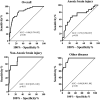
Results: We included 214 patients (mean age 60 years, 55% male). EEG tracings were categorized as: mild (n = 111, 52%), moderate (n = 65, 30%) or severe (n = 16, 8%) encephalopathy; burst-suppression (n = 19, 9%) or suppression background (n = 3, 1%); a total of 38 (18%) EEG were classified as "unreactive". We found a significant difference in all pupillometry variables among different EEG categories. Moreover, an unreactive EEG was associated with lower NPi, pupil size, pupillary reactivity, CV and DV and a higher latency than reactive recordings. Low DV (Odds ratio 0.020 [95% confidence intervals 0.002-0.163]; p < 0.01) was independently associated with an unreactive EEG, together with the use of analgesic/sedative drugs and high lactate concentrations. In particular, DV values had an area under the curve (AUC) of 0.86 [0.79-0.92; p < 0.01] to predict the presence of unreactive EEG. In subgroups analyses, AUC of DV to predict unreactive EEG was lower (0.72 [0.56-0.87]; p < 0.01) in anoxic brain injury than Non-anoxic brain injury (0.92 [0.85-1.00]; p < 0.01) and other diseases (0.96 [0.90-1.00]; p < 0.01).
Conclusions: This study suggests that low DV measured by the AP might effectively identify an unreactive EEG background, in particular in critically ill patients without anoxic brain injury.
Keywords: EEG; autonomic dysfunction; brain injury; encephalopathy; intensive care unit; pupillary function.
Copyright © 2022 Peluso, Ferlini, Talamonti, Ndieugnou Djangang, Gouvea Bogossian, Menozzi, Annoni, Macchini, Legros, Severgnini, Creteur, Oddo, Vincent, Gaspard and Taccone.
Conflict of interest statement
FT and MO are scientific advisors for NeurOptics Inc. The remaining authors declare that the research was conducted in the absence of any commercial or financial relationships that could be construed as a potential conflict of interest.
Figures


References
LinkOut - more resources
Full Text Sources

