Review
. 2022 Mar 31;3(2):e127.
doi: 10.1002/mco2.127. eCollection 2022 Jun.

Redox signaling at the crossroads of human health and disease

Affiliations
Review

Redox signaling at the crossroads of human health and disease

Jing Zuo et al. MedComm (2020). .

Abstract

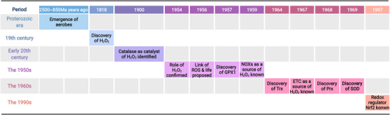
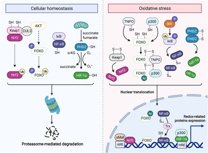
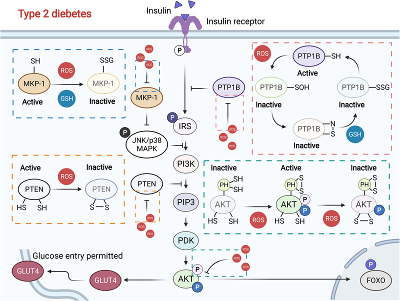
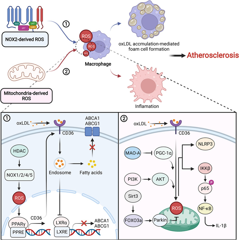
Redox biology is at the core of life sciences, accompanied by the close correlation of redox processes with biological activities. Redox homeostasis is a prerequisite for human health, in which the physiological levels of nonradical reactive oxygen species (ROS) function as the primary second messengers to modulate physiological redox signaling by orchestrating multiple redox sensors. However, excessive ROS accumulation, termed oxidative stress (OS), leads to biomolecule damage and subsequent occurrence of various diseases such as type 2 diabetes, atherosclerosis, and cancer. Herein, starting with the evolution of redox biology, we reveal the roles of ROS as multifaceted physiological modulators to mediate redox signaling and sustain redox homeostasis. In addition, we also emphasize the detailed OS mechanisms involved in the initiation and development of several important diseases. ROS as a double-edged sword in disease progression suggest two different therapeutic strategies to treat redox-relevant diseases, in which targeting ROS sources and redox-related effectors to manipulate redox homeostasis will largely promote precision medicine. Therefore, a comprehensive understanding of the redox signaling networks under physiological and pathological conditions will facilitate the development of redox medicine and benefit patients with redox-relevant diseases.

Keywords: hydrogen peroxide; oxidative stress; reactive oxygen species; redox signaling; redox therapy; redox‐relevant disease.


Conflict of interest statement

Canhua Huang is an editorial board member of MedComm. Author Canhua Huang was not involved in the journal's review of, or decisions related to, this manuscript. The other authors have no conflicts of interest to declare.

Figures

FIGURE 1
FIGURE 1
Key events in the history of redox biology
FIGURE 2
FIGURE 2
Redox regulation of Keap1‐Nrf2, FOXO, NF‐κB, and HIF. Keap1‐Nrf2: Keap1 binds to Nrf2 and CUL3, inducing the proteasomal degradation of Nrf2. Keap1 as a redox sensor is tightly regulated by H2O2, which helps to form an intramolecular disulfide and releases NRF2. FOXO: Under moderate levels of H2O2, FOXO engages proteasomal degradation through AKT signaling. However, pathological levels of H2O2 facilitate the formation of an intermolecular disulfide between TNPO and FOXO, promoting the nuclear translocation of FOXO. Intriguingly, FOXO can also form an intermolecular disulfide with acetyltransferase p300, enhancing the activity of FOXO. NF‐κB: Normally, NF‐κB remains inactive in the cytoplasm by interacting with IκB that can be phosphorylated and inhibited by IKK. Under OS, H2O2‐mediated IKK activation drives the proteasomal degradation of IκB and subsequent nuclear translocation of NF‐κB. Notably, NF‐κB is also oxidized by H2O2, which needs to be reversed by Trx in the nucleus. HIF: The redox sensor PHD2 inactivates HIF‐1α via hydroxylation, which can be prevented by forming intermolecular disulfide between PHD2 under H2O2 regulation. As a result, HIF‐1α enters the nucleus and prompts the transcription of target genes
FIGURE 3
FIGURE 3
Redox regulation of the insulin signaling pathway. Normally, moderate ROS levels inactivate PTPB1 and PTEN and activate AKT, prompting glucose uptake through GLUT4. However, excessive ROS accumulation leads to glutathionylation modification and proteasomal degradation of MKP‐1, which prevents insulin signaling. Simultaneously, ROS burden also inactivates AKT, inhibiting GLUT4 and subsequent glucose uptake, which eventually causes type 2 diabetes
FIGURE 4
FIGURE 4
The roles of NOX and mitochondria‐derived ROS in atherosclerosis. NOX‐derived ROS promote CD36 expression, which facilitates the transportation of oxLDL and inhibition of ABCA1 and ABCG1 transcription. Under these conditions, fatty acids accumulate in the cytoplasm that engenders macrophage transforming into foam cells. On the other hand, mitochondria‐derived ROS elicit the production of NLRP3 and IL‐1β through multiple signaling pathways, accounting for an inflammatory phenotype in atherosclerosis pathogenesis. Both of them are important inducers of atherosclerosis
FIGURE 5
FIGURE 5
Exogenous and endogenous factor‐induced ROS mediate COPD pathogenesis. Exogenous sources (i.e., air pollution, biomass smoke, and CS) and endogenous sources (i.e., mitochondria and NOX) are important inducers of COPD, which accelerate various ROS accumulation in the lung through multiple pathways including TGF‐β signaling, Fenton reaction and mitochondrial dysfunction, stimulating cell senescence and death, autophagy defects, inflammation and airway, and vascular remodeling
FIGURE 6
FIGURE 6
ROS accelerates Aβ accumulation and p‐tau formation in AD. Phosphorylated tau protein (p‐tau) and Aβ are essential risk factors in AD, which originate from the downregulated Nrf2 and increased ROS burden. Dysregulated Nrf2 is controlled by different proteins, promoting ROS overproduction and BACE1 expression which elicits Aβ accumulation. Aβ accumulation further causes ROS generation and activates RACN1, phosphorylating tau protein through calcineurin and GSK‐3β
FIGURE 7
FIGURE 7
ROS promote the EMT of epithelial cells and survival of CTCs during cancer metastasis. During cancer metastasis, extracellular and intracellular ROS activate TGF‐β signaling and trigger an EMT phenotype through canonical and noncanonical pathways. CTCs entering the blood stream also face ROS stress, which protects them from apoptosis and immune surveillance
FIGURE 8
FIGURE 8
The positive feedback loop of the aging‐ROS‐mitochondrial dysfunction‐ROS axis. Aging generally accompanies ROS overproduction, which contributes to mitochondrial dysfunction via different mechanisms. Notably, mitochondrial dysfunction further induces ROS generation and accelerates aging, suggesting that scavenging ROS could efficiently attenuate aging

References

    1. Herrmann JM, Dick TP. Redox biology on the rise. Biol Chem. 2012;393(9):999‐1004. - PubMed
    1. Sies H. Hydrogen peroxide as a central redox signaling molecule in physiological oxidative stress: oxidative eustress. Redox Biol. 2017;11:613‐619. - PMC - PubMed
    1. Mantzaris MD, Bellou S, Skiada V, Kitsati N, Fotsis T, Galaris D. Intracellular labile iron determines H2O2‐induced apoptotic signaling via sustained activation of ASK1/JNK‐p38 axis. Free Radic Biol Med. 2016;97:454‐465. - PubMed
    1. Lloyd D, Aon MA, Cortassa S. Why homeodynamics, not homeostasis? Scientific World J. 2001;1:133‐145. - PMC - PubMed
    1. Santolini J, Wootton SA, Jackson AA, Feelisch M. The redox architecture of physiological function. Curr Opin Physiol. 2019;9:34‐47. - PMC - PubMed