. 2021 Oct 4:2:754501.
doi: 10.3389/falgy.2021.754501. eCollection 2021.

Potent Anti-Inflammatory Effects of Tetracyclines on Human Eosinophils

Affiliations

Potent Anti-Inflammatory Effects of Tetracyclines on Human Eosinophils

Manuela Gehring et al. Front Allergy. .

Abstract

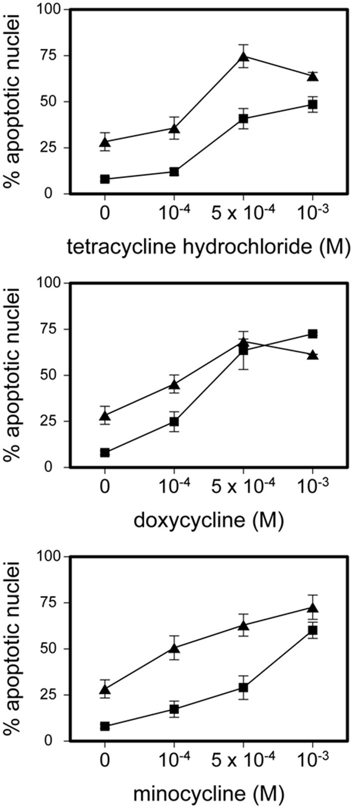
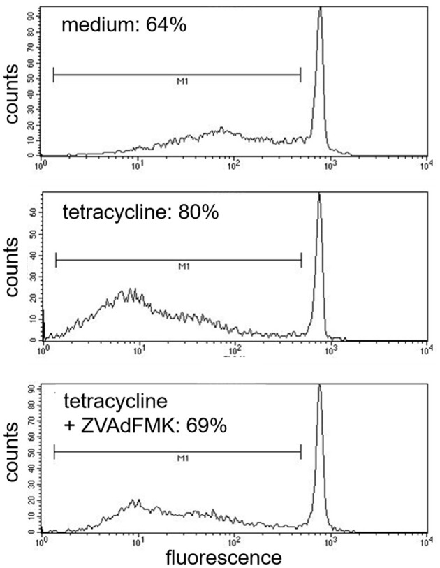
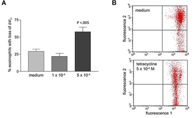
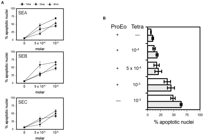
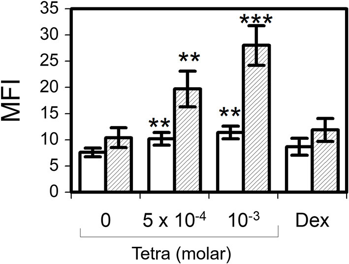
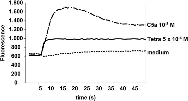
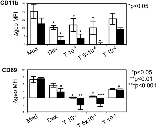
Eosinophils are potent pro-inflammatory cells. Not only in allergic diseases but also in other diseases there is a need for treatment strategies to induce resolution of eosinophil-mediated inflammation. During the last years beneficial non-antibiotic activities of tetracyclines (TCNs) have been shown in different diseases in which eosinophils play a role, for example, asthma and bullous pemphigoid. The working mechanism of these effects remains to be clarified. Aim of the present study was to investigate the effects of TCNs on eosinophils. Flow cytometry analysis of apoptosis, mitochondrial membrane potential, activation of caspases, intracellular H2O2 and calcium, surface expression of eosinophil activation markers was performed in highly purified peripheral blood eosinophils of non-atopic donors. Tetracycline hydrochloride, minocycline and doxycycline significantly induced eosinophil apoptosis. All TCNs were able to significantly overcome the strong survival enhancing effects of pro-eosinophilic cytokines and staphylococcus aureus enterotoxins. Tetracycline hydrochloride induced eosinophil apoptosis was accompanied by intracellular production of hydrogen peroxide, loss of mitochondrial membrane potential and activation of caspases. Moreover, tetracycline hydrochloride significantly down regulated eosinophil surface expression of CD9 and CD45, and of the activation markers CD11b and CD69, but not of CD54, CD63, or CD95. Our data, propably for the first time, point to a potent anti-inflammatory role of TCNs on eosinophils.

Keywords: Staphylococcus enterotoxin; allergic inflammation; apoptosis; eosinophils; tetracycline.


Conflict of interest statement

The authors declare that the research was conducted in the absence of any commercial or financial relationships that could be construed as a potential conflict of interest.

Figures

Figure 1
Figure 1
Tetracycline hydrochloride, minocycline and doxycycline induced eosinophil apoptosis. Stimulation of eosinophils with indicated TCNs at 10−4 M up to 10−3 M for 24 h (■) and 48 h (▲) (p < 0.05 up to p < 0.001 vs. medium control = 0). Mean percentage of apoptotic nuclei ± SEM (n = 9). Representative histogram analysis.
Figure 2
Figure 2
Inhibition of tetracycline-induced apoptosis by pan-capase inhibitor (zVAD-fmk). Cells co-incubated for 48 h with pan-caspase inhibitor (zVAD-fmk) and tetracycline (5 × 10⁻⁴ M) showed a similar amount of apoptotic nuclei (cells under the bar M1) like medium control (69%). One representative experiment out of five is shown.
Figure 3
Figure 3
Loss of mitochondrial membrane potential (ΔΨm) after 48 h stimulation with tetracycline hydrochloride. (A) Percentage of eosinophils with loss of ΔΨm cells is significantly increased after 48 h stimulation with 5 × 10⁻⁴ M tetracycline hydrochloride compared to medium (p < 0.005, n = 5). (B) Representative dot plot (five independent experiments) of red fluorescence 2 vs. green fluorescence 1. Compared to medium stimulation with tetracycline hydrochloride 5 × 10⁻⁴ resulted in a significant loss of ΔΨm. Live cells with intact ΔΨm are in the upper right quadrant, apoptotic cells with decreased fluorescence 2 are in the lower right quadrant (loss of ΔΨm).
Figure 4
Figure 4
TCNs overcame the survival prolonging effects of SEA, SEB, SEC, and of key pro-survival eosinophil cytokines, i.e., a mixture of IL-3, IL-5, and GM-CSF (ProEo). (A) Co-incubation of 5 × 10⁻⁴ M or 10−3 M tetracycline hydrochloride (Tetra), doxycycline (Doxy), or minocycline (Mino) wit SEA, SEB, and SEC (5 μg/ml each; p < 0.05 up to p < 0.001 vs. medium control = 0). Data are presented as percentage of apoptotic nuclei ± SEM (n = 6). (B) Tetra partly abrogated the effect of ProEo. Mean percentage of apoptotic nuclei ± SEM (n = 5) after stimulation for 24 h (unfilled bars) and 48 h (striped bars). P < 0.05 up to p < 0.001.
Figure 5
Figure 5
Tetracycline hydrochloride induced intracellular hydrogen peroxide production. In contrast to dexamethasone 10−6 M (Dex) tetracycline hydrochloride (Tetra) induced apoptosis was accompanied by a significant intracellular production of H2O2 after 2 h (unfilled bars) and 24 h (striped bars). Dihydrorhodamine assay, mean fluorescence intensity (MFI) ± SEM (n = 7). **p < 0.01, ***p < 0.001.
Figure 6
Figure 6
Tetracycline hydrochloride induced calcium influx. Tetracycline hydrochloride 5 × 10⁻⁴ M and C5a (10−8 M, positive control) but not medium induced a significant calcium influx in eosinophils at a very early stage. Cellular fluorescence was recorded per second for 50 s. Fluo4-AM assay.
Figure 7
Figure 7
Tetracycline hydrochloride downregulated eosinophil activation markers. Tetracycline hydrochloride (T) at indicated concentrations (in molar) downregulated CD11b and CD69 after 24 h (unfilled bars) and 48 h (filled bars) stimulation. P as indicated vs. control medium = Med). Dexamethasone (10−6 M, Dex) was less effective. Δ geometric mean fluorescence intensity (Δgeo MFI) ± SEM (n = 4).

References

    1. Hogan SP, Rosenberg HF, Moqbel R, Phipps S, Foster PS, Lacy P, et al. Eosinophils: biological properties and role in health and disease. Clin Exp Allergy. (2008) 38:709–50. 10.1111/j.1365-2222.2008.02958.x - DOI - PubMed
    1. Huang L, Appleton JA. Eosinophils in helminth infection: defenders and dupes. Trends Parasitol. (2016) 32:798–807. 10.1016/j.pt.2016.05.004 - DOI - PMC - PubMed
    1. Kapp A, Czech W, Krutmann J, Sch pf E. Eosinophil cationic protein in sera of patients with atopic dermatitis. J Am Acad Dermatol. (1991) 24:555–8. 10.1016/0190-9622(91)70081-C - DOI - PubMed
    1. Wedi B, Raap U, Lewrick H, Kapp A. Delayed eosinophil programmed cell death in vitro: a common feature of inhalant allergy and extrinsic and intrinsic atopic dermatitis. J Allergy Clin Immunol. (1997) 100:536–43. 10.1016/S0091-6749(97)70147-7 - DOI - PubMed
    1. Vignola AM, Chanez P, Chiappara G, Siena L, Merendino A, Reina C, et al. Evaluation of apoptosis of eosinophils, macrophages, and T lymphocytes in mucosal biopsy specimens of patients with asthma and chronic bronchitis. J Allergy Clin Immunol. (1999) 103:563–73. 10.1016/S0091-6749(99)70225-3 - DOI - PubMed