. 2022 Mar 21:13:863133.
doi: 10.3389/fmicb.2022.863133. eCollection 2022.

Novel Secreted Peptides From Rhizopus arrhizus var. delemar With Immunomodulatory Effects That Enhance Fungal Pathogenesis

Affiliations

Novel Secreted Peptides From Rhizopus arrhizus var. delemar With Immunomodulatory Effects That Enhance Fungal Pathogenesis

Sameh S M Soliman et al. Front Microbiol. .

Abstract

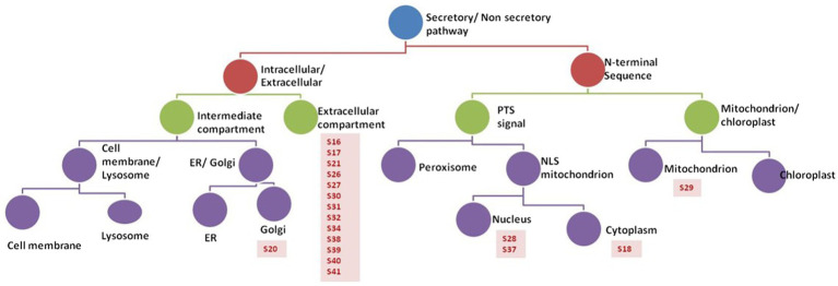
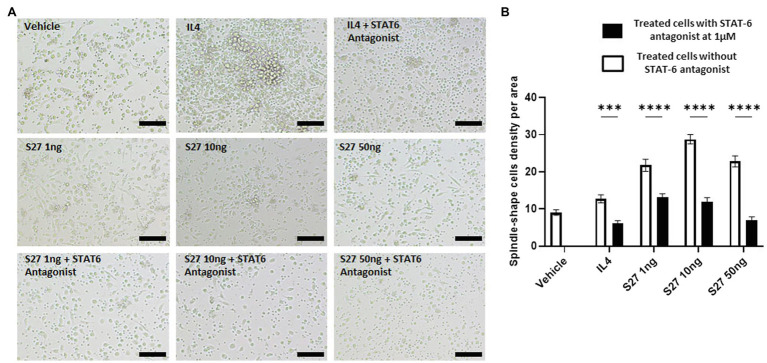
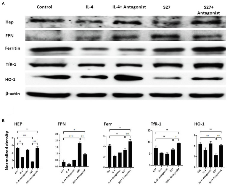
Secreted fungal peptides are known to influence the interactions between the pathogen and host innate immunity. The aim of this study is to screen and evaluate secreted peptides from the fungus Rhizopus arrhizus var. delemar for their immunomodulatory activity. By using mass spectrometry and immuno-informatics analysis, we identified three secreted peptides CesT (S16), Colicin (S17), and Ca2+/calmodulin-dependent protein kinase/ligand (CAMK/CAMKL; S27). Culturing peripheral blood-derived monocytic macrophages (PBMMs) in the presence of S16 or S17 caused cell clumping, while culturing them with S27 resulted in the formation of spindle-shaped cells. S27-treated PBMMs showed cell cycle arrest at G0 phase and exhibited alternatively activated macrophage phenotype with pronounced reduction in scavenger receptors CD163 and CD206. Homology prediction indicated that IL-4/IL-13 is the immunomodulatory target of S27. Confirming this prediction, S27 initiated macrophage activation through phosphorylation of STAT-6; STAT-6 inhibition reversed the activity of S27 and reduced the formation of spindle-shaped PBMMs. Lastly, S27 treatment of PBMMs was associated with altered expression of key iron regulatory genes including hepcidin, ferroportin, transferrin receptor 1, and ferritin in a pattern consistent with increased cellular iron release; a condition known to enhance Rhizopus infection. Collectively, R. arrhizus var. delemar secretes peptides with immunomodulatory activities that support fungal pathogenesis. Targeting the IL-4/IL-13R/STAT-6 axis is a potential therapeutic approach to enhance the PBMM-mediated fungal phagocytosis. This represents a potential new approach to overcome lethal mucormycosis.

Keywords: Rhizopus arrhizus var. delemar; immunomodulation; iron metabolism; macrophages; secreted peptides.


Conflict of interest statement

AI owns shares in Vitalex Biosciences, a start-up company that is developing immunotherapies and diagnostics for mucormycosis. The remaining authors declare that the research was conducted in the absence of any commercial or financial relationships that could be construed as a potential conflict of interest.

Figures

Figure 1
Figure 1
R. arrhizus var. delemar secretome analysis in relation to initiation of infection. SDS gel electrophoresis of culture supernatants collected from mammalian cell cultures and co-cultures of R. arrhizus var. delemar and lung epithelial cells.
Figure 2
Figure 2
Model representing R. arrhizus var. delemar secretome analysis in response to growth at two different environmental conditions (initiation- and dissemination-like conditions).
Figure 3
Figure 3
Activity of R. arrhizus var. delemar cultures supernatants. Mammalian cell damage, pH values and anti-E. coli activities of concentrated Rhizopus PD (dissemination-like) versus F12K (initiation-like) cultures. All values are relative to control culture media without the fungus.
Figure 4
Figure 4
Analysis of peptides secreted by R. arrhizus var. delemar. Prediction analysis of the sub-cellular localization of the R. arrhizus var. delemar small peptides was carried out using DeepLoc1.0 (Almagro Armenteros et al., 2017; http://www.cbs.dtu.dk/services/DeepLoc/instructions.php).
Figure 5
Figure 5
PBMM response to S16, S17, and S27 peptides. (A) Changes in the morphology of macrophages in response to peptides. Scale bar = 100 um. (B) XTT proliferation assay. The results were analyzed using one-way ANOVA and presented as mean ± SEM. * p < 0.05, considered as significantly different from other groups. (C) Flow cytometry histogram indicating the shift in cells population from G1 phase to sub G0 phase (dashed circle marked with arrow). (D) Cell cycle analysis of macrophages incubated with the peptides. The results were analyzed using two-way ANOVA and presented as means ± SEM of three independent experiments. * p < 0.05, considered as significantly different from other groups.
Figure 6
Figure 6
Flow cytometry analysis of PBMM treated with LPS and IL-4 positive controls, and S27 at 1, 10, and 50 ng compared to untreated control. Phenotypic profiling of macrophages following treatment with secreted peptides was examined by assessing the differential expression of key macrophage-related markers including CD14, CD16, CD86, CD163, and CD206. The results were analyzed using one-way ANOVA and presented as mean ± SEM. * p < 0.05, ** p < 0.01, *** p < 0.001, and **** p < 0.0001, which is significantly different from another group.
Figure 7
Figure 7
Homology studies between S16, S17, S27, and IL-13 and IL-4. (A) Sequence alignment of tested proteins using ClustalOmega and MuscleWS. The colors represent the level of conservation between protein sequences. The blue colored residues are conserved between all proteins (high conservation), while the green and pink are more conserved between S27 and IL-4/IL-13, and the yellow residues are more conserved between S27 and IL-4 (low conservation). Consensus sequence is the most frequent amino acid residues in the alignment, while the occupancy represents the level of the primary consensus binding site motif. (B) Neighbor-joining tree using BLOSUM62. (C) Pairwise alignment to calculate the identity percentage. Jalview 2.11.1.4 software was used for visualization.
Figure 8
Figure 8
Western blot analysis of the expression of STAT-6 and pSTAT-6 in PBMMs treated with IL-4 or S27. Cell lysates were prepared from PBMMs treated with IL-4 (15 ng/ml) or S27 (100 ng/ml) for 15 min, 1 h and 4 h. Cell lysates of untreated PBMMs or PBMMs treated with LPS (100 ng/ml) for 15 min served as negative controls. B-actin was used as a loading control. Data shown is representative of 3 replicates.
Figure 9
Figure 9
Morphological changes of macrophages in response to different treatments. (A) microscopy-based detection of treated macrophages. Cells were treated with different concentrations of S27 (1, 10, and 50 ng/ml), S27 mixed with STAT-6 antagonist. Untreated cells and IL-4 were used as negative and positive controls, respectively. Scale bar = 50 um, magnification 10×, Olympus microscope. (B) Counts of macrophages with spindle shape, represented as count per area for more than three fields. White bars representing vehicle, S27 1, 10 and 50 ng. Black bars represent same treatments with the presence of STAT-6 antagonist. The results were analyzed using two-way ANOVA and presented as means ± SEM of three independent experiments. *** p < 0.001 and **** p < 0.0001, which is significantly different from the other group.
Figure 10
Figure 10
Western blot analysis of the expression of Hep, FPN, Ferritin, TfR-1, and HO-1 in PBMMs cells treated with S27 alone or in the presence of STAT-6 antagonist. (A) Cell lysates were prepared from PBMMs treated with IL-4 at 15 ng/ml and in addition to STAT-6 antagonist at 1 μM and S27 at 10 ng/ml/STAT-6 antagonist at 1 μM. As they were cultured for 8 days. The results shown are representative of three experiments. (B) The band intensity was measured by densitometry using the Image J software. The results were analyzed using one-way ANOVA and presented as means ± SEM of three independent experiments. *p < 0.05, **p < 0.01 and ***p < 0.001, which is significantly different from other groups.
Figure 11
Figure 11
The mechanism by which S27 peptide caused anti-inflammatory modulation to PBMMs. Upon dissemination, secreted S27 interact with IL-4/IL-13R complex causing and early phosphorylation of STAT6. Macrophages will polarize into M2-like phenotype and modulation in iron handling proteins that favors the release of iron into the milieu of mucormycosis that support further persistence and dissemination.

References

    1. Agrawal G. K., Jwa N.-S., Lebrun M.-H., Job D., Rakwal R. (2010). Plant secretome: unlocking secrets of the secreted proteins. Proteomics 10, 799–827. doi: 10.1002/pmic.200900514, PMID: - DOI - PubMed
    1. Alexander J., Limaye A. P., Ko C. W., Bronner M. P., Kowdley K. V. J. L. T. (2006). Association of hepatic iron overload with invasive fungal infection in liver transplant recipients. Liver Transpl. 12, 1799–1804. doi: 10.1002/lt.20827, PMID: - DOI - PubMed
    1. Allavena P., Chieppa M., Monti P., Piemonti L. (2004). From pattern recognition receptor to regulator of homeostasis: the double-faced macrophage mannose receptor. Crit. Rev. Immunol. 24, 179–192. doi: 10.1615/CritRevImmunol.v24.i3.20, PMID: - DOI - PubMed
    1. Almagro Armenteros J. J., Sønderby C. K., Sønderby S. K., Nielsen H., Winther O. (2017). DeepLoc: prediction of protein subcellular localization using deep learning. Bioinformatics 33, 3387–3395. doi: 10.1093/bioinformatics/btx431, PMID: - DOI - PubMed
    1. Andrianaki A. M., Kyrmizi I., Thanopoulou K., Baldin C., Drakos E., Soliman S. S. M., et al. (2018). Iron restriction inside macrophages regulates pulmonary host defense against Rhizopus species. Nat. Commun. 9:3333. doi: 10.1038/s41467-018-05820-2, PMID: - DOI - PMC - PubMed